

Abstract
Background:
Anomalies in primary dentition are often found to be associated with anomalies in permanent dentition.
Aims:
This study was performed to evaluate the prevalence of supernumerary teeth, hypodontia, double teeth, and talon cusp in the primary dentition, and their effect on succedaneous permanent teeth.
Materials and Methods:
In this prospective cross-sectional study, we clinically investigated 2757 Bengali speaking nursery children (1474 girls and 1283 boys), of age four to six years, at their respective schools, and the presence of supernumerary teeth, hypodontia, double teeth, and talon cusp in the primary dentition were recorded. Children with anomalous primary teeth were further subjected to periapical and panoramic radiographic examination, to determine the status of the underlying permanent teeth.
Results:
The total prevalence of all anomalies in this study was 1.8%. A total of 38 children (21 girls and 17 boys) had anomalies. The prevalence of anomalies was as follows: Supernumerary teeth (0.4%), hypodontia (0.5%), double teeth (0.4%), and talon cusp (0.07%), in both sexes combined. Radiographic examination showed 50% of the patients (19 children) had anomalies in the permanent dentition.
Conclusions:
Anomalies in primary dentition exhibited a correlation with anomalies in permanent dentition.
Keywords: Correlation, dental anomaly, primary dentition, permanent dentition
INTRODUCTION
Anomalies in the number and shape of teeth can occur in both primary and permanent dentitions. Their incidence and degree of expression can provide important information for genetic and phylogenetic studies.[1] The exact etiology of dental anomalies is not clearly known. Both genetic and environmental factors are responsible for its development. The anomalous teeth are often asymptomatic, and may be discovered during clinical and radiographic examination of the oral cavity.
Numerical anomalies include supernumerary teeth or hyperdontia, and, hypodontia or congenitally missing teeth.[2] A supernumerary tooth is one that is present in addition to the normal number of teeth.[3,4] Hypodontia describes the absence of a tooth both clinically and radiographically.[5,6] Double teeth and talon cusps are morphological variations of teeth. Double teeth represent the union of two normally separated tooth germs, or an abortive attempt of a single tooth germ to divide.[6,7,8] Talon cusps are accessory cusps projecting from the cementoenamel junction to the incisal edge of an anterior tooth.[9,10,11] Its frequency and degree of expression also vary among populations.
Complications associated with anomalous primary teeth result in an unsightly appearance of the affected teeth, increased susceptibility to caries, and malocclusion. More significantly, anomalies in the primary dentition exhibit anomalies in the permanent dentition.[10,11,12,13,14,15,16] Timely intervention would minimize complications in the permanent dentition.
Variations in the distribution and location of congenital dental anomalies have been reported across various ethnic groups.[1,2,6,7,12,13,14,15,16,17,18] To date, no study on anomalous primary teeth in the Bengali population has been performed.
The present study aims to document the prevalence of supernumerary teeth, hypodontia, double teeth, and talon cusp in the primary dentition, and their effect on the succedaneous permanent teeth, using periapical and panoramic radiographs.
MATERIALS AND METHODS
A descriptive cross-sectional study was performed on healthy Bengali subjects after obtaining permission from the Institutional Ethical Committee. We also obtained a written informed consent from the parents of the participants prior to the start of the investigation.
Inclusion criteria
Healthy Bengali children with primary dentition, having no history of tooth loss due to trauma or extraction, were enrolled in this study.
Exclusion criteria
Children with systemic diseases or syndromic backgrounds were not included in this study.
Data collection
We investigated a total of 2757 Bengali nursery children of age four to six years, comprising 1283 boys (46.5%) and 1474 girls (53.5%), for the present study. Children enrolled in this survey were selected using a two-stage random selection technique. At the outset, we had picked up 10 nursery schools from two districts of West Bengal by using a random sampling technique. Again children were selected by simple random sampling from the selected schools. One investigator examined the children at their respective schools, under portable light, using a dental mirror and explorer. The entire maxillary and mandibular arches were examined. Dental anomalies were determined by visual inspection of the dental arches. Four anomalies, supernumerary teeth, hypodontia or congenitally missing teeth, double teeth, and talon cusps were recorded. In this study, we determined number anomalies (supernumerary teeth and hypodontia) by counting the number of teeth present in the dental arch. Morphological anomalies (double teeth and talon cusps) were determined by their characteristic appearance. All children diagnosed as having anomalies in the primary dentition had a radiographic examination. Intraoral periapical radiographs of anomalous teeth and panoramic radiographs were taken. The patients’ records and radiographs were examined to investigate gender distribution, number, and location of the anomalies in the dental arch, as also the radiographic evidence of anomalies in the underlying permanent dentition.
Statistical analysis
Statistical analysis was performed using Epi Info software version 7. The Fisher exact test was used to compare the occurrence of dental anomalies between boys and girls. A P < 0.05 was considered statistically significant. This study has been designed and reported in accordance with the STROBE (Strengthening the Reporting of Observational Studies in Epidemiology) statement (http://www.strobe-statement.org).
RESULTS
Of the 2757 children examined, 38 children had anomalies in the primary dentition. Among them 21 were girls (55.3%) and 17 were boys (44.7%). In our study, hypodontia was the most common dental anomaly (n = 14, 36.8%), followed by supernumerary teeth and double teeth (n = 11, 28.9% each), and talon cusp (n = 2, 5.3%). The total prevalence of all anomalies in both the sexes combined was 1.8%.
Supernumerary teeth
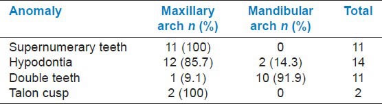
A total of 11 children had supernumerary teeth, with a prevalence of 0.4% [Table 1]. Most of the anomalies had a unilateral distribution (90.9%), and only 9.1% (n = 1) were present bilaterally [Table 2]. Among the 11 children with supernumeraries, eight patients (72.7%) had mesiodens, two children (18.2%) had supernumerary maxillary lateral incisors, and only one patient (9.1%) had a supernumerary maxillary canine. All primary supernumeraries were present in the maxillary region [Table 3].
Table 1.
Prevalence of anomalies in the primary dentition
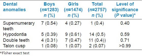
Table 2.
Distribution of anomalies by side
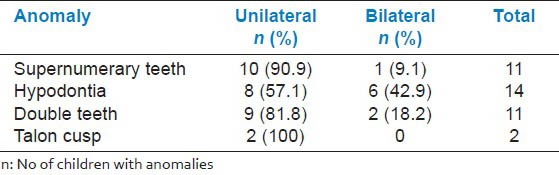
Table 3.
Arch-wise distribution of anomalies
Hypodontia
In our study, the girls showed a higher prevalence of hypodontia (0.61%) than boys (0.39%), although this difference was not statistically significant [Table 1]. Most of the hypodontia cases exhibited unilateral distribution (57.1%) [Table 2]. On the whole, in 14 subjects with congenitally missing teeth, 12 cases (85.7%) showed maxillary hypodontia [Table 3]. The maximum number of missing teeth in this study was two. In the maxillary arch, all the missing primary teeth were lateral incisors (n = 12, 85.7%). Among the two cases of mandibular hypodontia, one patient had a missing central incisor (7.1%) and the other one (7.1%) had a lateral incisor missing.
Double teeth
Table 1 shows the prevalence of double teeth; 81.8% (n = 9) had a unilateral distribution and 91.9% were located in the mandibular arch [Tables 2 and 3]. Only 18.2% of the cases exhibited a bilateral distribution involving the mandibular lateral incisors and canines. Seven double teeth (63.6%) involved mandibular lateral incisors and canines, three double teeth (27.3%) affected the mandibular central and lateral incisors, and only one (9.1%) involved the maxillary central and lateral incisors.
Talon cusp
In this study, only two subjects (a boy, and a girl) displayed primary talon cusps, with a prevalence of 0.07% [Table 1]. In both cases, the maxillary central incisors were affected [Tables 2 and 3].
Correlation with the permanent dentition
In this study, two subjects (18.2%) with primary supernumerary teeth also had anomalies in the permanent dentition [Table 4]. The anomalies included permanent mesiodens and a talon cusp on the permanent maxillary lateral incisor [Figure 1].
Table 4.
Anomalous primary teeth and its correlation with permanent teeth
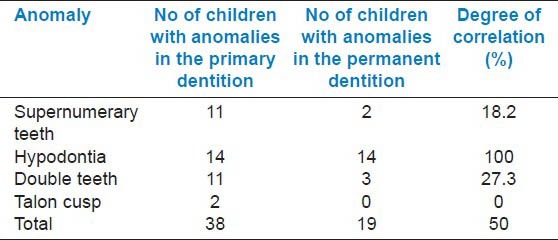
Figure 1.
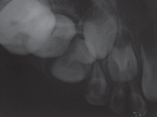
Supernumerary teeth in primary dentition associated with talon cusp on permanent lateral incisor
Patients with hypodontia in the primary dentition, in our study, displayed hypodontia in the permanent dentition in 100% (n = 14) of the cases.
Double teeth in the primary dentition exhibited anomalies in the permanent dentition in 27.3% (n = 3) of the cases. The anomalies in the permanent dentition were hypodontia in two cases (18.2%) and double teeth in one case (9.1%).
Talon cusp in the primary dentition, in this study, did not show any anomaly in the underlying permanent dentition [Table 4].
DISCUSSION
Worldwide, many studies have been published on the prevalence of anomalies in permanent dentition. Relatively few surveys have been reported on anomalous primary teeth.[1,6,14,15,16,17,18,19] These studies were mostly on Caucasians and Mongoloid populations.[1,6,14,15,16,17,18] Most of the reported data on dental anomalies in Indian populations are case reports of supernumerary teeth, hypodontia, double teeth, and talon cusps.
Supernumerary teeth
Supernumerary teeth in the primary dentition are rarer than in the permanent dentition.[20] Its prevalence in the primary dentition varies from 0.05 to 0.07% in the Japanese population,[17,18] 0.1% in Croats,[14] and 0.8% of each in British[6] and Belgian subjects.[19] In this study, Bengali subjects showed a 0.4% prevalence of primary supernumerary teeth. The primary supernumerary teeth have no significant sex distribution.[20,21] Although in the present study, boys displayed a higher prevalence of primary supernumeraries, the difference was not statistically significant.
In our study, mesiodens was the most common primary supernumerary tooth. This agrees with the results of Nik-Hussein et al.[13] But contrasts with the other studieswho reported that maxillary lateral incisors were the most frequently occurring supernumeraries in primary dentition.[14,18,21]
Hypodontia
The prevalence of hypodontia in the primary dentition ranges from 0.08 to 1.55% in various pediatric populations.[16,22] In our survey, Bengali children demonstrated a prevalence of 0.5%, which is lower than the results of Yonezu et al.,[17] who obtained 2.4% in Japanese children, but higher than the results of a Turkish study[23] (0.2%). In this investigation, children with hypodontia exhibited one or two teeth missing, and the maxillary lateral incisors were the most common missing teeth (85.7%). These results support the observations of Whitington et al.,[16] Arte and others,[5] and Ravn et al.,[24] but disagree with the findings of Yonezu and others,[17] who reported that mandibular lateral incisors were the most frequently missing teeth.
Double teeth
Double teeth describe both germination and fusion.[7] These anomalies are more common in the primary dentition than in the permanent dentition. The prevalence of primary double teeth varies from 4.1% in Japan,[17] 0.5% in Croatia,[14] in 0.4% in Belgium,[19] and 0.6% in Finland,[15] to 1.3% in Turkish children.[23] Tasa et al. have reported a prevalence of 1.5% in a group of children from western India.[25] In our investigation, Bengali subjects demonstrate a prevalence of 0.4%, with no significant gender distribution. Our results have shown that double teeth typically occur unilaterally (81.8%), are more common in the mandibular arch (91.9%), and all the cases involve anterior teeth. These observations support the findings of Yonezu et al.,[17] Cheng and others,[26] and Järvinen et al.,[15] but disagree with the reports of Aguiló and others,[27] who found statistically no significant difference between the maxilla and mandible.
Talon cusp
Talon cusps are uncommon dental anomalies affecting both primary and permanent dentitions. Its incidence is less common in the primary dentition, with three-fourth of all cases occurring in the permanent dentition.[10,11] To date, no prevalence study on primary talon cusp is available. The lack of precise criteria to classify an accessory cusp as a ‘talon’ has contributed to extensive variations in its prevalence.[28] Case reports on talon cusps in the primary dentition indicate that maxillary central incisors are more frequently affected than maxillary lateral incisors.[10,11] In the present study, talon cusps in the primary dentition have shown a prevalence of 0.07% (two patients) and the accessory cusps have occurred exclusively on the maxillary central incisor.
Correlation with permanent dentition
Anomalies in the primary dentition are positively correlated with anomalies in the permanent dentition.[1,10,12,13,14,15,16] Skrinjarić et al. reported that children with primary supernumerary teeth displayed anomalies in the primary dentition in 85.7% of the cases.[14] A Malaysian survey reported anomalies of the permanent dentition in 50% of the subjects with primary supernumerary teeth.[13] In our study, only 18.2% of the children (two patients) with primary supernumerary teeth had anomalies in the permanent dentition.
In the present study, all children (14 subjects) with hypodontia in the primary dentition demonstrated hypodontia in the permanent dentition. This was in agreement with the reports of Whittington and others,[16] Skrinjarić et al.,[14] and Nik- Hussein et al.,[13] who observed it in 100% of the cases.
Various investigators have reported a correlation between double teeth in the primary dentition and a numerical variation of teeth in the permanent dentition.[13,14,15,16] In this study, we observed it in 27.3% of the cases, which is lower than what Skrinjarić et al.[14] (61.1%) and Nik-Hussein and others[13] (59%) reported, but comparable to those obtained by Järvinen et al.[15] (37.5%).
In most of the cases, talon cusps on primary central incisors have no effect on the permanent successors. However, talon cusps on maxillary lateral incisors have a high proportion (78%) of underlying permanent successors displayed in dental anomalies.[10] In our study, two patients had talon cusps on the maxillary central incisors, without exhibiting odontogenic abnormalities in the permanent dentition.
The differences in prevalence of various anomalies among populations could be due to differences in sample size, ethnicity, and genetic differences of the subjects studied.
CONCLUSION
Anomalies in the primary dentition are important because of their effect on the underlying permanent dentition. Early identification of these anomalies and intervention at the appropriate time would minimize complicated treatments in future.
Footnotes
Source of Support: Nil
Conflict of Interest: None declared.
REFERENCES
- 1.Gellin ME. The distribution of anomalies of primary anterior teeth and their effect on the permanent successors. Dent Clin North Am. 1984;28:69–89. [PubMed] [Google Scholar]
- 2.Stecker SS, Beiraghi S, Hodges JS, Peterson VS, Myers SL. Prevalence of dental anomalies in a South East Asian population in the Minneapolis/Saint Paul metropolitan area. North West Dent. 2011;90:25–8. [PubMed] [Google Scholar]
- 3.Garvey MT, Barry HJ, Blake M. Supernumerary teeth: An overview of classification, diagnosis and management. J Can Dent Assoc. 1999;65:612–6. [PubMed] [Google Scholar]
- 4.Liu JF. Characteristics of premaxillary supernumerary teeth: A survey of 112 cases. ASDC J Dent Child. 1995;62:262–5. [PubMed] [Google Scholar]
- 5.Arte S, Pirinen S. Hypodontia. [Last accessed on 01.03.2013]. Available from: http://www.orphanet/data/patho/GB/UK-hypodontia.pdf .
- 6.Brook AH. Dental anomalies of number, form, and size: Their prevalence in British school children. J Int Assoc Dent Child. 1974;5:37–53. [PubMed] [Google Scholar]
- 7.Brook AH, Winter GB. A retrospective study of ‘geminated’ and ‘fused’ teeth in children. Br Dent J. 1970;129:123–30. doi: 10.1038/sj.bdj.4802533. [DOI] [PubMed] [Google Scholar]
- 8.McDonald RE, Avery DR. 5th ed. St Louis: CV Mosby Co; 1983. Fusion of teeth .In dentistry for child and adolescent; pp. 121–2. [Google Scholar]
- 9.Liu J, Chen L. Talon cusp affecting the primary maxillary central incisors in two sets of female twins: Report of two cases. Pediatr Dent. 1995;17:362–4. [PubMed] [Google Scholar]
- 10.Lee CK, King NM, Lo EC, Cho SY. The relationship between a primary maxillary incisor with a talon cusp and the permanent successors: A study of 57 cases. Int J Ped Dent. 2007;17:178–85. doi: 10.1111/j.1365-263X.2007.00823.x. [DOI] [PubMed] [Google Scholar]
- 11.Lee CK, King NM, Lo EC, Cho SY. Management of supplemental permanent maxillary lateral incisors in association with talon cusp on the primary predecessors: A report of 3 cases. J Dent Child. 2008;75:59–63. [PubMed] [Google Scholar]
- 12.Grahnen H, Granath LE. Numerical variations in primary in primary dentition and their correlation with the permanent dentition. Odontol Revy. 1961;12:348–57. [Google Scholar]
- 13.Nik-Hussein, Majid ZA. Dental anomalies in the primary dentition; distribution and correlation with the permanent dentition. J Clin Pediatr Dent. 1996;21:15–9. [PubMed] [Google Scholar]
- 14.Skrinjarić I, Barac-Furtinović V. Anomalies of deciduous teeth and findings in permanent dentition. Acta Stomatol Croat. 1991;25:151–6. [PubMed] [Google Scholar]
- 15.Järvinen S, Lehtinen L, Milén A. Epidemiologic study of joined primary teeth in Finnish children. Community Dent Oral Epidemiol. 1980;8:201–2. doi: 10.1111/j.1600-0528.1980.tb01286.x. [DOI] [PubMed] [Google Scholar]
- 16.Whittington BR, Durward CS. Survey of anomalies in primary teeth and their correlation with the permanent dentition. NZ Dent J. 1996;92:4–8. [PubMed] [Google Scholar]
- 17.Yonezu T, Hayashi Y, Sasaki J, Machida Y. Prevalence of congenital dental anomalies of the deciduous dentition in Japanese children. Bull Tokyo Dent Coll. 1997;38:27–32. [PubMed] [Google Scholar]
- 18.Miyoshi S, Tanaka S, Kunimatsu H, Murakami Y, Fukami Y, Fujisawa S. An epidemiological study of supernumerary primary teeth in Japanese children: A review of racial differences in the prevalence. Oral Dis. 2000;6:99–102. doi: 10.1111/j.1601-0825.2000.tb00108.x. [DOI] [PubMed] [Google Scholar]
- 19.Carvalho JC, Vinker F, Declerck D. Malocclusion, dental injuries, and dental anomalies in the primary dentition of Belgian children. Int J Paediatr Dent. 1998;8:137–41. doi: 10.1046/j.1365-263x.1998.00070.x. [DOI] [PubMed] [Google Scholar]
- 20.Mukhopadhyay S. Mesiodens: A clinical and radiographic study in children. J Indian Soc Pedod Prev Dent. 2011;29:34–8. doi: 10.4103/0970-4388.79928. [DOI] [PubMed] [Google Scholar]
- 21.Luten JR. The prevalence of supernumerary teeth in primary and mixed dentition. J Dent Child. 1967;34:346–53. [PubMed] [Google Scholar]
- 22.McNamara C, Foley T, McNamara CM. Multidisplinary management of hypodontia in adolescents: Case report. J Can Dent Assoc. 2006;72:740–6. [PubMed] [Google Scholar]
- 23.Kapdan A, Kustarci A, Buldur B, Arslan D, Kapdan A. Dental anomalies in the primary dentition of Turkish children. Eur J Dent. 2012;6:178–83. [PMC free article] [PubMed] [Google Scholar]
- 24.Ravn JJ. Aplasia, supernumerary teeth, and fused teeth in the primary dentition. Scand J Dent Res. 1971;79:1–6. doi: 10.1111/j.1600-0722.1971.tb01986.x. [DOI] [PubMed] [Google Scholar]
- 25.Tasa GL, Lukacs JR. The prevalence and expression of primary double teeth in Western India. J Dent Child. 2000;68:196–200. [PubMed] [Google Scholar]
- 26.Cheng R, Chen Xu, Liu S, Pan L, Wu X. An epidemiological survey on fusion of deciduous teeth of 4286 kindergarten children in Shenyang city. Shanghai Kou Qiang Yi Xue. 2003;12:424–6. [PubMed] [Google Scholar]
- 27.Aguuiló L, Gandia JL, Cibrian R, Catala M. Primary double teeth. A retrospective clinical study of their morphological characteristics and associated anomalies. Int J Pediatr Dent. 1999;9:175–83. doi: 10.1046/j.1365-263x.1999.00131.x. [DOI] [PubMed] [Google Scholar]
- 28.Hattab FN, Yassin OM, Al-Nimri KS. Talon cusps in permanent dentition associated with other dental anomalies: Review of literature and reports of seven cases. J Dent Child. 1996;63:368–76. [PubMed] [Google Scholar]