Abstract
We present a case of a young man with severe mucositis following an upper respiratory tract infection limited to the ophthalmic and oral mucosa while sparing the rest of the skin, genitalia and perianal regions. Investigations revealed that the mucositis was a rare extrapulmonary manifestation of Mycoplasma pneumoniae infection. He had progressive vision-threatening symptoms despite antibiotics and best supportive care and thus was treated with intravenous corticosteroids, immunoglobulins, temporary ocular amniotic membrane grafts and tarsorrhaphy. The patient made an almost complete recovery over 6 weeks.
Background
Mycoplasma pneumonia is a common cause of bacterial pneumonia in the community. However, mycoplasma pneumonia-associated mucositis (MPAM) is a rare extrapulmonary manifestation of Mycoplasma pneumoniae infection, involving the oral and ophthalmic mucosa. Unlike Mycoplasma-associated Steven Johnson's syndrome (MASJS), there is little to no cutaneous involvement in MPAM making it less morbid than MASJS. It is important for clinicians to differentiate between these two entities to direct appropriate management strategies.
Case presentation
A 20-year-old man was in a normal state of health until 10 days prior to admission when he developed a non-productive cough, nasal congestion, malaise and fever after exposure to his room-mate who recently recovered from an upper respiratory tract infection. An evolving conjunctivitis ensued (figure 1) which prompted treatment with oral azithromycin and topical gentamicin eye drops. The following day, he developed lip swelling, oral sores, worsening conjunctivitis and thick copious oral secretions (figure 2). He was admitted to a local hospital where a drug allergy was suspected, and azithromycin was stopped. A CT of the chest revealed patchy airspace infiltrates in the lingula and the right upper lobe. He was administered methylprednisolone, clindamycin, acyclovir and levofloxacin, and was transferred to our institution for further management.
Figure 1.
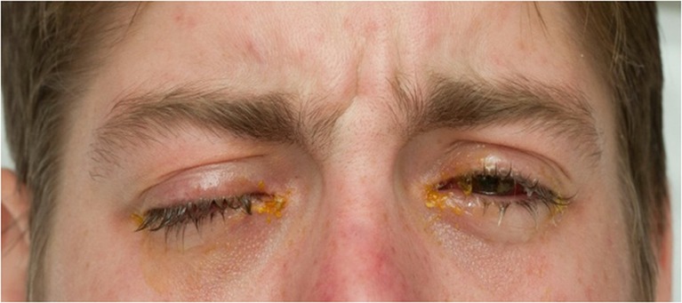
Initial presentation showing severe signs of conjunctivitis.
Figure 2.
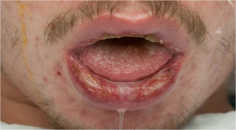
Initial presentation with lip swelling, oral sores, copious oral secretions and odynophagia.
On examination, the patient was afebrile with normal vital signs. He appeared uncomfortable due to difficulty in clearing secretions, and odynophagia. He reported irritation in his eyes with blurring of vision (figure 3). There was swelling, erythema and sloughing of his lips with superficial ulcerations of the buccal mucosa (figure 4). An eye examination demonstrated bilateral injected conjunctiva with significant corneal abrasions and symblepharon. Vision and extraocular motions were intact and fundus was normal. Genitalia, perianal regions and skin did not show desquamation or ulceration.
Figure 3.
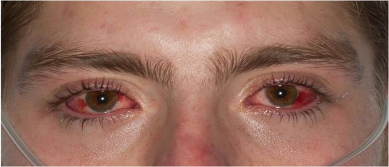
Day 16 since onset of symptoms. The figure showing bilateral injected conjunctiva with significant corneal abrasions. An eye examination demonstrated symblepharon.
Figure 4.
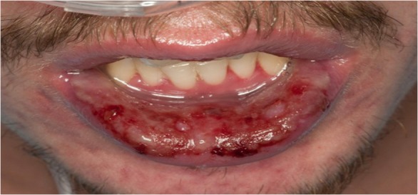
Day 16 since onset of symptoms. The figure illustrates swelling, erythema and sloughing of the patient's lips with superficial ulcerations of the buccal mucosa.
Investigations
Investigations revealed a leukocytosis to 17×109/L with a neutrophilic predominance. His erythrocyte sedimentation rate and C reactive protein were elevated at 41 mm/h and 80.7 mg/L, respectively. Screening for antinuclear antibodies, desmogleins 1 and 3, legionella and leptospira antibodies, HIV1 and 2, Epstein-Barr and adenovirus PCR were negative. Mycoplasma pneumoniae IgM and IgG were positive and confirmed by immunofluorescence antibody assay.
Treatment
Levofloxacin and clindamycin were continued. However, given his severe and non-resolving ocular lesions despite antibiotic therapy for several days, intravenous immunoglobulins (IVIG) at 2 g/kg divided over 3 days along with intravenous methylprednisolone 1 g/day for 5 days were initiated. To prevent permanent corneal damage from dryness, he received erythromycin eye ointments and carboxymethylcellulose eye drops. In addition, he underwent temporary bilateral amniotic membrane grafts and tarsorrhaphy to promote corneal healing and protection.
During his hospitalisation, he also required intravenous pain medications and peripheral parenteral nutrition due to severe odynophagia. He was slowly transitioned to an oral diet. He was placed on a 6-week steroid taper and completed a 10-day course of levofloxacin and clindamycin.
Outcome and follow-up
Within 6 weeks of admission, the patient made a remarkable recovery. The only symptoms remaining after 6 weeks were mild burning of his right eye and mild discomfort in the throat when eating spicy food (figures 5 and 6).
Figure 5.
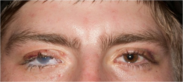
Day 27 since onset of symptoms. Almost complete resolution of eye symptoms except for mild burning of the patient's right eye.
Figure 6.
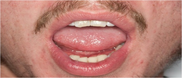
Day 27 since onset of symptoms. Complete resolution of mucositis and oral ulcerations.
Discussion
Mycoplasma pneumonia usually presents as a self-limited upper respiratory tract infection characterised by cough, pharyngitis, fever and malaise. Evolution into pneumonia occurs in about 3–10% of infected individuals.1 Various extrapulmonary manifestations of M pneumoniae infection have been described including dermatological, central nervous system, haematological, cardiac and rheumatological involvement.
Dermatological manifestations associated with M pneumoniae range from mild erythematous maculopapular/vesicular rash to frank Steven-Johnson's syndrome (SJS) reported in about 1–5% of M pneumoniae infections.2 However, M pneumoniae infection associated with ocular and/or oral mucositis with little to no skin involvement is rare. In the past, this isolated pathology without skin involvement was classified as atypical SJS; however, given that SJS requires skin involvement by definition, it is now described as MPAM.3 Unlike MASJS, MPAM carries a more favourable prognosis.3 The mechanism of MPAM is thought to occur through direct cytotoxic damage and through cross-reacting autoantibody formation.4 It is thought that these cross-reacting autoantibodies, originally aimed at the glycolipid antigens of M pneumoniae, attack the mucosal cells that share a similar epitope.4
Most cases of MPAM described in the literature occur in the paediatric population with a male predominance.5 The diagnosis of MPAM requires high clinical suspicion. Positive IgM and IgG M pneumoniae antibodies in a clinical setting of mucositis without dermal involvement should alert the clinician to include MPAM in the differential diagnosis.
The optimal treatment of MPAM is unknown. While there are cases in which patients have recovered with antibiotic therapy, others demonstrate the need for anti-inflammatory treatment.5 6 In fact, one series of 32 MPAM cases reported relapse in one-third of patients treated with macrolides alone.7 Our patient had progressive vision-threatening symptoms despite antibiotics and best supportive care; therefore, he was additionally treated with IVIG and corticosteroids. The role of IVIG and corticosteroids in patients presenting with MPAM will need to be further investigated.
Learning points.
Mycoplasma pneumoniae can cause a mucous membrane-limited disease with little to no skin involvement called M pneumoniae-associated mucositis (MPAM).
MPAM has a more favourable prognosis than M pneumoniae-associated Stevens Johnson syndrome (MASJS).
The treatment for MPAM includes antibiotics and supportive care.
The addition of IVIG and corticosteroids can be considered in severe cases of MPAM if there is concern for permanent ocular damage.
Footnotes
Contributors: CV, KS, JS and KR contributed to the collection of data, information, writing and editing of the manuscript.
Competing interests: None.
Patient consent: Obtained.
Provenance and peer review: Not commissioned; externally peer reviewed.
References
- 1.Mansel JK, Rosenow EC, III, Smith TF, et al. Mycoplasma pneumoniae pneumonia. Chest 1989;95:639–46 [DOI] [PubMed] [Google Scholar]
- 2.Schalock PC, Dinulos JG. Mycoplasma pneumoniae-induced cutaneous disease. Int J Dermatol 2009;48:673–80 [DOI] [PubMed] [Google Scholar]
- 3.Schalock PC, Dinulos JG. Mycoplasma pneumoniae-induced Stevens-Johnson syndrome without skin lesions: fact or fiction? J Am Acad Dermatol 2005;52:312–15 [DOI] [PubMed] [Google Scholar]
- 4.Waites KB, Talkington DF. Mycoplasma pneumonia and its role as a human pathogen. Clin Microbiol Rev 2004;17:697–728 [DOI] [PMC free article] [PubMed] [Google Scholar]
- 5.Bressan S, Mion T, Andreola B, et al. Severe Mycoplasma pneumonia-associated mucositis treated with immunoglobulins. Acta Paediatrica 2011; 100:e238–40 [DOI] [PubMed] [Google Scholar]
- 6.Trapp LW, Schrantz SJ, Joseph-Griffin MA, et al. A 13-year-old boy with pharyngitis, oral ulcers, and dehydration. Pediatr Ann 2013; 42:148–50 [DOI] [PubMed] [Google Scholar]
- 7.Sauteur PM, Goetschell P, Lautenschalager S. Mycoplasma pneumoniae and mucositis—part of the Stevens-Johnson syndrome spectrum. J Dtsch Dermatol Ges 2012;10:740–5 [DOI] [PubMed] [Google Scholar]


