Abstract
A 41-year-old Canadian man presented with a 1-week history of new bizarre behavior including agoraphobia. Nested PCR confirmed the presence of rabies in both nuchal snips and saliva. An initial MRI with gadolinium enhancement was normal. On day 8, repeat neuroimaging revealed new cranial nerve enhancement and slight hyperintensities of the caudate nuclei bilaterally (figure). This is consistent with cranial nerve involvement reported in animal studies1 and with a remote report of electron microscopic evidence of rabies virus in trigeminal ganglion cells reported in 2 human cases.2
A 41-year-old Canadian man presented with a 1-week history of new bizarre behavior including agoraphobia. Nested PCR confirmed the presence of rabies in both nuchal snips and saliva. An initial MRI with gadolinium enhancement was normal. On day 8, repeat neuroimaging revealed new cranial nerve enhancement and slight hyperintensities of the caudate nuclei bilaterally (figure). This is consistent with cranial nerve involvement reported in animal studies1 and with a remote report of electron microscopic evidence of rabies virus in trigeminal ganglion cells reported in 2 human cases.2
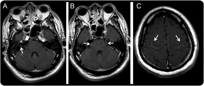
Figure. Cranial nerve enhancement in a patient with rabies encephalitis.

MRI of the brain. Axial contrast-enhanced T1-weighted images demonstrate enhancement of the right seventh and eighth cranial nerves in the internal auditory canal (arrow in A) and of the fifth cranial nerves bilaterally (arrows in B). The third and ninth cranial nerves on the left were enhanced as well (not shown in the images). Abnormal faint enhancement involving the cortex of the precentral and postcentral gyri was also seen (arrows in C). Light hyperintensity involving the heads of the caudate nuclei bilaterally was also noted (not in images).
Footnotes
Author contributions: Dr. Wilcox wrote the first draft of the manuscript. Dr. Poutanen provided feedback on the manuscript and approved the final version. Dr. Krajden provided feedback on the manuscript and approved the final version. Dr. Agit provided feedback on the manuscript, wrote the figure legend, and approved the final version. Dr. Kiehl provided feedback on the manuscript and approved the final version. Dr. Tang-Wei provided feedback on the manuscript and approved the final version.
Study funding: No targeted funding reported.
Disclosures: The authors report no disclosures relevant to the manuscript. Go to Neurology.org for full disclosures.
References
- 1.Umoh JU, Blenden DC. The dissemination of rabies virus into cranial nerves and other tissues of experimentally infected goats and dogs and naturally infected skunks. Int J Zoonoses 1982;9:1–11 [PubMed] [Google Scholar]
- 2.Garcia-Tamayo J, Avila-Mayor A, Anzola-Perez E. Rabies virus neuronitis in humans. Arch Pathol 1972;94:11–15 [PubMed] [Google Scholar]


