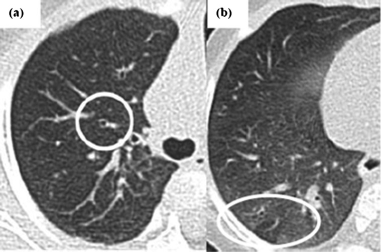
Figure 2.
Examples of CT images from infants with cystic fibrosis (CF) showing mild abnormalities in bronchial dilatation and air trapping leading to discrepancy in scoring. (A) An example of thin section CT of the left lung in an infant with CF taken at 1 year of age showing discrepancies in scoring bronchial dilatation (circled). This was scored as normal by scorer A, but mild by scorer B during the initial study round, whereas during the subsequent rescoring round ∼ 8 months later, scorer A scored this as mild bronchial dilatation, while scorer B scored it as normal. (B) Subtle tiny areas of hyperlucency in some of the scattered secondary pulmonary lobules of the lower lobes in keeping with air trapping (ringed by oval). During the initial scoring round, scorer A scored this as mild air trapping while scorer B labelled it as no air trapping. During the rescoring round, both scorers allocated mild air trapping.

