Abstract
Aims/hypothesis
Chronic inflammation in type 2 diabetes is proposed to affect islets as well as insulin target organs. However, the nature of islet inflammation and its effects on islet function in type 2 diabetes remain unclear. Moreover, the immune cell profiles of human islets in healthy and type 2 diabetic conditions are undefined. We aimed to investigate the correlation between proinflammatory cytokine expression, islet leucocyte composition and insulin secretion in type 2 diabetic human islets.
Methods
Human islets from organ donors with or without type 2 diabetes were studied. First and second phases of glucose-stimulated insulin secretion were determined by perifusion. The expression of inflammatory markers was obtained by quantitative PCR. Immune cells within human islets were analysed by FACS.
Results
Type 2 diabetic islets, especially those without first-phase insulin secretion, displayed higher CCL2 and TNFa expression than healthy islets. CD45+ leucocytes were elevated in type 2 diabetic islets, to a greater extent in moderately functional type 2 diabetic islets compared with poorly functional ones, and corresponded with elevated ALOX12 but not with CCL2 or TNFa expression. T and B lymphocytes and CD11c+ cells were detectable within both non-diabetic and type 2 diabetic islet leucocytes. Importantly, the proportion of B cells was significantly elevated within type 2 diabetic islets.
Conclusions/interpretation
Elevated total islet leucocyte content and proinflammatory mediators correlated with islet dysfunction, suggesting that heterogeneous insulitis occurs during the development of islet dysfunction in type 2 diabetes. In addition, the altered B cell content highlights a potential role for the adaptive immune response in islet dysfunction.
Keywords: B cell, CCL2, Dendritic cells, Flow cytometry, Insulin secretion, 12 Lipoxygenase, Macrophages, Perifusion, T cell, TNF-α
Introduction
The pathogenesis of type 2 diabetes consists of insulin resistance and islet dysfunction [1]. Metabolic stress from excessive nutrition contributes to insulin resistance by provoking chronic inflammation in insulin target organs [1–3]. It is plausible that islet dysfunction is also related to global chronic inflammation in type 2 diabetes. Indeed, some clinical studies targeting IL-1β- and nuclear factor κB-related inflammatory pathways improved aspects of type 2 diabetic beta cell function [4–6]. IL-1β and IL-12, as well as the chemokine (C-C motif) ligand 2 (CCL2) and CCL13, are increased in type 2 diabetic islets and circulation [2, 7, 8]. Similarly, some alterations in circulating leucocyte subsets have been reported in individuals with type 2 diabetes [3, 9]. Peripheral blood T helper (Th)17 and Th1 cells are increased, but T regulatory cells are diminished in obese individuals with type 2 diabetes compared with obese controls [10, 11]. Circulating T cells reactive to islets are detectable in type 2 diabetic patients [12]. Similarly, peripheral blood circulating B cells from individuals with type 2 diabetes secrete more IL-8 but less IL-10, and support contact-dependent T cell activation [13]. However, it is unclear whether B or T cells are present and participate in inducing islet dysfunction. Thus, the mechanisms that connect adipose and islet tissue inflammation and dysfunction in type 2 diabetes are unclear [14].
In this respect, there is limited information concerning the impact of local inflammation on the deterioration of human islet function or the profile of islet-associated leucocytes during the development of type 2 diabetes. While most knowledge about the potential role of leucocytes in islet dysfunction is derived from mouse models of type 1 diabetes [15], several recent studies have aimed to examine the immune cell content of human type 2 diabetic islets. CD68+ leucocytes, which are mostly macrophages, were increased in histological studies of humans with type 2 diabetes [16, 17], suggesting that islet macrophages may play a role in type 2 diabetes. However, a comprehensive analysis is required to determine whether other leucocyte subsets exist within human islets, where they might orchestrate inflammation during type 2 diabetes.
Immune cell accumulation has been reported in multiple rodent models of type 2 diabetes, including ob/ob mice, high-fat-fed mice, Goto-Kakizaki rats and Zucker diabetic fatty rats, supporting the notion that inflammation may contribute to islet dysfunction [16, 18, 19]. Although animal models offer valuable insight into islet biology, human islets are known to differ from rodent islets in morphology [20, 21] and functionality [22], highlighting the importance of studying human islets. The scarcity and difficulty of procuring human islets has been a major hurdle in understanding the pathogenesis of islet failure during type 2 diabetes. In the present study, we applied a flow cytometry-based approach to examine the distribution of leucocyte subsets in non-diabetic and type 2 diabetic human islets, in combination with assessments of islet function and proinflammatory marker expression, to determine the relationship between inflammation and islet function.
Methods
Human islet culture
Human islets were acquired from the Integrated Islet Distribution Program (IIDP; Duarte, CA, USA, for 40 donors, see electronic supplementary material [ESM] Methods) and Beta-Pro (Charlottesville, VA, USA, for three donors), with approval from the institutional review board at the Eastern Virginia Medical School. Islets were incubated overnight in CMRL-1066 containing 10% FBS and 1% penicillin–streptomycin at 37°C and 5% CO2 to recover from shipment. For cytokine treatments, a mixture of 0.57 mmol/l TNF-α, 5.9 mmol/l IFN-γ and 0.29 mmol/l IL-1β (all from BD Bioscience, San Jose, CA, USA) were added to the culture overnight.
Ex vivo perifusion assay
A total of 500 islet equivalents (IEQ) of human islets were perifused at 3 or 23 mmol/l glucose (between 45 and 65 min) [23]. The samples were collected at 1 ml/min for human insulin measurement by ELISA (Mercodia, Winston Salem, NC, USA). The islet insulin content was measured by ELISA after extraction by acidified ethanol [24]. Variables used to compare glucose-stimulated insulin secretion (GSIS) are detailed in ESM Methods.
Gene expression analyses
cDNA was prepared from 500 IEQ of human islets, as described in ESM Methods. Gene expression was analysed using the TaqMan gene-expression assay (Invitrogen, Carlsbad, CA, USA), normalised against β actin expression.
Flow cytometry
A total of 5,000–7,000 IEQ islets digested with 0.025% trypsin and dispersed into single-cell suspensions were used for flow cytometry experiments (detailed in ESM Methods).
Statistics
The data are presented as mean ± SEM. Differences in numeric values between two groups were assessed using an unpaired Student’s t test or Mann–Whitney test. Categorical variables (Table 1) were compared with Fisher’s exact test. Spearman’s rank correlation coefficiency was obtained using GraphPad Prism version 5.00 (GraphPad Software, La Jolla, CA, USA). p<0.05 was considered significant.
Table 1.
Characteristics of human islets
| Non-diabetes (n=21)a | Type 2 diabetes (n=18)a | Cytokine treatment (Fig. 4g, n=4) | |
|---|---|---|---|
| Age, years | 44.8±2.9 (47) | 51.2±1.7 (54) | 31.3±4.2 (33.5) |
| BMI, kg/m2 | 28.5±1.2 (29.1) | 34.5±1.9* (33.3) | 24.0±3.7 (22.8) |
| Sex, male/female | 14/7 (66.7/33.3) | 10/8 (55.6/44.4) | 2/2 (50/50) |
| Race W/B/H/A | 15/3/3/0 (71.4/14.2/14.2/0) | 10/3/4/1 (55.6/16.7/22.2/5.6) | 2/2/0/0 (50/50/0/0) |
| Cause of death | |||
| CVA | 8 (38.1) | 12 (66.7) | 0 |
| Head trauma | 10 (47.6) | 3 (16.7) | 3 (75) |
| Anoxia | 3 (14.3) | 2 (11.1) | 0 |
| Other | 0 | 1b (5.5) | 1 (25) |
| % Purity | 85.1±1.6 (90) | 86.3±1.4 (90) | 86.3±3.2 (85) |
| Time in culture, days | 3.3±0.3 (3) | 3.4± 0.3 (3) | 3±0 (3) |
| Isolation centre | |||
| A/B/C/D/E/othersc n | 2/4/3/4/6/2 | 0/1/2/8/4/3 | 0/2/0/0/1/1 |
Data are means ± SEM (median) or n (%), unless otherwise stated
Quantitative PCR, perifusion and flow cytometry analyses were performed using a selection of donor islets due to the limited quantity of islets. The sample size for each assay is shown in the figures
Excluded from flow cytometry
The list of isolation centres is available at http://iidp.coh.org/center_info.aspx
p<0.01 compared with the non-diabetic cohort
A, Asian; B, black; CVA, cerebrovascular accident; H, Hispanic; W, white
Results
Impairment of GSIS in human type 2 diabetic islets
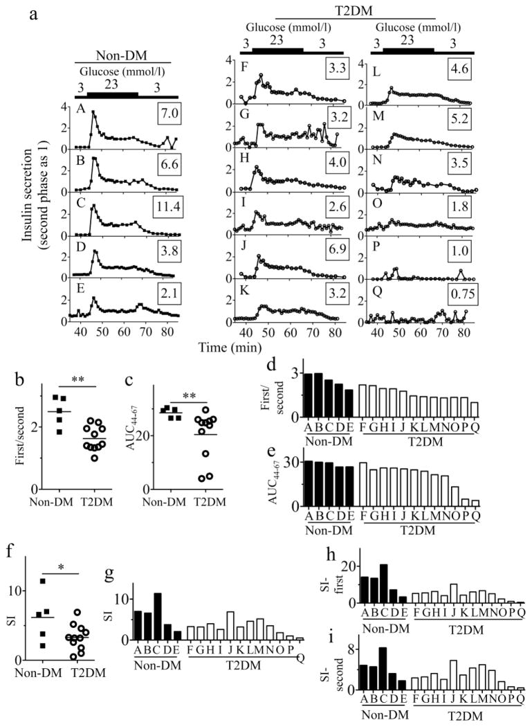
GSIS was measured during perifusion to assess the functionalities of human islets (Fig. 1a). The characteristics of the islets from both cohorts were comparable, aside from a higher BMI in the type 2 diabetic cohort (Table 1). GSIS corrected for total insulin content varied widely in both cohorts (ESM Fig. 1a). Therefore, we focused on the sizes of the first- and second-phase responses in a GSIS profile from each donor, as these variables are not affected by variations in total insulin content and are important indices of islet function (Fig 1a, ESM Methods). The impairment in first-phase response is the earliest functional impairment of beta cells in type 2 diabetes [25, 26]. A prominent first phase followed by a sustained second phase was typically seen in perifused non-diabetic islets (Fig. 1a, A–E). In contrast, type 2 diabetic islets displayed a spectrum of GSIS impairment (Fig. 1a, F–Q), some with a weaker first-phase response (Fig. 1a, F–K) and others without a clear first-phase response but a retained second-phase response (Fig. 1a, L–O). In addition, some type 2 diabetic islets lacked GSIS (Fig. 1a, P and Q). Although the quality of islets could be affected by the isolation process, the range of GSIS impairment is reminiscent of progressive beta cell failure in type 2 diabetes [25, 26]. Based on Fig. 1a, the GSIS profile was expressed using two variables (Fig. 1b–e): the first/second ratio, denoting the average first-phase over second-phase insulin secretion; and AUC44–67, denoting insulin secretion during entire high-glucose perifusion period (ESM Methods). Both variables were significantly reduced in type 2 diabetic islets compared with non-diabetic islets (Fig. 1b,c). The stimulation index (SI), a widely used GSIS metric in batch assays, was calculated for comparisons (Fig. 1f,g). Although the SI correlated with the first/second ratio and AUC44–67 (ESM Fig. 1b,c), it showed substantial overlaps between non-diabetic and type 2 diabetic groups (Fig. 1f,g). The magnitude of the first-phase response (Fig. 1a) was better represented by the first/second ratio (Fig. 1d) than by SI during the entire high-glucose perifusion period (Fig. 1g) or SI calculated for the first phase only (Fig. 1h). For those without a clear first-phase response (samples L-Q), AUC44–67 and SI represented GSIS similarly well (Fig. 1e,g). Collectively, the perifusion of human islets permitted the characterisation of GSIS within the islet cohorts by providing the first/second ratio as a valuable variable.
Fig. 1.
Impairment in GSIS of human islets from type 2 diabetic donors. (a) Ex vivo perifusion of human islets from non-diabetic (non-DM, A–E) and type 2 diabetic (T2DM, F–Q) donors in response to the indicated concentration of glucose given for time periods shown on the horizontal line above. A total of 23 mmol/l glucose was given between 45 and 65 min. Each graph indicates an individual donor. Numbers in rectangles indicate the SI (the ratio of average insulin secretion during 23 mmol/l glucose over 3 mmol/l glucose). (b–e) Two variables of GSIS determined based on (a) were compared between human islets from non-diabetic (non-DM; black squares, A–E) and type 2 diabetic (T2DM; open circles, F–Q) donors. The ratio of the first peak and second-phase response (b) and the AUC between time 44 and 67 min (AUC44–67) (c), as determined in ESM Methods, are shown with the means indicated by horizontal lines, along with the data for individual donors in (d) and (e). The SI from the non-diabetic and type 2 diabetic groups (f) and each individual donor (g) is shown, with the means indicated by horizontal lines in (f). The SI during the first-phase response (time 46–48 min) (h) and the second-phase response (time 55–60 min) (i) from an individual donor. *p<0.05; **p<0.01
The correlation between GSIS and proinflammatory gene expression in type 2 diabeticislets
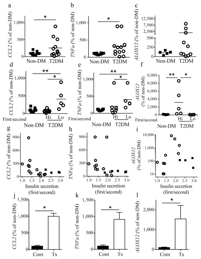
The association of inflammation with islet pathology was addressed by comparing GSIS with the expression of proinflammatory genes. TNFa and CCL2 expression levels were elevated overall in type 2 diabetic islets (Fig. 2a,b). 12 Lipoxygenase (12LO) reacts with arachidonic acid and is associated with inflammation in adipose tissues and islet dysfunction [27]. The expression of ALOX12 (the human gene encoding 12LO) was markedly increased only in some type 2 diabetic islets (Fig. 2c). The expression levels of CCL2 and TNFa were significantly elevated in type 2 diabetic islets with markedly reduced first-phase insulin secretion (<1.45, ‘Lo’), but unchanged in mildly impaired type 2 diabetic islets (>1.45, ‘Hi’, Fig. 2d,e). Interestingly, elevated ALOX12 expression was seen in mildly impaired type 2 diabetic islets (Fig. 2f). The relationship between GSIS and ALOX12 levels was distinct from that of CCL2 and TNFa (Fig. 2g–i). Both CCL2 and TNFa expression showed a significant negative correlation with GSIS (Fig. 2g,h), with tight correlation between the two (ESM Fig. 2a). Thus, CCL2 and TNFa expression levels closely reflect severe impairment in islet function, but ALOX12 expression was uniquely increased in type 2 diabetic islets with moderate GSIS impairment. BMI did not correlate with any islet inflammatory marker or GSIS score (ESM Fig. 1f, 2b–d). In contrast to the discrepancy between TNFa/CCL2 and ALOX12 expression patterns in type 2 diabetic islets, all three genes were acutely upregulated when non-diabetic islets were treated with a mixture of TNF-α, IL-β and IFN-γ (Fig. 2j–l), Thus, the mechanisms responsible for the increase of these genes in type 2 diabetic islets are different from those in acutely treated islets, a commonly used model to mimic type 1 diabetes-associated inflammation [28].
Fig. 2.
The expression of proinflammatory genes was increased in human islets from type 2 diabetic donors. Quantitative PCR compared the expression levels of CCL2 (a, d, g), TNFa (b, e, h) and ALOX12 (c, f, i) between islets from non-diabetic (non-DM, black squares) and type 2 diabetic (T2DM, open circles) donors. Each symbol represents an individual donor with the means indicated as horizontal lines (n=5–10 for non-DM islets and n=11–13 for T2DM islets). The data were normalised to the expression of β actin. (a–c) Comparisons were made between non-DM and T2DM islets. (d–f) Islets from T2DM donors were divided into high GSIS response (≥1.45, Hi) and low GSIS response (<1.45, Lo) based on the ratio of first over second phase of insulin secretion (Fig. 1b). (g–i) The correlation between expression levels of proinflammatory genes and the ratio of first over second phase of insulin secretion (Fig. 1b) was examined. The expression of CCL2 and TNFa correlated with insulin secretion in (g) (p<0.05) and (h) (p<0.01), while no correlation was seen between ALOX12 expression and insulin secretion in (i). (j–l) Overnight treatment of non-diabetic islets with TNF-α, IL-1β and IFN-γ combined markedly increased the expression of CCL2 (j), TNFa (k) and ALOX12 (l). Cont, control; Tx, treatment. *p<0.05; **p<0.01
Increased number of leucocytes in human type 2 diabetic islets
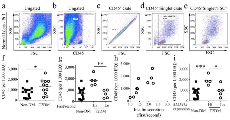
M1-like macrophage recruitment into islets has been reported in rodent models of type 2 diabetes and the number of CD68+ cells within islets has been found to be elevated in histological studies of pancreases from type 2 diabetic humans [16, 17]. However, it is unknown if there are changes in the number or subsets of islet leucocytes in type 2 diabetes. Thus, we adapted a flow cytometry-based method to assess whether leucocyte subsets are present within human islets and if their numbers correlate with islet dysfunction. Flow cytometry offers an advantage over standard histological analyses by permitting the analysis of heterogeneous immune cell populations through the use of multiple surface antigen markers [29]. Non-lymphoid tissues contain a small number of resident leucocytes, which can expand during inflammation. Because of the low frequency and heterogeneous nature of tissue leucocytes, and the high degree of autofluorescence obtained from some non-lymphoid tissues, CD45, the common leucocyte antigen, is typically used to distinguish total leucocytes from tissue cells for subsequent analyses of leucocyte subsets [30–32]. Thus, we used CD45 as a marker to initially identify and examine the abundance of leucocytes within islet cell suspensions (Fig. 3a–f). To rule out possible contamination of the islet preparation with circulating leucocytes, we measured islet cell suspensions for CD235a+ erythrocytes, as previously described [31]. We estimate that 0.0007% of the islet leucocytes represent blood circulating leucocytes (data not shown). In addition, we performed simultaneous staining for endocrine cells and islet leucocytes, to confirm that CD45 cleanly distinguishes leucocytes from endocrine cells (ESM Fig. 3). We also used Live/Dead Aqua dye to assess potential differences in the viability of islet leucocytes between non-diabetic and type 2 diabetic islets (ESM Fig. 4). The majority of CD45+ leucocytes (>90%), as well as major leucocyte subsets, were viable within the islet microenvironment, and the percentage of viable CD45+ leucocytes was equivalent between the two cohorts (ESM Fig. 4). Finally, we detected no differences in analysed islet leucocyte antigen expression between mechanically and enzymatically dispersed islets, suggesting that the enzymatic digestion of human islets has no effect on the stability of the surface antigens used in these studies (ESM Fig. 5).
Fig. 3.
Human islet leucocytes could be detected by flow cytometry and were elevated in type 2 diabetic human islets. Examination of non-diabetic (non-DM, black squares) and type 2 diabetic islet (T2DM, open circles) CD45+ leucocyte content normalised to 1,000 IEQ. (a–e) Representative flow cytometry gating scheme for human islet leucocytes (patient 1, non-DM, ESM Table 1). First, all of the acquired human islet data were gated on leucocytes using CD45 (b), the common leucocyte antigen. Next, cellular aggregates and events smaller than 50,000 FSC or larger than 250,000 SSC were excluded from the analyses, based on FSC-H/FSC-A (c) and FSC/SSC variables (d). K indicates thousands, i.e. 50K = 50,000 etc. (f) The number of normalised CD45+ leucocytes within the non-DM and T2DM islet cohorts and (g) the number of normalised CD45+ leucocytes within non-DM, high GSIS T2DM islets (first/second ≥1.45, Hi) and low GSIS T2DM islets (first/second <1.45, Lo). (h) Correlation between insulin secretion scores (GSIS) and the number of IEQ-normalised leucocytes. There was a positive correlation between the two (p<0.01). Each symbol represents an individual donor, with the group means indicated as horizontal lines. (i) The number of CD45+-normalised leucocytes within non-DM, T2DM with high ALOX12 expression (≥168% of control, Hi) and low ALOX12 expression (<168% of control, Lo) by quantitative PCR. The percentage 168% was chosen based on the 95% CI of ALOX12 expression in the non-DM cohort. *p<0.05; **p<0.01; ***p<0.005
We next estimated the overall leucocyte cellularity within human islets based on CD45-positive leucocytes normalised to 1,000 IEQ. After correcting for cell loss during sample preparation, the abundance of CD45+ leucocytes per 1,000 IEQ obtained by flow cytometry was similar to that previously reported in histological estimates of islet leucocyte cellularity [16, 33]. The number of CD45+ leucocytes was increased within type 2 diabetic islets (Fig. 3f). Since the degree of islet functionality varied widely within the type 2 diabetic islet cohort (Fig. 1), the number of islet leucocytes was compared between high and low insulin-secreting type 2 diabetic islets (Fig. 3g). In contrast to the negative relationship between proinflammatory CCL2 expression and insulin secretion, the number of CD45+ leucocytes was elevated within type 2 diabetic islets with preserved insulin secretion, in comparison with dysfunctional type 2 diabetic islets (Fig. 3g). A positive correlation was observed between GSIS (first/second-phase ratio) and the number of CD45+ leucocytes within type 2 diabetic islets (Fig. 3h); however, BMI and CCL2 and TNFa expression did not correlate with CD45+ leucocytes (ESM Fig. 3b–d). These data suggest that the accumulation of leucocytes within islets is associated with the pathology of type 2 diabetes, although the peak accumulation of leucocytes did not coincide with CCL2 expression. Altogether, other factors may affect the recruitment of immune cells, resulting in variable amounts of islet inflammation within both high- and low-insulin-secreting type 2 diabetic islets. Interestingly, the increase in CD45+ leucocytes was strongly associated with elevated expression of ALOX12 in type 2 diabetic islets (‘Hi’ vs ‘Lo’, Fig. 3i), suggesting that islet leucocytes may be an important source of 12LO production within type 2 diabetic islets. Alternatively, 12LO, which is also produced by beta cells [34], may support the recruitment of leucocytes to the islets.
Type 2 diabetic islets contain elevated levels of B cells
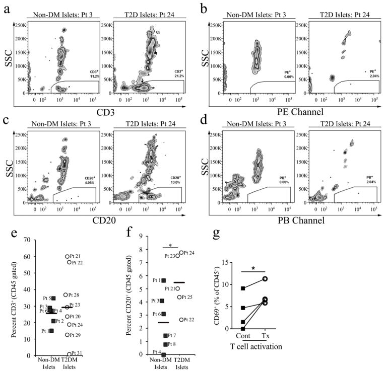
Next, we further characterised the composition of CD45+ leucocytes within human islets. The donor populations are described in ESM Tables 1 and 2. Two major populations of CD45+ leucocytes could be distinguished based on their forward scatter (FSC) and side scatter (SSC) profiles (Fig. 3e): a smaller (50,000–120,000 FSC; 0–50,000 SSC) and a larger population of leucocytes (100,000–200,000 FSC; 70,000–220,000 SSC), reminiscent of lymphocytes and myeloid cells, respectively. To date, limited information is available about the presence and functions of the immune cells in type 2 diabetic islets. Therefore, we examined the presence of the major leucocyte subsets, including T cells, B cells and myeloid cells, within islets. We assessed human islets for the presence of T lymphocytes using T cell marker CD3(ε) [35] and B lymphocytes with CD20 expression (Fig. 4) [36], based on fluorescence minus one and/or isotype control staining. Overall, T cells were the predominate lymphocytes within the human islets; however, the percentage of CD3+ T cells between type 2 diabetic and non-diabetic islets (Fig. 4c) was similar. Interestingly, while the percentage of CD3+ T cells distributed tightly within non-diabetic islets, the percentage varied widely within type 2 diabetic islets, with two patients (patients 21 and 22, ESM Table 2) having up to 60% and one patient (patient 31) with 1% CD3+ T cells (Fig. 4e).
Fig. 4.
T and B lymphocytes were both present within non-diabetic and type 2 diabetic islets, and B lymphocytes were elevated within type 2 diabetic islets. (a) Representative flow cytometry plots of CD3+ islet T lymphocytes from non-diabetic (non-DM, patient [Pt] 3) and type 2 diabetic (T2DM, Pt 24) islet donors, gated on CD45+singlet+FSC+ events that were below 50,000 SSC (a). Corresponding human islet fluorescence minus one for the phycoerythrin (PE) channel (b) or isotype controls (not shown) were used to set the CD3+ gates (a). (c) Representative flow cytometry plots of CD20+ B lymphocytes from non-DM (Pt 2) and T2DM (Pt 23) islet donors, as conducted in panel (a). The corresponding representative Pacific Blue (PB) fluorescence minus one controls are also shown (d). K indicates thousands, i.e. 50K = 50,000 etc. (e) The percentage of islet CD3+ T lymphocytes within CD45+singlet+FSC+ gated non-DM (black squares) and T2DM (open circles) islet donors. Each symbol represents an individual donor, with the group means indicated as horizontal lines. (f) The percentage of gated CD20+ B lymphocytes, as represented in (c). (g) The effects of overnight treatment of non-DM islets with of TNF-α, IL-1β and IFN-γ on the expression of the activation marker CD69 within CD45+CD3+CD44+CD45RA− memory T cells. The symbols depict non-treated (black squares) and treated (open circles) islets. *p<0.05
An important role for B cells in the development of systemic and adipose inflammation has been reported in a mouse model of insulin resistance [37]. In parallel, a recent study with peripheral blood circulating B cells from type 2 diabetic individuals highlighted the unique phenotype and functions of these lymphocytes [38]. To test whether B cells are present within islets, we performed staining for CD20, a marker that is expressed by the majority of B cells, including pre-, resting and memory B cells. A small but distinguishable resident population of CD20+ B cells was present within non-diabetic and type 2 diabetic islets (Fig. 4c). Overall, the percentage of B cells was increased up to 2.2-fold in type 2 diabetic islets compared with non-diabetic controls (Fig. 4f). To assess whether human islet T cells may be activated in response to cytokines, we assessed TNF-α-, IL-1β - and IFN-γ -treated non-diabetic islets for upregulation of the early T cell activation marker CD69 on intraislet memory T cells (CD3+CD44+CD45RA− T cells). In this ex vivo system, memory T cells significantly upregulated CD69 in comparison to vehicle-treated controls (Fig. 4g). These results suggest that islet T cells are functional and can be activated in response to proinflammatory cytokines, likely through islet antigen presenting myeloid or B cells.
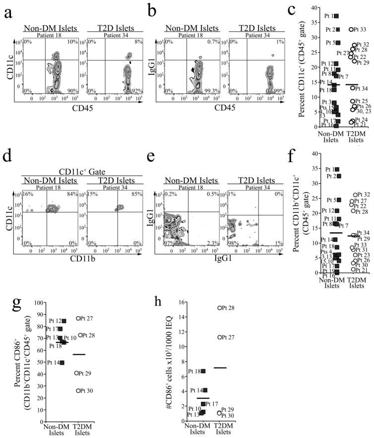
Next, we examined whether myeloid cells were altered between type 2 diabetic and non-diabetic islets (Fig. 5). As there are no completely unique markers for dendritic cells (DCs) or macrophages, we initially characterised islet myeloid cells based on CD11c and CD11b expression [39]. A noticeable population of CD11c+ cells was detected within islets from both non-diabetic (patient 18, Fig. 5a) and type 2 diabetic (patient 34, Fig. 5a) donors; however, the percentage of CD11c+ cells between non-diabetic and type 2 diabetic islets was similar (Fig. 5c). To further examine the phenotype of CD11c+ cells, we analysed the expression of CD11b by CD11c+ cells. The majority of CD11c+ myeloid cells also expressed CD11b+ (Fig. 5d–f), suggesting that these myeloid cells may represent CD11b+CD11c+ macrophages or DCs. The frequency of CD11b+CD11c+ cells was unchanged between non-diabetic and type 2 diabetic islets and varied greatly within both cohorts (Fig. 5f). To characterise the nature of islet CD11b+CD11c+ myeloid cells the expression of CD86, a T cell costimulatory molecule, was assessed (Fig. 5g,h). The majority of islet CD11b+CD11c+ cells from non-diabetic and type 2 diabetic individuals expressed CD86 (Fig. 5g,h). Together, our data demonstrate the presence of T and B lymphocytes, and myeloid cell subsets within non-diabetic human islets, that likely reflect constitutive homing of leucocytes and significant alterations in the immune content of type 2 diabetic islets. These data also highlight the potential utility of flow cytometry for analysing islet leucocytes and inflammation during the development of type 2 diabetes.
Fig. 5.
Resident islet CD86-expressing CD11b+CD11c+ myeloid cells were present within non-diabetic and type 2 diabetic human islets. (a) Representative flow cytometry plots of CD45+singlet+FSC+ gated CD11c+ myeloid cells from non-diabetic (non-DM, patient 18) and type 2 diabetic (T2DM, patient 34) islet donors. The gates were set based on corresponding islet isotype control specimens (b). (c) Quantification of the percentage of CD11c+ myeloid cells within CD45+singlet+FSC+ gated leucocytes. (d) Representative flow cytometry plots of CD11b+CD11c+ myeloid cells gated on CD45+singlet+FSC+CD11c+ leucocytes, and the corresponding isotype controls (e). (f) The back-calculated percentage of CD11b+CD11c+ myeloid cells among CD45+singlet+FSC+ gated leucocytes. The percentage (g) and number (h) of CD86 expressing CD11b+CD11c+CD45+ myeloid cells. (c, f–h) Symbols depict individual non-DM (black squares) and T2DM (open circles) islet donors, while the horizontal lines represent group means. Pt, patient
Discussion
In this study, we have established a strong correlation between the proinflammatory cytokine TNF-α and the chemokine CCL2 with GSIS in type 2 diabetic human islets. We found an increased content of CD45+ leucocytes within type 2 diabetic islets, specifically within moderately functional type 2 diabetic islets. Importantly, CD20+ B cells were increased in type 2 diabetic human islets in comparison with non-diabetic islets. This study is the first to attempt simultaneous examination of both physiological and immunological variables in human islets, in order to determine if intraislet immune cells are associated with islet dysfunction in human type 2 diabetes.
Low first-phase insulin secretion upon i.v. glucose injection is a pathognomonic feature of human type 2 diabetes [25, 26]. It is noteworthy that the perifusion of human islets from clinically labelled type 2 diabetic donors unanimously showed blunting or loss of first-phase insulin secretion, suggesting that islets from type 2 diabetic donors retain features of islet dysfunction ex vivo. In comparison with previous studies that used glucose-ramp or batch assays [40, 41], we determined both the first- and second-phase GSIS responses in a large cohort of type 2 diabetic islets. The wide spectrum of impairment in first- and second-phase insulin secretion that we observed may reflect a gradual decline in functional beta cell mass, which is considered to be responsible for the progression of type 2 diabetes in humans [26]. The IIDP in the USA is an invaluable resource that facilitates access to a large number of islet donors. However, the medical history details available to researchers are currently limited (see ESM Methods). Future studies should determine whether the clinical history of a donor, including the duration and severity of type 2 diabetes, correlates with GSIS and islet leucocyte content ex vivo.
Several lines of evidence suggest that the levels of proinflammatory cytokines such as IL-β 1, IL-8 and IL-12 and chemokines CCL2, CCL13 and chemokine (C-X-C motif) ligand 10 (CXCL10) are elevated in type 2 diabetic islets [7, 8, 14, 42]. However, it is unclear whether proinflammatory cytokines and chemokines are definitively associated with the development of islet dysfunction in type 2 diabetes. Our study provides compelling evidence for a negative correlation between the levels of CCL2 and TNF-α within type 2 diabetic islets and their functions, as determined by GSIS. CCL2 and TNF-α levels were significantly elevated in dysfunctional type 2 diabetic islets in comparison with moderately functional type 2 diabetic islets, indicating the existence of several subgroups with different levels of inflammation within type 2 diabetic individuals. Surprisingly, ALOX12, which is strongly associated with inflammation in adipose and other tissues [27], was upregulated within moderately functional but not dysfunctional type 2 diabetic islets, indicating that 12LO may play a role in islet dysfunction at early certain stages of the pathogenesis and that beta cell production of 12LO might participate in the early homing of leucocytes to islets. Alternatively, increased ALOX12 expression may originate from non-beta cells, considering the correlation between ALOX12 expression and the abundance of CD45+ leucocytes.
M1-like macrophages are recruited into islets in rodent type 2 diabetes models and, similarly, CD68+ macrophages are elevated within human T2D islets [16, 17, 43]. To further determine if the immune system is associated with the pathology of type 2 diabetes, we examined the overall abundance of leucocytes and leucocyte subsets within human islets from non-diabetic and type 2 diabetic individuals. Importantly, we observed evidence of increased accumulation of CD45+ leucocytes within type 2 diabetic islets, which strongly correlated with islet function. To our surprise, individuals with moderately functional islets possessed significantly more leucocytes than markedly dysfunctional type 2 diabetic islets. Based on these results, initial steps in the development of type 2 diabetes may be accompanied by an influx of CD45+ leucocytes, which likely supports active islet inflammation. In contrast, at an advanced stage of type 2 diabetes, significant beta cell damage and apoptosis due to oxidative stress, endoplasmic reticulum stress and mitochondrial dysfunction may provide less support for the recruitment of leucocytes to type 2 diabetic islets. Together, these results suggest that the accumulation of leucocytes within islets is associated with the pathology of type 2 diabetes, but the recruitment of leucocytes may occur in a temporal, stage-dependent manner. Considering the variable degrees of islet inflammation within high- and low-insulin-secreting type 2 diabetic islets, other factors such as the management of the individual’s diabetes and end-of-life treatment may modify the recruitment of leucocytes.
Chronic low-grade inflammation resulting from changes in lymphocyte and myeloid cell subsets promotes and exacerbates insulin resistance, which, together with islet dysfunction, defines type 2 diabetes. We detected an increased accumulation of B cells within type 2 diabetic islets. Recent reports suggest that circulating B cells are hyperactivated in type 2 diabetic patients and elicit T cell-derived proinflammatory TNF-α, IL-17A and IL-6 production in T cell/B cell cocultures [9]. Thus, an elevated proportion of islet B lymphocytes may work together with costimulatory molecule-expressing myeloid cells (CD86+ cells) to affect islet T cell activation or serve as “accessory cells” to promote islet-specific T cell proliferation, as shown for islet B lymphocytes in the non-obese diabetic mouse model [44].
Interestingly, comparable levels of CD3+ T cells and CD11c+ myeloid cells were present in both non-diabetic and type 2 diabetic islets. Islets treated with TNF-α, IL-1β and IFN-γ demonstrated that islet memory T cells are responsive to cytokines and may become activated during inflammation. Importantly, subtle changes in the proportion of T and B cell subsets may exist in type 2 diabetic islets, in addition to changes in the overall abundance of T and B cells within the islet. Future studies are required to determine if the overall abundance and differentiation of T cell subsets are affected by the elevated number of islet B cells.
Recent studies have demonstrated that several DC subsets are present within rodent pancreatic islets, where they may play proinflammatory or tolerogenic roles in the context of type 1 diabetes [32, 45]. Two subsets of islet DCs with distinct functions are found within healthy murine islets. CD11b+CD103−CX3CR1+ DCs represent the majority of islet DCs with high phagocytic, but low antigen presentation activity [32]. In contrast, a small subset of CD11blow CD103+CX3CR1− DCs is able to migrate to the draining lymph node and cross-present antigens. In addition, suppressive CD11b+CD11c+ tolerogenic DCs have been described in a non-obese diabetic mouse model [45]. Multiple myeloid cell subsets may be similarly present within human islets, where they may participate in T cell activation and the development of islet dysfunction in type 2 diabetes. Indeed, sizable populations of CD11c+ cells are present in non-diabetic and type 2 diabetic human islets, and further characterisation of CD11c+ cells will be required to determine their roles in type 2 diabetes.
Collectively, our results demonstrate that insulitis is associated with and may participate in the deterioration of islet function in human type 2 diabetes. CCL2 and TNFa expression were strongly correlated with severe GSIS impairment, while the abundance of ALOX12 and CD45+ leucocytes correlated with moderate impairment in GSIS, providing evidence for a complex interplay between islets and the immune system. Moreover, the current study highlights the potential importance of B lymphocytes in human islet biology. The detection of leucocyte populations in non-diabetic and type 2 diabetic human islets may not only provide insight into the physiologic role of immune cells associated with islets, but also highlight new directions for studying inflammation in type 2 diabetes. This could lead to new, more targeted therapies to prevent the decline of functional beta cell mass in patients with type 2 diabetes.
Supplementary Material
Acknowledgments
Human islets were provided to J. Nadler and Y. Imai by the IIDP. We thank J. Kaddis and B. Olack of the IIDP for assisting with documentation on the IIDP and donor data.
Funding
This work was supported by the Juvenile Diabetes Research Foundation (JN), grants from the National Institutes of Health to JN (R01-HL112605) and YI (R01-DK090490), and by the IIDP pilot programme (YI), a BD Pharmingen Research Grant (EVG), AstraZeneca (JN) and start-up funds from the Eastern Virginia Medical School (EVG).
Abbreviations
- CCL
Chemokine (C-C motif) ligand
- CXCL
Chemokine (C-X-C motif) ligand
- DC
Dendritic cell
- GSIS
Glucose-stimulated insulin secretion
- IEQ
Islet equivalent
- IIDP
Integrated Islet Distribution Program
- 12LO
12 Lipoxygenase
- SI
Stimulation index
- SSC
Side scatter
- Th
T helper
Footnotes
Duality of interest
The authors declare that there is no duality of interest associated with this manuscript.
Contribution statement
JN, EVG and YI contributed to the study conception and design. MJB, EVG (flow cytometry), DH, EG, YM (GSIS, quantitative PCR), SC (quantitative PCR) and YI (GSIS, quantitative PCR, flow cytometry) were responsible for the acquisition and analysis of the data. MJB, EVG and YI interpreted the data, drafted the manuscript and critically revised the manuscript for important intellectual content. DH, EG, YM, SC and JN also revised the manuscript. All authors approved the final version of the manuscript.
References
- 1.Nolan CJ, Damm P, Prentki M. Type 2 diabetes across generations: from pathophysiology to prevention and management. Lancet. 2011;378:169–181. doi: 10.1016/S0140-6736(11)60614-4. [DOI] [PubMed] [Google Scholar]
- 2.Donath MY, Shoelson SE. Type 2 diabetes as an inflammatory disease. Nature reviews Immunology. 2011;11:98–107. doi: 10.1038/nri2925. [DOI] [PubMed] [Google Scholar]
- 3.Lumeng CN, Saltiel AR. Inflammatory links between obesity and metabolic disease. The Journal of clinical investigation. 2011;121:2111–2117. doi: 10.1172/JCI57132. [DOI] [PMC free article] [PubMed] [Google Scholar]
- 4.Cavelti-Weder C, Babians-Brunner A, Keller C, et al. Effects of gevokizumab on glycemia and inflammatory markers in type 2 diabetes. Diabetes care. 2012;35:1654–1662. doi: 10.2337/dc11-2219. [DOI] [PMC free article] [PubMed] [Google Scholar]
- 5.Donath MY, Dalmas E, Sauter NS, Boni-Schnetzler M. Inflammation in obesity and diabetes: islet dysfunction and therapeutic opportunity. Cell metabolism. 2013;17:860–872. doi: 10.1016/j.cmet.2013.05.001. [DOI] [PubMed] [Google Scholar]
- 6.Goldfine AB, Fonseca V, Jablonski KA, Pyle L, Staten MA, Shoelson SE. The effects of salsalate on glycemic control in patients with type 2 diabetes: a randomized trial. Annals of internal medicine. 2010;152:346–357. doi: 10.1059/0003-4819-152-6-201003160-00004. [DOI] [PMC free article] [PubMed] [Google Scholar]
- 7.Igoillo-Esteve M, Marselli L, Cunha DA, et al. Palmitate induces a proinflammatory response in human pancreatic islets that mimics CCL2 expression by beta cells in type 2 diabetes. Diabetologia. 2010;53:1395–1405. doi: 10.1007/s00125-010-1707-y. [DOI] [PubMed] [Google Scholar]
- 8.Taylor-Fishwick DA, Weaver JR, Grzesik W, et al. Production and function of IL-12 in islets and beta cells. Diabetologia. 2013;56:126–135. doi: 10.1007/s00125-012-2732-9. [DOI] [PMC free article] [PubMed] [Google Scholar]
- 9.Nikolajczyk BS, Jagannathan-Bogdan M, Denis GV. The outliers become a stampede as immunometabolism reaches a tipping point. Immunological reviews. 2012;249:253–275. doi: 10.1111/j.1600-065X.2012.01142.x. [DOI] [PMC free article] [PubMed] [Google Scholar]
- 10.Jagannathan-Bogdan M, McDonnell ME, Shin H, et al. Elevated proinflammatory cytokine production by a skewed T cell compartment requires monocytes and promotes inflammation in type 2 diabetes. J Immunol. 2011;186:1162–1172. doi: 10.4049/jimmunol.1002615. [DOI] [PMC free article] [PubMed] [Google Scholar]
- 11.Zeng C, Shi X, Zhang B, et al. The imbalance of Th17/Th1/Tregs in patients with type 2 diabetes: relationship with metabolic factors and complications. J Mol Med (Berl) 2012;90:175–186. doi: 10.1007/s00109-011-0816-5. [DOI] [PubMed] [Google Scholar]
- 12.Brooks-Worrell BM, Reichow JL, Goel A, Ismail H, Palmer JP. Identification of autoantibody-negative autoimmune type 2 diabetic patients. Diabetes care. 2011;34:168–173. doi: 10.2337/dc10-0579. [DOI] [PMC free article] [PubMed] [Google Scholar]
- 13.Jagannathan M, McDonnell M, Liang Y, et al. Toll-like receptors regulate B cell cytokine production in patients with diabetes. Diabetologia. 2010;53:1461–1471. doi: 10.1007/s00125-010-1730-z. [DOI] [PMC free article] [PubMed] [Google Scholar]
- 14.Imai Y, Dobrian AD, Morris MA, Nadler JL. Islet inflammation: a unifying target for diabetes treatment? Trends in endocrinology and metabolism: TEM. 2013;24:351–360. doi: 10.1016/j.tem.2013.01.007. [DOI] [PMC free article] [PubMed] [Google Scholar]
- 15.Lehuen A, Diana J, Zaccone P, Cooke A. Immune cell crosstalk in type 1 diabetes. Nature reviews Immunology. 2010;10:501–513. doi: 10.1038/nri2787. [DOI] [PubMed] [Google Scholar]
- 16.Ehses JA, Perren A, Eppler E, et al. Increased number of islet-associated macrophages in type 2 diabetes. Diabetes. 2007;56:2356–2370. doi: 10.2337/db06-1650. [DOI] [PubMed] [Google Scholar]
- 17.Richardson SJ, Willcox A, Bone AJ, Foulis AK, Morgan NG. Islet-associated macrophages in type 2 diabetes. Diabetologia. 2009;52:1686–1688. doi: 10.1007/s00125-009-1410-z. [DOI] [PubMed] [Google Scholar]
- 18.Homo-Delarche F, Calderari S, Irminger JC, et al. Islet inflammation and fibrosis in a spontaneous model of type 2 diabetes, the GK rat. Diabetes. 2006;55:1625–1633. doi: 10.2337/db05-1526. [DOI] [PubMed] [Google Scholar]
- 19.Jones HB, Nugent D, Jenkins R. Variation in characteristics of islets of Langerhans in insulin-resistant, diabetic and non-diabetic-rat strains. International journal of experimental pathology. 2010;91:288–301. doi: 10.1111/j.1365-2613.2010.00713.x. [DOI] [PMC free article] [PubMed] [Google Scholar]
- 20.Cabrera O, Berman DM, Kenyon NS, Ricordi C, Berggren PO, Caicedo A. The unique cytoarchitecture of human pancreatic islets has implications for islet cell function. Proceedings of the National Academy of Sciences of the United States of America. 2006;103:2334–2339. doi: 10.1073/pnas.0510790103. [DOI] [PMC free article] [PubMed] [Google Scholar]
- 21.Brissova M, Fowler MJ, Nicholson WE, et al. Assessment of human pancreatic islet architecture and composition by laser scanning confocal microscopy. The journal of histochemistry and cytochemistry : official journal of the Histochemistry Society. 2005;53:1087–1097. doi: 10.1369/jhc.5C6684.2005. [DOI] [PubMed] [Google Scholar]
- 22.MacDonald MJ, Longacre MJ, Stoker SW, et al. Differences between human and rodent pancreatic islets: low pyruvate carboxylase, atp citrate lyase, and pyruvate carboxylation and high glucose-stimulated acetoacetate in human pancreatic islets. The Journal of biological chemistry. 2011;286:18383–18396. doi: 10.1074/jbc.M111.241182. [DOI] [PMC free article] [PubMed] [Google Scholar]
- 23.Imai Y, Patel HR, Doliba NM, Matschinsky FM, Tobias JW, Ahima RS. Analysis of gene expression in pancreatic islets from diet-induced obese mice. Physiological genomics. 2008;36:43–51. doi: 10.1152/physiolgenomics.00050.2008. [DOI] [PMC free article] [PubMed] [Google Scholar]
- 24.Imai Y, Patel HR, Hawkins EJ, Doliba NM, Matschinsky FM, Ahima RS. Insulin secretion is increased in pancreatic islets of neuropeptide Y-deficient mice. Endocrinology. 2007;148:5716–5723. doi: 10.1210/en.2007-0404. [DOI] [PubMed] [Google Scholar]
- 25.Brunzell JD, Robertson RP, Lerner RL, et al. Relationships between fasting plasma glucose levels and insulin secretion during intravenous glucose tolerance tests. The Journal of clinical endocrinology and metabolism. 1976;42:222–229. doi: 10.1210/jcem-42-2-222. [DOI] [PubMed] [Google Scholar]
- 26.Kahn SE, Zraika S, Utzschneider KM, Hull RL. The beta cell lesion in type 2 diabetes: there has to be a primary functional abnormality. Diabetologia. 2009;52:1003–1012. doi: 10.1007/s00125-009-1321-z. [DOI] [PMC free article] [PubMed] [Google Scholar]
- 27.Dobrian AD, Lieb DC, Cole BK, Taylor-Fishwick DA, Chakrabarti SK, Nadler JL. Functional and pathological roles of the 12- and 15-lipoxygenases. Progress in lipid research. 2011;50:115–131. doi: 10.1016/j.plipres.2010.10.005. [DOI] [PMC free article] [PubMed] [Google Scholar]
- 28.Delaney CA, Pavlovic D, Hoorens A, Pipeleers DG, Eizirik DL. Cytokines induce deoxyribonucleic acid strand breaks and apoptosis in human pancreatic islet cells. Endocrinology. 1997;138:2610–2614. doi: 10.1210/endo.138.6.5204. [DOI] [PubMed] [Google Scholar]
- 29.Beare A, Stockinger H, Zola H, Nicholson I. Monoclonal antibodies to human cell surface antigens. Current protocols in immunology. 2008:4A. doi: 10.1002/0471142735.ima04as53. Appendix 4. [DOI] [PMC free article] [PubMed] [Google Scholar]
- 30.Butcher MJ, Gjurich BN, Phillips T, Galkina EV. The IL-17A/IL-17RA axis plays a proatherogenic role via the regulation of aortic myeloid cell recruitment. Circulation research. 2012;110:675–687. doi: 10.1161/CIRCRESAHA.111.261784. [DOI] [PMC free article] [PubMed] [Google Scholar]
- 31.Galkina E, Kadl A, Sanders J, Varughese D, Sarembock IJ, Ley K. Lymphocyte recruitment into the aortic wall before and during development of atherosclerosis is partially L-selectin dependent. The Journal of experimental medicine. 2006;203:1273–1282. doi: 10.1084/jem.20052205. [DOI] [PMC free article] [PubMed] [Google Scholar]
- 32.Yin N, Xu J, Ginhoux F, et al. Functional specialization of islet dendritic cell subsets. J Immunol. 2012;188:4921–4930. doi: 10.4049/jimmunol.1103725. [DOI] [PMC free article] [PubMed] [Google Scholar]
- 33.Pisania A, Weir GC, O’Neil JJ, et al. Quantitative analysis of cell composition and purity of human pancreatic islet preparations. Laboratory investigation; a journal of technical methods and pathology. 2010;90:1661–1675. doi: 10.1038/labinvest.2010.124. [DOI] [PMC free article] [PubMed] [Google Scholar]
- 34.Weaver JR, Holman TR, Imai Y, et al. Integration of pro-inflammatory cytokines, 12-lipoxygenase and NOX-1 in pancreatic islet beta cell dysfunction. Molecular and cellular endocrinology. 2012;358:88–95. doi: 10.1016/j.mce.2012.03.004. [DOI] [PubMed] [Google Scholar]
- 35.Gold DP, Puck JM, Pettey CL, et al. Isolation of cDNA clones encoding the 20K non-glycosylated polypeptide chain of the human T cell receptor/T3 complex. Nature. 1986;321:431–434. doi: 10.1038/321431a0. [DOI] [PubMed] [Google Scholar]
- 36.Tedder TF, Streuli M, Schlossman SF, Saito H. Isolation and structure of a cDNA encoding the B1 (CD20) cell-surface antigen of human B lymphocytes. Proceedings of the National Academy of Sciences of the United States of America. 1988;85:208–212. doi: 10.1073/pnas.85.1.208. [DOI] [PMC free article] [PubMed] [Google Scholar]
- 37.Winer DA, Winer S, Shen L, et al. B cells promote insulin resistance through modulation of T cells and production of pathogenic IgG antibodies. Nature medicine. 2011;17:610–617. doi: 10.1038/nm.2353. [DOI] [PMC free article] [PubMed] [Google Scholar]
- 38.DeFuria J, Belkina AC, Jagannathan-Bogdan M, et al. B cells promote inflammation in obesity and type 2 diabetes through regulation of T cell function and an inflammatory cytokine profile. Proceedings of the National Academy of Sciences of the United States of America. 2013;110:5133–5138. doi: 10.1073/pnas.1215840110. [DOI] [PMC free article] [PubMed] [Google Scholar]
- 39.Geissmann F, Gordon S, Hume DA, Mowat AM, Randolph GJ. Unravelling mononuclear phagocyte heterogeneity. Nature reviews Immunology. 2010;10:453–460. doi: 10.1038/nri2784. [DOI] [PMC free article] [PubMed] [Google Scholar]
- 40.Del Guerra S, Lupi R, Marselli L, et al. Functional and molecular defects of pancreatic islets in human type 2 diabetes. Diabetes. 2005;54:727–735. doi: 10.2337/diabetes.54.3.727. [DOI] [PubMed] [Google Scholar]
- 41.Deng S, Vatamaniuk M, Huang X, et al. Structural and functional abnormalities in the islets isolated from type 2 diabetic subjects. Diabetes. 2004;53:624–632. doi: 10.2337/diabetes.53.3.624. [DOI] [PubMed] [Google Scholar]
- 42.Boni-Schnetzler M, Thorne J, Parnaud G, et al. Increased interleukin (IL)- 1beta messenger ribonucleic acid expression in beta-cells of individuals with type 2 diabetes and regulation of IL-1beta in human islets by glucose and autostimulation. The Journal of clinical endocrinology and metabolism. 2008;93:4065–4074. doi: 10.1210/jc.2008-0396. [DOI] [PMC free article] [PubMed] [Google Scholar]
- 43.Eguchi K, Manabe I, Oishi-Tanaka Y, et al. Saturated fatty acid and TLR signaling link beta cell dysfunction and islet inflammation. Cell metabolism. 2012;15:518–533. doi: 10.1016/j.cmet.2012.01.023. [DOI] [PubMed] [Google Scholar]
- 44.Carrillo J, Puertas MC, Alba A, et al. Islet-infiltrating B cells in nonobese diabetic mice predominantly target nervous system elements. Diabetes. 2005;54:69–77. doi: 10.2337/diabetes.54.1.69. [DOI] [PubMed] [Google Scholar]
- 45.Kriegel MA, Rathinam C, Flavell RA. Pancreatic islet expression of chemokine CCL2 suppresses autoimmune diabetes via tolerogenic CD11c+ CD11b+ dendritic cells. Proceedings of the National Academy of Sciences of the United States of America. 2012;109:3457–3462. doi: 10.1073/pnas.1115308109. [DOI] [PMC free article] [PubMed] [Google Scholar]
Associated Data
This section collects any data citations, data availability statements, or supplementary materials included in this article.


