Abstract
The aim of this study was to quantify internal joint moments of the lower limb during vertical jumping and the weightlifting jerk in order to improve awareness of the control strategies and correspondence between these activities, and to facilitate understanding of the likely transfer of training effects. Athletic males completed maximal unloaded vertical jumps (n=12) and explosive push jerks at 40 kg (n=9). Kinematic data were collected using optical motion tracking and kinetic data via a force plate, both at 200 Hz. Joint moments were calculated using a previously described biomechanical model of the right lower limb. Peak moment results highlighted that sagittal plane control strategies differed between jumping and jerking (p<0.05) with jerking being a knee dominant task in terms of peak moments as opposed to a more balanced knee and hip strategy in jumping and landing. Jumping and jerking exhibited proximal to distal joint involvement and landing was typically reversed. High variability was seen in non-sagittal moments at the hip and knee. Significant correlations were seen between jump height and hip and knee moments in jumping (p<0.05). Whilst hip and knee moments were correlated between jumping and jerking (p<0.05), joint moments in the jerk were not significantly correlated to jump height (p>0.05) possibly indicating a limit to the direct transferability of jerk performance to jumping. Ankle joint moments were poorly related to jump performance (p>0.05). Peak knee and hip moment generating capacity are important to vertical jump performance. The jerk appears to offer an effective strategy to overload joint moment generation in the knee relative to jumping. However, an absence of hip involvement would appear to make it a general, rather than specific, training modality in relation to jumping.
Keywords: musculoskeletal modelling, joint moments, weightlifting, jumping, correspondence
INTRODUCTION
Vertical jumping is a common assessment tool in strength and power programmes and frequently a primary marker used to describe general enhancements in leg extension force production capabilities. The jerk is a component of techniques taken from Olympic style weightlifting (OL) utilised with a view to enhance leg extension force production in time constrained stretch-shorten cycle conditions. Understanding the relationships between the mechanics of such training, monitoring and performance skills offers insight into the correspondence and likely transferability of adaptations between these skills and further to the effective selection of training and monitoring modalities within a programme.
There has been a plethora of studies exploring the biomechanics of vertical jumping1,4,18,28,33,39,40, landing3,13,24,29,36,51 and Olympic weightlifting6,19,20,21,25,43. These studies have used a range of techniques to evaluate the kinematics and kinetics of these activities. One technique in biomechanics is to reduce the description of the body to a series of linked rigid segments. Inverse dynamics techniques can then be used to calculate the inter-segmental forces and moments (moments refer to internal moments unless otherwise stated) expressed at each joint14,49. It is common to make the assumption that the moments calculated in an inverse dynamics analysis represent the resultant muscle moments12,16. In reality, the inter-segmental moments are likely to be a result of the combination of both muscular forces and the restraint provided by the passive tissues27. In the sagittal plane it is likely that a large proportion of the observed moment is a result of muscular actions and thus a consideration of sagittal plane moments can be very useful in understanding performance. However, in the frontal and transverse plane it is likely that the passive structures provide a more significant proportion of the calculated moment, especially at the knee where the musculature is not well equipped to resist non-sagittal plane loading. An analysis of moments in 3 dimensions (3D) is therefore instructive in ascertaining the loading of passive tissues, and consequently frontal and transverse plane moments may be revealing as to the potential mechanisms of injury.
In contrast to the large number of studies that have employed inverse dynamics to study jumping and landing, there is a paucity studies that have used the technique to study OL11,53. Those studies that have employed inverse dynamics have not presented comprehensive details as to the inter-segmental moments calculated. The profiles of joint moments in these activities are therefore relatively unknown. Certainly there have been no studies that have compared joint moment production between jumping and OL, and thus the kinetic similarities and differences between the two activities in terms of joint moments has yet to be determined. Despite this, it is common for strength and conditioning coaches to contend that there is a high degree of mechanical similarity between the two exercises, and to use this observation as the basis of training choices.
Although comparisons of joint moment profiles, either between athletic skills or between differing athletic groups are not common in the strength and conditioning literature they can be illuminating when analysing performance and prescribing training interventions26,46. For instance, Vanezis and Lees46 analysed the differences in moment production between good and poor vertical jumpers. They found a non-significant trend that the peak moments produced by the good jumpers were greater than those of the poor jumpers. Equally, the good jumpers developed joint moments more rapidly during jumping. The authors did not however, present details on the correlation between peak moment production and jump height leaving the predictive power of peak moments as to jump height unresolved. Nonetheless, such analysis can be used to inform coaches as to the differing strategies adopted by athletes such that their performance might be better understood and either replicated or altered. In particular this might include understanding the differing emphasis athletes place on the utilisation of joint moments at the hip, knee or ankle.
Equally, impulse generation across a range of propulsive and landing movements is likely to be optimally created by different strategies of lower limb muscle and joint involvement. Due to general issues of specificity of adaptation, the transfer of adaptation benefits between different athletic tasks is likely to be limited by their degree of mechanical correspondence; an idea longstanding in the coaching48. It is therefore informative for coaches to consider issues of mechanical similarity between different movement tasks. Whilst the use of electromyography for this purpose has been popular7 the application of inverse dynamics does not appear common in coaching research literature. This incorporates consideration of both the relative amplitudes of peak moments as well as their temporal arrangement in the moment profile. Importantly an improvement in understanding from coaches of the way in which constraining particular movements (such as demanding a near vertical line of travel with body segments closely connected to a barbell) changes the pattern of moment production required by the athlete is likely to result in an enhanced ability to make appropriate exercise selection choices and movement modifications.
The purpose of this study was therefore to perform a 3D optimization based approach to the inverse dynamics analysis of vertical jumping, landing and jerking. The primary aim of the study was to provide a detailed description of the 3D moments experienced at the joints in the local coordinate frame for each of the activities using an optimisation approach9,10. A secondary aim was then to use these descriptions to facilitate a better understanding of the relationship between vertical jumping and jerking. Finally, a tertiary aim was to determine whether peak joint moments produced during jumping and jerking were predictive of vertical jump height.
METHODS
Experimental approach to the problem
To determine jump height and related jump moments subjects (n=12) completed uninstructed vertical jumps with arms isolated. To allow comparison to the jerk those with weightlifting experience (n=9) (classified as athletes who reported previously using jerks as part of the sports conditioning programme) also completed a series of weightlifting jerks in the same session following the jump trials in a repeated measures design. Activities were subject to kinetic and kinematic data collection to allow calculation of 3D moments at the ankle knee and hip.
Subjects
12 athletic males from varied sports (mean ± SD age 27.1 ± 4.3 years; body mass 83.7 ± 9.9 kg) volunteered the study and were informed of the procedures and risks prior to providing written consent for participation. The study was approved by the institutional research ethics committee for St Mary’s University College. Nine of the volunteers had been previously familiarised with the weightlifting jerk through its utilisation as part of their sport training and only these subjects jerked in the study. The data therefore comprise jumps from all 12 subjects and push jerks from 9 of the subjects.
Procedures
Following a standardised dynamic warm-up (A skips, straight leg run, A run, walking lunge, sumo squat, backward walking lunge and vertical jumps) subjects completed 5 unloaded vertical jumps with the arms held on the hips, with the highest jump (determined from air time using standard classical mechanics) utilised for analysis. Nine of the subjects then also performed an explosive weightlifting push jerk with a load of 40 kg following a self selected number of practice repetitions. Recovery between all repetitions was self selected by subjects with an instruction to take full recovery.
18 retro-reflective markers were placed on anatomical landmarks44,45 defining the right lower limb and pelvis (Table 1) and kinematic data for the markers collected using an 8 camera automated motion tracking system (Vicon MX System, Nexus 1.2 software, Vicon Motion Systems Ltd, Oxford, UK) and filtered using a 5th order Woltring filter50 with a low-pass cut-off frequency of 10Hz. Kinetic data were collected from the right lower limb using a portable force plate (Kistler Type 9286AA, Bioware 3.24 software, Kistler Instruments AG, Winterthur, Switzerland). All data were collected synchronously at 200Hz.
Table 1.
Retroreflectice marker locations for kinematic data capture
| Location |
|---|
| Right anterior superior iliac spine |
| Left anterior superior iliac spine |
| Right posterior superior iliac spine |
| Left posterior superior iliac spine |
| Lateral femoral epicondyle |
| Medial femoral epicondyle |
| 3 non-colinear markers placed on a rigid plastic plate attached to the midthigh |
| Apex of the lateral malleolus |
| Apex of the medial malleolus |
| 3 non-colinear markers placed on the anterior tibial shaft |
| Posterior aspect of calcaneus |
| Tuberosity of the fifth metatarsal |
| Head of the second metatarsal |
| Additional marker placed on top the foot |
The musculoskeletal model consisting of a series of 4 linked rigid segments (foot, leg, thigh and pelvis) connected by ball and socket joints at the ankle, knee and hip and the inverse optimization process utilised to calculate joint moments in 3D was implemented in C++ using Microsoft Visual Studio (Professional Edition; Microsoft Corporation, 2005) and has been previously described9,10. Musculoskeletal geometry was based on cadaveric data23 and linearly scaled individually based on subject anthropometry. Patellar rotation through knee flexion range was calculated by spline interpolating the data of Nha and colleagues31. The model was used to generate lines of action and moment arms for 163 muscle elements. Inter-segmental forces were calculated iteratively from the ground reaction forces and linear accelerations of segments based upon standard Newtonian mechanics49,52. Following this the moment part of the wrench equations of Dumas et al.15 was used to formulate the rotational equations of motion incorporating an explicit description as to the effect of each muscle element. This represents an indeterminate problem involving 9 equations and 163 unknowns which was solved using optimization techniques and a cost function based on the imperative to minimise muscle stress12.
Finally, the internal joint moments were calculated by summing the individual moments created by each muscle element. The details as to the modelling approach are described in more detail elsewhere8,9,10. All moments were calculated in the local coordinate frame of the proximal body segment of each joint and normalized by body mass.
Statistical analyses
Data are presented as mean ± SDs. 2 × 2 analysis of variance (joint × activity), with post hoc t-tests, was used to assess the hypothesis that there would be differences in peak sagittal joint moments between the jump and jerk. The hip:knee ratio in the jump and jerk were compared, to support coaches interpretation of interactions, with paired t-tests. Non sagittal plane moments are presented descriptively. Pearson correlation was used to consider relationships between jump performances and joint moments in the jump and the jerk. Pearson correlation was also used to consider relationships between the joint moments presented during the jerk and jump. A significance level of p<0.05 was set a priori. Data for subjects who only jumped were excluded from statistical analyses that related to the jerk. They were included in correlations between jump height and moments during jumping and also in the characterisation of different jump patterns
RESULTS
Peak (and 95% likely range for the difference) sagittal plane moments observed during jumping, landing and jerking are shown along with hip:knee ratios in Table 2. Sagittal plane joint extension moments are represented for the hip, knee and ankle by negative, positive and negative values respectively. Analysis of variance revealed significant main effects for activity (F(2,16)=49.20, p<0.05) and joint (F(2,16)=340.83, p<0.05). The sphericity assumption was violated for the interaction, however Greenhouse-Geisser corrected statistic still highlighted a significant interaction (F(2.18,17.42)=6.16, p<0.05). Post hoc analysis highlighted no differences in the moments generated at the ankle across the three tasks (p>0.05). Knee moments generated during the jerk were higher than those in both the jump and land (p<0.05). At the hip all three activities differed from one another with the jump generating the highest and the jerk the lowest hip moments (p<0.05). Hip:knee ratios were different (p<0.05) between all three tasks with jumping being typically the most balanced and landing and then jerking seeing respectively lower ratios and therein hip involvement.
Table 2.
Peak normalized sagittal plane moments (Nm/kg) for the ankle, knee and hip during jumping, landing and jerking and hip:knee peak torque ratio (%) along with differences and 95% likely range for the difference. * = p<0.05
| Ankle | Knee | Hip | Hip:Knee Ratio | |
|---|---|---|---|---|
| Jump | −1.56 0.16 | 1.65 0.21 | −1.56 0.21 | 96.02 16.21 |
| Land | −1.59 0.34 | 1.75 0.51 | −1.05 0.43 | 70.44 46.46 |
| Jerk | −1.48 0.35 | 2.34 0.46 | −0.61 0.48 | 25.99 17.52 |
| Jump to land difference | 0.03 (−0.18 to 0.24) | −0.10 (−0.37 to 0.17) | −0.52 * (−0.74 to −0.29) | 25.58 * (3.95 to 47.21) |
| Jump to jerk difference | −0.09 (−0.35 to 0.17) | −0.72 * (−0.98 to −0.46) | −0.99 * (−1.26 to −0.72) | 73.71 * (60.46 to 86.97) |
| Land to jerk difference | −0.07 (−0.46 to 0.32) | −0.60 * (−0.95 to −0.25) | −0.50 * (−0.80 to −0.21 | 50.51 * (21.34 to 79.7) |
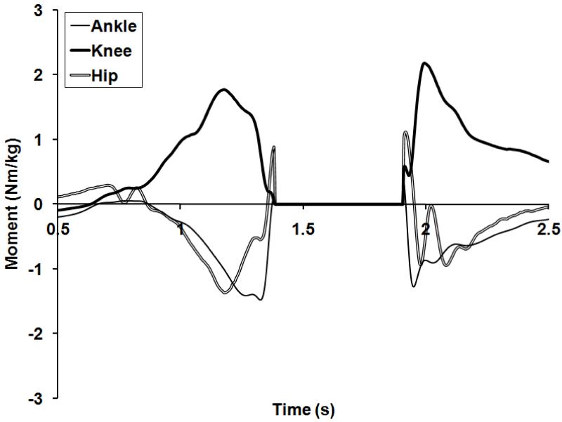
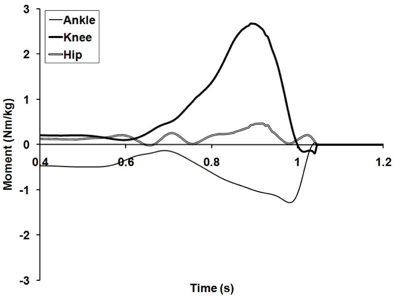
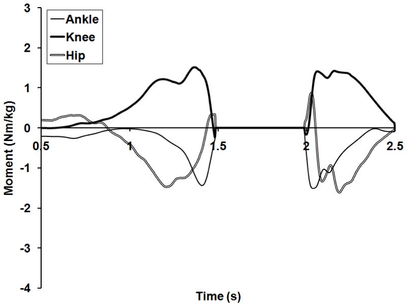
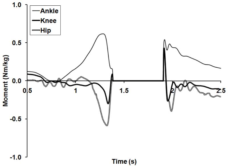
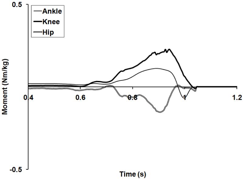
Single subject example profiles (selected qualitatively as being the most typical in pattern) are presented as opposed to ensemble averages as variability in the frontal and transverse planes, along with different jumping styles being evident, meant that ensemble average profiles lost the capacity to describe any movement strategy evident in the population. Figure 1 presents a typical sagittal plane moment curve for a jump and landing and Figure 2 presents a jerk trial for the same subject. In jumping peak hip moment was achieved prior to peak knee moment, which in turn was followed by peak ankle moment. Similarly, during jerking peak knee moment was achieved prior to peak ankle moment. This ordering of moments in the jump and jerk, evaluated qualitatively, was consistent for all subjects. There was less hip involvement in jerking (p<0.05), and the timing of peak hip moment was less consistent in relation to the peak knee moment. During landing, the reverse sequence of peak moment development was observed with peak ankle moment occurring prior to peak knee, and then hip moment. There was some variation in the hip moment developed during landing as two subjects developed very little hip moment, whereas the majority developed a large hip moment in effect replicating the jump takeoff in reverse.
Figure 1.
Sagittal plane moments during vertical jumping and landing (Subject 3). Extension moments are negative at the hip, positive at the knee and negative at the ankle.
Figure 2.
Sagittal plane moments during jerking (Subject 3).
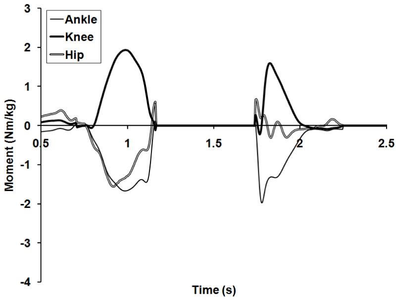
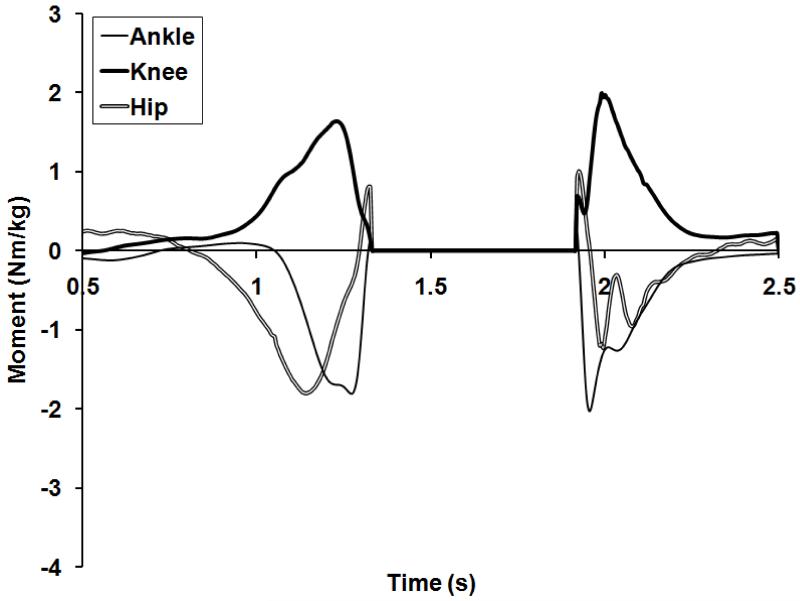
Three jump strategies were described in the group. Where subjects hip:knee ratio exceeded 1.1 (hip torque 10% higher than knee torque) jumpers were classed as hip dominant. Vice versa where the ratio dropped below 0.9 jumpers were classified as knee dominant. Ratios between 0.9 and 1.1 were classed as balanced. 3 athletes were hip dominant, 4 were balanced and 5 were knee dominant. Figures 3, 4 and 5 highlight example profiles knee, hip, and balanced jumpers respectively.
Figure 3.
Sagittal plane moments during vertical jumping and landing for a knee dominant jumper (Subject 5).
Figure 4.
Sagittal plane moments during vertical jumping and landing for a hip dominant jumper (Subject 10).
Figure 5.
Sagittal plane moments during vertical jumping and landing for a balanced jumper (Subject 12).
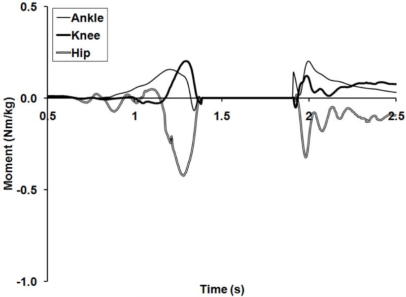
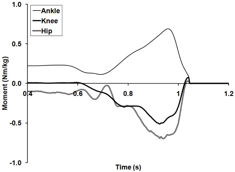
Tables 2 and 3 present the mean peak frontal and transverse plane moments observed during the three activities. Figure 6 presents the frontal and Figure 7 the transverse plane moments for an example subject during jumping and landing. Figure 8 presents the frontal and Figure 9 the transverse plane moments for the same subject during jerking. Positive frontal plane moments represent adduction moments in a proximal frame and positive transverse plane moments represent internal rotation in a proximal frame.
Table 3.
Mean peak frontal plane moments (Nm/kg) during jumping, landing and jerking. * indicates mean peak ankle moment was not an eversion moment – minimum inversion moment is presented instead.
| Ankle | Knee | Hip | ||||
|---|---|---|---|---|---|---|
| Inversion | Eversion | Adduction | Abduction | Adduction | Abduction | |
| Jump | 0.49 0.17 | 0.03 0.09 | 0.33 0.13 | 0.34 0.18 | 0.48 0.31 | 0.36 0.17 |
| Land | 0.61 0.23 | 0.01 0.10 | 0.48 0.14 | 0.35 0.37 | 0.49 0.24 | 0.37 0.34 |
| Jerk | 0.57 0.14 | 0.05* 0.03 | 0.21 0.10 | 0.29 0.17 | 0.29 0.21 | 0.40 0.23 |
Figure 6.
Frontal plane moments during jumping and landing (Subject 3).
Figure 7.
Transverse plane moments during jumping and landing (Subject 3).
Figure 8.
Frontal plane moments during jerking (Subject 3).
Figure 9.
Transverse plane moments during jerking (Subject 3).
There was a wide degree of variation in the non-sagittal plane moments observed during jumping and landing. The most consistent pattern evident was that the ankle joint experienced an inversion moment throughout jumping and landing for all subjects. The ankle joint was generally subjected to an internal rotation moment, although for most subjects there was a brief external rotation moment immediately prior to takeoff, and some subjects experienced an external rotation moment at the point of landing. The majority of subjects experienced principally knee abduction during jumping with a brief adduction loading just before takeoff. During landing there was a high degree of variation in adduction and abduction loading of the knee. Similarly the internal/external rotation moment at the knee was highly variable during both takeoff and landing, and in particular it was not uncommon for the knee to experience rotation moments in both directions during takeoff. There was a trend to experience adduction followed by abduction moments at the hip during takeoff, with some subjects then experiencing a further adduction moment just before takeoff. The rotation moment at the hip tended to rapidly switch from internal to external multiple times during takeoff. There was a high degree of variability in both the adduction/abduction and internal/external rotation moments experienced during landing although around half the cohort experienced a consistent pattern of adduction loading at landing.
During jerking the non-sagittal plane moments at the ankle were similar to those displayed during jumping. All subjects experienced inversion coupled with internal rotation moments at the ankle, with some subjects experiencing external rotation at the ankle at the point of take-off. The knee tended towards abduction loading although this pattern was not clear, whereas the rotation loading was highly variable. Frontal plane loading at the hip was variable, whereas in the transverse plane most subjects experienced some external rotation loading.
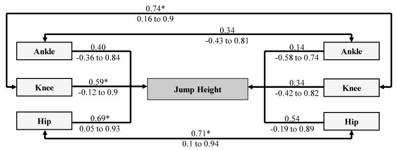
Figure 10 presents the correlation between peak ankle, knee and hip moments and vertical jump height. It is clear that there was no correlation between jump height and peak ankle moment (p>0.05). In contrast there were moderately-strong positive correlations between jump height and peak knee and hip moments which were significant (p<0.05). There was no correlation between peak moments during jerking and jump height (p>0.05). There was a significant and strong positive correlation between peak knee and hip moments between jumping and jerking (p<0.05).
Figure 10.
Correlations (Pearson’s r and 95% likely range) between jump height and peak joint moments for the jump and jerk. (* p < 0.05)
DISCUSSION
In this study the inter-segmental moments produced during vertical jumping and the weightlifting jerk were evaluated. The mean sagittal plane moments during vertical jumping were found to be consistent with previous research26,46,47. For instance, Vanezis and Lees computed the peak moments produced by the combined action of both legs in vertical jumping, and found that good jumpers produced peak moments at the ankle, knee and hip of 3.06 Nm/kg, 3.40 Nm/kg and 3.50 Nm/kg respectively, whereas poorer jumpers produced moments of 2.75 Nm/kg, 3.13 Nm/kg and 3.09 Nm/kg respectively. In this study, the peak moments for the right limb alone were calculated, however if these figures are doubled the values for ankle, knee and hip are 3.13 Nm/kg, 3.29 Nm/kg and 3.12 Nm/kg respectively. These values are in close agreement with those produced by the jumpers in the Vanezis and Lees study. Further, a visual inspection of the timing of peak moments, showed a proximal to distal pattern in all cases. This was replicated during jerking, whereas during landing it was reversed. Bobbert and colleagues4 have argued that a proximal to distal recruitment strategy allows the attainment of maximum vertical jump height by preventing a premature takeoff during vertical jumping. The pattern of rotations facilitates the joints of the lower limb to extend maximally, allowing the ankle, knee and hip to perform the greatest amount of work. It seems likely that during jerking the same proximal to distal recruitment strategy allows the torso segment to impart the greatest velocity to the bar prior to lift off from the shoulders. This similarity in extension sequencing in the jump and jerk likely also serves to allow effective energy transfer from proximal to distal joints and is common to other tasks such as running37. Such commonality of the extension strategy for the lower limb increases the likelihood of transfer of performance gains across tasks.
The correlation analysis revealed a moderately-strong and significant positive correlation between vertical jump height and jump peak knee and hip moments. There have been a number of studies that demonstrated a relationship between muscular strength and jump performance32,35,38,41. The findings of this study suggest one mechanism by which this relationship could be mediated, as athletes with higher levels of muscular strength would be expected to be capable of expressing higher peak hip and knee moments. In contrast the relationships between jump height and jump ankle moments and all jerk moments were substantially weaker and not statistically significant here. The weak relationship between ankle moments and jumping is in agreement with previous work and is intuitive if the primary input via the ankle is late and restricted to providing the final thrust when the majority of velocity creation is complete4. More importantly to coaches the meagre relationships apparent between jerking and jump performance illustrate the limited influence of jerk performance in a group that is homogenous on jump performance but heterogeneous technically. Coaches therefore need to consider carefully when selecting an exercise such as jerking for performance enhancement, precisely what adaptation is likely to be promoted and how is it proposed this will transfer to, for example, jumping performance for a specific athlete.
In contrast, there was a strong and significant correlation between the peak knee and hip moments achieved in jumping and jerking. This seems intuitive considering the strongest athletes are likely to be able to produce higher moments in both test conditions providing they can demonstrate competent levels of skill in each task. It is interesting to note that the weights jerked in this study were sub maximal whereas the jumps were maximal. It may be that the relationship between knee and hip moments becomes stronger as the weights jerked approach maximal loads. Equally the strength of the relationship may fall since athletes were still exerting maximal effort on those sub maximal loads, and as load increases the force-time characteristics of the jerk skill progress further away from jumping and the correspondence between the two skills may well fall. This point requires further investigation. Nevertheless the results of this study suggest that those subjects who are capable of expressing for example higher knee joint moments are able to express these knee joint moments during both jumping and jerking. The ability to express high knee joint moments is therefore at least in some part independent of the skill performed and represents a characteristic of transferability.
Rousanoglou and colleagues38 found that measures of knee extensor strength correlated to jump height in volleyball players but not with track and field jumpers. Their work supports a proposition that different groups of athletes select different movement strategies to perform maximal height vertical jumps and that as such different strength adaptations might be required to further enhance performance. In the present study, the cohort of jumpers considered was a heterogeneous group in which both peak knee and hip moment was correlated with jump height. It may be that in ‘knee dominant’ jumpers there is a stronger relationship between peak knee moment and jump height and vice versa. If such a relationship could be established this would potentially provide an explanation as to the findings of Rousanoglou and co-workers. The track and field jumpers may select a more hip dominant strategy and thus their performance may be more correlated with peak hip moment production more so than peak knee moment. Conversely, the volleyball players may be more knee dominant, and thus their performance may be more strongly influenced by their ability to produce peak knee moments. This may be in part due to the relative differences in horizontal components of motion between these sports and points further to the importance of more fully understanding the correspondence between skills and likely transferability of strength qualities. Further it raises potential questions as to whether a knee dominant jumper’s performance would benefit most from increases in hip moment or alternately further increases in knee moment capability.
In common with previous research46, a variation in jumping strategy was identified here. Subjects selected a knee dominant, hip dominant or more balanced movement strategy although taken as a cohort the production of hip moment was greater than that of knee moment. In contrast, the mean moments produced upon landing favoured the production of knee moment but still two different landing strategies were observed in this study. Most subjects developed peak moments in a similar but reversed pattern to takeoff, using both the hip and knee joints, whereas two subjects relied heavily upon the development of knee joint moment to absorb the landing forces. These two subjects were also knee dominant jumpers, which may represent a consistent preferential recruitment of the knee over the hip. Cohort size restricted further correlational analysis of these subgroups which might inform a stronger characterisation of hip and knee dominant jumpers. Wider investigation is needed to determine what characteristics might cause athletes to select a particular joint dominant strategy, whether this is optimal for performance outcome and importantly whether this increases risk of injury at the same joint, particularly in the case of the knee. It may also be important to understand if these strategies transfer to other movements and further investigation to characterise movement strategy preference is necessary in jumping and other sports skills.
Comparative analysis of jumping and jerking reveals that the ankle joint appears to serve a similar function during the two activities, that of providing the final thrust, and that the magnitude of this contribution is the same in the two activities. Equally, during landing the moment produced at the ankle is similar. In contrast, the knee and hip are used very differently in jumping and jerking. Jerking is a knee dominant activity with a much greater emphasis on knee moment development as demonstrated in the substantially higher knee:hip moment ratios demonstrated, whereas jumping represents a much more balanced recruitment of knee and hip averaged across the cohort. This variation in movement strategy is likely linked to the restriction of trunk motion enforced with the use of the barbell in the jerk. The apparent disassociation in the relative magnitude of contribution of hip and knee moments further points to the generality of jerking in relation to jumping tasks. Whilst this, in conjunction with previously described correlations, might lead to a conclusion that jerking lacks potential to transfer to jumping it should be considered that only 40kg was lifted in the jerk trials. Assuming larger jerk loads would further increase knee moments, then jerking would appear to offer an effective strategy to provide overload for peak knee moment generation and would also likely offer greater correspondence to tasks which are similarly dominated by knee function or have benefit where athletes are perceived to be lacking in torque capability at the knee such as to say the knee represents a ‘weak link’. Jump squatting, or likewise any training activity where trunk motion is similarly constrained, might be subject to a similar “disconnect” from free jumping and requires further investigation.
The non-sagittal plane moments reported in this study also show agreement with previous work2,22. The ankle experienced a consistent pattern of inversion and internal rotation loading in all three activities in this study which suggests both that the ankle joint is utilized in a fairly consistent pattern in the studied activities and that non-sagittal plane moments are well controlled at the ankle. In contrast, the non-sagittal plane moments experienced at the hip and knee were much more variable. The mechanism of injury to the knee during non-contact landings is generally due to a multiplane loading at the knee, often at small knee flexion angles, when a large extensor moment is coupled with either frontal or transverse plane loading17,30,42. Thus knee injuries during landing may be due in a large part to the development of non-sagittal plane moments, especially unanticipated moments. Both the hip and knee exhibited variability in the joint moments observed during landing. The hip joint is structurally a very stable joint and is more likely to be able to withstand variability in loading. In contrast, a potential explanation for the high incidence of non-contact knee injuries during jumping16 could be the variability of moments in this relatively unstable joint.
In this study, two contrasting landing styles were observed as discussed earlier. It could be argued that the balanced landing strategy that equally recruited the knee and hip represents the safest option, as the development of extensor moment is evenly distributed between the hip and knee. However, no significant correlations were found between the magnitudes of the sagittal plane knee moments and the non-sagittal plane moments (not presented). That said the abduction moment correlation of 0.53 (95% likely range −0.06 to 0.85; p=0.07) would suggest that a meaningful positive relationship is might exist between these variables. Thus although the knee may be more heavily loaded in the sagittal plane by a knee dominant landing strategy, further investigation is necessary to elucidate the potential for this style to result in increased non-sagittal plane loading. This relationship should be explored further particularly in populations known to be at higher risk of knee injury such as females5,34 using designs with higher statistical power than was achieved here.
This study provides further evidence as to the utility of inverse dynamics analysis in understanding movement correspondence. The existence of distinct jumping strategies (knee or hip dominant) has been reinforced and the constraint of vertical bar motion in the jerk shown to result in the activity being clearly knee dominant. Despite the direct relationship between jerk moments and jump performance being weak an avenue for correspondence is highlighted in the relationship between hip and knee peak moments in the jump and jerk. Sagittal plane control has been shown to be variable and the implications of this for injury risk along with the potential to reduce variability needs consideration. Future research should attempt to further describe variations in jumping and landing strategies, whether these strategies transfer across a wider set of sports skills and their impact on non-sagittal plane moments. Additionally the causes of jumping strategy variability require investigation along with the implications for training interventions centred at reinforcing or changing athletes existing strategy through general strength, flexibility or technical training. Equally the understanding of the characteristics of OL activities could be further improved and the relationship between weightlifting and jumping, as well as other fundamental sports skills, clarified.
PRACTICAL APPLICATIONS
The results of this study suggest that in order to improve vertical jump performance a key consideration should be to improve an athlete’s ability to produce both peak knee and hip moments during the jump. Coaches should be aware that their athletes may select to jump and land with hip or knee dominant strategies and understand that an athlete’s particular movement strategy may be a function of their relative strengths and weaknesses, and predicate the type of training that has potential for improving their performance, either through an approach to change this strategy (by enhancing hip moment capabilities in a knee dominant jumper) or to reinforce it (by increasing knee moment capabilities in a knee dominant jumper). Finally this study indicates that the jerk exercise can be classified as knee dominant and may be effective in overloading and therein increasing an athlete’s ability to generate peak knee moments more so than ankle or hip, although it is unable to substantiate whether this will in turn lead directly to increases in vertical jump height.
Table 4.
Mean peak transverse plane moments (Nm/kg) during jumping, landing and jerking.
| Ankle | Knee | Hip | ||||
|---|---|---|---|---|---|---|
| Int Rot | Ext Rot | Int Rot | Ext Rot | Int Rot | Ext Rot | |
| Jump | 0.12 0.07 | 0.09 0.08 | 0.17 0.12 | 0.10 0.09 | 0.35 0.24 | 0.30 0.14 |
| Land | 0.23 0.09 | 0.08 0.13 | 0.14 0.10 | 0.10 0.05 | 0.25 0.15 | 0.24 0.17 |
| Jerk | 0.12 0.07 | 0.06 0.05 | 0.14 0.09 | 0.7 0.7 | 0.07 0.06 | 0.21 0.14 |
Acknowledgements
The authors would like to thank Hayley Legg for her assistance with data collection during this study.
Footnotes
The authors have no conflict of interests nor received funding from any external agency for this project.
References
- 1.Anderson FC, Pandy MG. A dynamic optimization solution for vertical jumping in three dimensions. Computer Methods in Biomechanics and Biomedical Engineering. 1999;2:201–231. doi: 10.1080/10255849908907988. [DOI] [PubMed] [Google Scholar]
- 2.Bisseling RW, Hof AL, Bredeweg SW. Are the take-off and landing phase dynamics of the volleyball spike jump related to patellar tendinopathy? British Journal of Sports Medicine. 2008;42:483–489. doi: 10.1136/bjsm.2007.044057. [DOI] [PubMed] [Google Scholar]
- 3.Blackburn JT, Padua DA. Influence of trunk flexion on hip and knee joint kinematics during a controlled drop landing. Clinical Biomechanics. 2008;23:313–319. doi: 10.1016/j.clinbiomech.2007.10.003. [DOI] [PubMed] [Google Scholar]
- 4.Bobbert MF, van Soest AJ. Why do people jump the way they do? Exercise and Sport Sciences Reviews. 2001;29:95–102. doi: 10.1097/00003677-200107000-00002. [DOI] [PubMed] [Google Scholar]
- 5.Borowski LA, Yard EE, Fields SK, Comstock RD. The epidemiology of US high school basketball injuries, 2005-2007. American Journal of Sports Medicine. 2008;36:2328–2336. doi: 10.1177/0363546508322893. [DOI] [PubMed] [Google Scholar]
- 6.Campos J, Poletaev P, Cuesta A, Pablos C, Carratala V. Kinematical analysis of the snatch in elite male junior weightlifters of different weight categories. Journal of Strength and Conditioning Research. 2006;20:843–850. doi: 10.1519/R-55551.1. [DOI] [PubMed] [Google Scholar]
- 7.Clarys JP. Electromyography in sports and occupational settings: an update of its limits and possibilities. Ergonomics. 2000;43:1750–62. doi: 10.1080/001401300750004159. [DOI] [PubMed] [Google Scholar]
- 8.Cleather DJ, Bull AMJ. Lower extremity musculoskeletal geometry effects the calculation of patellofemoral forces in vertical jumping and weightlifting. Proceedings of the Institution of Mechanical Engineers. Part H, Journal of Engineering in Medicine. 2010;224:1073–83. doi: 10.1243/09544119JEIM731. [DOI] [PubMed] [Google Scholar]
- 9.Cleather DJ, Goodwin JE, Bull AMJ. An optimization approach to inverse dynamics provides insight as to the function of the biarticular muscle during vertical jumping. Annals of Biomedical Engineering. 2011;39:147–60. doi: 10.1007/s10439-010-0161-9. [DOI] [PubMed] [Google Scholar]
- 10.Cleather DJ, Goodwin JE, Bull AMJ. Erratum to: An optimization approach to inverse dynamics provides insight as to the function of the biarticular muscle during vertical jumping. Annals of Biomedical Engineering. 2011;39:2476–8. doi: 10.1007/s10439-010-0161-9. [DOI] [PubMed] [Google Scholar]
- 11.Collins JJ. Antagonistic-synergistic muscle action at the knee during competitive weightlifting. Medical and Biological Engineering and Computing. 1994;32:168–174. doi: 10.1007/BF02518914. [DOI] [PubMed] [Google Scholar]
- 12.Crowninshield RD, Brand RA. A physiologically based criterion of muscle force prediction in locomotion. Journal of Biomechanics. 1981;14:793–801. doi: 10.1016/0021-9290(81)90035-x. [DOI] [PubMed] [Google Scholar]
- 13.Decker MJ, Torry MR, Wyland DJ, Sterett WI, Steadman JR. Gender differences in lower extremity kinematics, kinetics and energy absorption during landing. Clinical Biomechanics. 2003;18:662–669. doi: 10.1016/s0268-0033(03)00090-1. [DOI] [PubMed] [Google Scholar]
- 14.Dumas R. A 3D Generic Inverse Dynamic Method using Wrench Notation and Quaternion Algebra. Computer methods in biomechanics and biomedical engineering. 2004;7:159–166. doi: 10.1080/10255840410001727805. [DOI] [PubMed] [Google Scholar]
- 15.Dumas R, Aissaoui R, de Guise JA. A 3D generic inverse dynamic method using wrench notation and quaternion algebra. Computer methods in biomechanics and biomedical engineering. 2004;7:159–166. doi: 10.1080/10255840410001727805. [DOI] [PubMed] [Google Scholar]
- 16.Erdemir A, McLean S, Herzog W, van den Bogert AJ. Model-based estimation of muscle forces exerted during movements. Clinical Biomechanics. 2007;22:131–154. doi: 10.1016/j.clinbiomech.2006.09.005. [DOI] [PubMed] [Google Scholar]
- 17.Ferretti A, Papandrea P, Conteduca F, Mariani PP. Knee ligament injuries in volleyball players. American Journal of Sports Medicine. 1992;20:203–207. doi: 10.1177/036354659202000219. [DOI] [PubMed] [Google Scholar]
- 18.Fukashiro S, Komi PV. Joint Moment and Mechanical Power Flow of the Lower-Limb During Vertical Jump. International Journal of Sports Medicine. 1987;8:15–21. doi: 10.1055/s-2008-1025699. [DOI] [PubMed] [Google Scholar]
- 19.Garhammer J. A review of power output studies of olympic and powerlifting: Methodology, performance prediction, and evaluation tests. Journal of Strength and Conditioning Research. 1993;7:76–89. [Google Scholar]
- 20.Garhammer J, Gregor R. Propulsion forces as a function of intensity for weightlifting and vertical jumping. Journal of Applied Sport Science Research. 1992;6:129–134. [Google Scholar]
- 21.Gourgoulis V, Aggeloussis N, Kalivas V, Antoniou P, Mavromatis G. Snatch lift kinematics and bar energetics in male adolescent and adult weightlifters. Journal of Sports Medicine and Physical Fitness. 2004;44:126–131. [PubMed] [Google Scholar]
- 22.Hass CJ, Schick EA, Tillman MD, Chow JW, Brunt D, Cauraugh JH. Knee biomechanics during landings: Comparison of pre- and postpubescent females. Medicine and science in sports and exercise. 2005;37:100–107. doi: 10.1249/01.mss.0000150085.07169.73. [DOI] [PubMed] [Google Scholar]
- 23.Klein Horsman MD, Koopman HFJM, van der Helm FCT, Prose LP, Veeger HEK. Morphological muscle and joint parameters for musculoskeletal modelling of the lower extremity. Clinical Biomechanics. 2007;22:239–247. doi: 10.1016/j.clinbiomech.2006.10.003. [DOI] [PubMed] [Google Scholar]
- 24.Kernozek TW, Ragan RJ. Estimation of anterior cruciate ligament tension from inverse dynamics data and electromyography in females during drop landing. Clinical Biomechanics. 2008;23:1279–1286. doi: 10.1016/j.clinbiomech.2008.08.001. [DOI] [PubMed] [Google Scholar]
- 25.Lauder MA, Lake JP. Biomechanical comparison of unilateral and bilateral power snatch lifts. Journal of Strength and Conditioning Research. 2008;22:653–660. doi: 10.1519/JSC.0b013e3181660c89. [DOI] [PubMed] [Google Scholar]
- 26.Lees A, Vanrenterghem J, de Clercq D. The maximal and submaximal vertical jump: Implications for strength and conditioning. Journal of Strength and Conditioning Research. 2004;18:787–791. doi: 10.1519/14093.1. [DOI] [PubMed] [Google Scholar]
- 27.Li G, Kaufman KR, Chao EYS, Rubash HE. Prediction of antagonistic muscle forces using inverse dynamic optimization during flexion/extension of the knee. Journal of Biomechanical Engineering. 1999;121:316–322. doi: 10.1115/1.2798327. [DOI] [PubMed] [Google Scholar]
- 28.Luhtanen P, Komi PV. Segmental Contribution to Forces in Vertical Jump. European Journal of Applied Physiology and Occupational Physiology. 1978;38:181–188. doi: 10.1007/BF00430076. [DOI] [PubMed] [Google Scholar]
- 29.McNitt-Gray JL. Kinetics of the Lower-Extremities During Drop Landings from 3 Heights. Journal of Biomechanics. 1993;26:1037–1046. doi: 10.1016/s0021-9290(05)80003-x. [DOI] [PubMed] [Google Scholar]
- 30.Meyer EG, Haut RC. Anterior cruciate ligament injury induced by internal tibial torsion or tibiofemoral compression. Journal of Biomechanics. 2008;41:3377–3383. doi: 10.1016/j.jbiomech.2008.09.023. [DOI] [PubMed] [Google Scholar]
- 31.Nha KW, Papannagari R, Gill TJ, Van de Velde SK, Freiberg AA, Rubash HE, Li G. In vivo patellar tracking: Clinical motions and patellofemoral indices. Journal of Orthopaedic Research. 2008;26:1067–1074. doi: 10.1002/jor.20554. [DOI] [PMC free article] [PubMed] [Google Scholar]
- 32.Nuzzo JL, McBride JM, Cormie P, McCaulley GO. Relationship between countermovement jump performance and multijoint isometric and dynamic tests of strength. Journal of Strength and Conditioning Research. 2008;22:699–707. doi: 10.1519/JSC.0b013e31816d5eda. [DOI] [PubMed] [Google Scholar]
- 33.Pandy MG, Zajac FE. Optimal muscular coordination strategies for jumping. Journal of Biomechanics. 1991;24:1–10. doi: 10.1016/0021-9290(91)90321-d. [DOI] [PubMed] [Google Scholar]
- 34.Parkkari J, Pasanen K, Mattila VM, Kannus P, Rimpela A. The risk for a cruciate ligament injury of the knee in adolescents and young adults: A population-based cohort study of 46500 people with a 9 year follow-up. British Journal of Sports Medicine. 2008;42:422–426. doi: 10.1136/bjsm.2008.046185. [DOI] [PubMed] [Google Scholar]
- 35.Peterson MD, Alvar BA, Rhea MR. The contribution of maximal force production to explosive movement young collegiate athletes. Journal of Strength and Conditioning Research. 2006;20:867–873. doi: 10.1519/R-18695.1. [DOI] [PubMed] [Google Scholar]
- 36.Pflum MA, Shelburne KB, Torry MR, Decker MJ, Pandy MG. Model prediction of anterior cruciate ligament force during drop-landings. Medicine and science in sports and exercise. 2004;36:1949–1958. doi: 10.1249/01.mss.0000145467.79916.46. [DOI] [PubMed] [Google Scholar]
- 37.Prilutsky BI, Zatsiorsky VM. Tendon action of two-joint muscles: transfer of mechanical energy between joints during jumping, landing and running. Journal of Biomechanics. 1994;27:25–34. doi: 10.1016/0021-9290(94)90029-9. [DOI] [PubMed] [Google Scholar]
- 38.Rousanoglou EN, Georgiadis GV, Boudolos KD. Muscular strength and jumping performance relationships in young women athletes. Journal of Strength and Conditioning Research. 2008;22:1375–1378. doi: 10.1519/JSC.0b013e31816a406d. [DOI] [PubMed] [Google Scholar]
- 39.Schenau GJV, Bobbert MF, deHaan A. Does elastic energy enhance work and efficiency in the stretch-shortening cycle? Journal of Applied Biomechanics. 1997;13:389–415. [Google Scholar]
- 40.Sell TC, Ferris CM, Abt JP, Tsai YS, Myers JB, Fu FH, Lephart SM. Predictors of proximal tibia anterior shear force during a vertical stop-jump. Journal of Orthopaedic Research. 2007;25:1589–1597. doi: 10.1002/jor.20459. [DOI] [PubMed] [Google Scholar]
- 41.Sheppard JM, Cronin JB, Gabbett TJ, McGuigan MR, Etxebarria N, Newton RU. Relative importance of strength, power, and anthropometric measures to jump performance of elite volleyball players. Journal of Strength and Conditioning Research. 2008;22:758–765. doi: 10.1519/JSC.0b013e31816a8440. [DOI] [PubMed] [Google Scholar]
- 42.Shimokochi Y, Shultz SJ. Mechanisms of noncontact anterior cruciate ligament injury. Journal of Athletic Training. 2008;43:396–408. doi: 10.4085/1062-6050-43.4.396. [DOI] [PMC free article] [PubMed] [Google Scholar]
- 43.Souza AL, Shimada SD. Biomechanical analysis of the knee during the power clean. Journal of Strength and Conditioning Research. 2002;16:290–297. [PubMed] [Google Scholar]
- 44.Van Sint Jan S. Skeletal landmark definitions: Guidelines for accurate and reproducible palpation. University of Brussels, Department of Anatomy; 2005. Www.Ulb.Ac.Be/~Anatemb [Google Scholar]
- 45.Van Sint Jan S, Croce UD. Identifying the location of human skeletal landmarks: Why standardized definitions are necessary - a proposal. Clinical Biomechanics. 2005;20:659–660. doi: 10.1016/j.clinbiomech.2005.02.002. [DOI] [PubMed] [Google Scholar]
- 46.Vanezis A, Lees A. A biomechanical analysis of good and poor performers of the vertical jump. Ergonomics. 2005;48:1594–1603. doi: 10.1080/00140130500101262. [DOI] [PubMed] [Google Scholar]
- 47.Vanrenterghem J, Lees A, Lenoir M, Aerts P, de Clercq D. Performing the vertical jump: Movement adaptations for submaximal jumping. Human movement science. 2004;22:713–727. doi: 10.1016/j.humov.2003.11.001. [DOI] [PubMed] [Google Scholar]
- 48.V Verkhoshansky, Y. Special Strength Training. Ultimate Athlete Concepts; Michigan: 2006. [Google Scholar]
- 49.Winter DA. Biomechanics and Motor Control of Human Movement. John Wiley & Sons; Hoboken, NJ: 2005. [Google Scholar]
- 50.Woltring HJ. A Fortran package for generalized, cross-validatory spline smoothing and differentiation. Advances in Engineering Software. 1986;8:104–113. [Google Scholar]
- 51.Yeow CH, Lee PVS, Goh JCH. Effect of landing height on frontal plane kinematics, kinetics and energy dissipation at lower extremity joints. Journal of Biomechanics. 2009;42:1967–1973. doi: 10.1016/j.jbiomech.2009.05.017. [DOI] [PubMed] [Google Scholar]
- 52.Zatsiorsky VM. Kinetics of Human Motion. Human Kinetics; Champaign, IL: 2002. [Google Scholar]
- 53.Zernicke RF, Garhammer J, Jobe FW. Human patellar-tendon rupture. Journal of Bone and Joint Surgery. American volume. 1977;59:179–183. [PubMed] [Google Scholar]


