Abstract
Multiple myeloma (MM) is an incurable hematological malignancy. Chimeric antigen receptor (CAR)-expressing T cells have been demonstrated successful in the clinic to treat B-lymphoid malignancies. However, the potential utility of antigen-specific CAR-engineered natural killer (NK) cells to treat MM has not been explored. In this study, we determined whether CS1, a surface protein that is highly expressed on MM cells, can be targeted by CAR NK cells to treat MM. We successfully generated a viral construct of a CS1-specific CAR and expressed it in human NK cells. In vitro, CS1-CAR NK cells displayed enhanced MM cytolysis and IFN-γ production, and showed a specific CS1-dependent recognition of MM cells. Ex vivo, CS1-CAR NK cells also showed similarly enhanced activities when responding to primary MM tumor cells. More importantly, in an aggressive orthotopic MM xenograft mouse model, adoptive transfer of NK-92 cells expressing CS1-CAR efficiently suppressed the growth of human IM9 MM cells and also significantly prolonged mouse survival. Thus, CS1 represents a viable target for CAR-expressing immune cells, and autologous or allogeneic transplantation of CS1-specific CAR NK cells may be a promising strategy to treat MM.
Keywords: CS1, Chimeric Antigen Receptor, NK Cells, Multiple Myeloma
INTRODUCTION
Multiple myeloma (MM) is a B-cell malignancy characterized by the aberrant clonal expansion of plasma cells (PCs) within the bone marrow (BM)1, with an estimated 21,700 new cases and 10,710 deaths from MM identified in the United States in 20122. Despite the use of proteasome inhibitors and immune-modulating drugs, which have improved overall survival3, MM remains an incurable malignancy4 for which novel therapeutic approaches are urgently needed.
Natural-killer (NK) cells are CD56+CD3− large granular lymphocytes that can kill virally infected and transformed cells, and constitute a critical cellular subset of the innate immune system5. Unlike cytotoxic CD8+ T lymphocytes, NK cells launch cytotoxicity against tumor cells without the requirement for prior sensitization, and can also eradicate MHC-I negative cells6. NK cells are safer effector cells, as they may avoid the potentially lethal complications of cytokine storms7, tumor lysis syndrome8, and on-target, off-tumor effects. Although NK cells have a well-known role as killers of cancer cells, and NK cell impairment has been extensively documented as crucial for progression of MM5,9, the means by which we might enhance NK cell-mediated anti-MM activity has been largely unexplored.
One intriguing approach for treating cancer involves the genetic modification of T cells or NK cells with chimeric antigen receptors (CARs) that directly target tumor-associated antigens. In fact, CAR T cells have been demonstrated successful in the clinic for treatment of CD19+ acute lymphoblastic leukemia (ALL) and chronic lymphocytic leukemia (CLL)8,10–12. However, treatment of patients with CAR T cells can result in cytokine storms7,10 and, in the setting of allogeneic transplantation, may induce graft-versus-host disease (GVHD). While CAR NK cells are thought to be safer effector cells, their therapeutic use has not been explored. In myeloma, unique surface antigens have been challenging to target13. Recent studies have suggested that the cell surface glycoprotein, CS1, may be an ideal target for the treatment of MM14–16. CS1 is highly, and nearly ubiquitously, expressed on MM cells, while expression remains very low on NK cells, some T-cell subsets, and normal B cells, and almost undetectable on myeloid cells and the majority of healthy tissues14. Importantly, CS1 is not expressed on hematopoietic stem cells14, which are often used in the form of autologous stem cell transplantation for treatment of MM. While the exact role of CS1 in normal plasma cells is unknown, CS1 co-localizes with CD138 on the surface of polarized MM cells, and may promote MM cell adhesion, clonogenic growth, and tumorigenicity via c-Maf-mediated interaction with bone marrow stromal cells (BMSC)1,16,17. Targeting CS1 with the humanized IgG1 monoclonal antibody (mAb), Elotuzumab (formerly Huluc63), can inhibit myeloma cell adhesion to BMSC, induce NK cell-mediated antibody-dependent cellular cytotoxicity (ADCC), and promote NK cell activation without killing autologous NK cells despite low levels of CS1 expression on the surface of normal NK cells16,18. Importantly, Elotuzumab has already been proven safe in phase 1 and 2 clinical trials, and phase 3 trials are ongoing19. This suggests that CS1-targeted cytotoxic leukocytes will not impair major immune cell subsets and hematopoietic stem cells. Using NK cells engineered to express CS1-CAR is promising as a means to augment the anti-MM capacity of NK cells.
In this study, we engineered human NK cells to express a CAR that was CS1-specific, and we incorporated a CD28-CD3 ζ costimulatory signaling domain. We evaluated the anti-MM function of these cells in vitro and in an in vivo orthotopic xenograft mouse model of MM. Our results showed that the expression of the CS1-CAR could redirect NK cells to specifically and efficiently eradicate CS1-expressing MM cells, both in vitro and in vivo, and this eradication is CS1-dependent. Our data suggest this CAR strategy may be suitable for development of an effective NK cell-based immunotherapy as a means to treat patients with refractory or relapsed MM. In addition, in contrast to CAR T cells, CAR NK cells allow us to use allogeneic NK cell sources, which are less likely to cause and may even help to suppress graft-versus-host disease (GVHD)20, while also potentiating an increase in cytotoxicity due to mismatched killer immunoglobulin-like receptors (KIR)21.
MATERIALS AND METHODS
Cell culture
Mice
Six- to eight-week-old NOD.Cg-prkdcscid IL2rgtm1Wjl/szJ (NSG) mice were obtained from Jackson Laboratories (Bar Harbor, Maine, USA). All animal work was approved by The Ohio State University Animal Care and Use Committee. Mice were monitored frequently for MM disease progression, and sacrificed when they were moribund with the symptoms of hind limb paralysis, lethargy, and obvious weight loss.
Generation of anti-CS1 CAR lentiviral construct
The CS1-scFv fragment, amplified from the hybridoma cell line Luc90, was fused with a sequence encoding a Myc tag immediately following the CS1-VL-encoding sequence. The fused DNA sequences were incorporated with CD28-CD3 ζ that was incised from a retroviral vector. The entire CS1-scFv-myc tag-CD28-CD3 ζ fragment was ligated into a lentiviral vector designated PCDH-CMV-MCS-EF1-copGFP (PCDH, System Biosciences, Mountain View, CA, USA) to generate a PCDH-CS1-scFv-myc tag-CD28-CD3 ζ (PCDH-CS1-CAR) construct.
Lentivirus production and transduction of NK cells
To produce lentivirus for infection of NK cells, 293T cells cultured in DMEM media (Life Technologies, Grand Island, NY, USA) were co-transfected with the aforementioned PCDH-CS1-scFv-CD28-CD3 ζ plasmid or a mock PCDH control vector together with the packaging constructs pCMV-VSVG and pCMV-dr9 using calcium phosphate transfection reagent (Promega, Madison, WI, USA). The transfection and infection procedures were modified from a previously published protocol22 and are detailed in Supplementary Information.
Generation of a U266 cell line stably expressing CS1
Human CS1 coding sequences were amplified from cDNA isolated from IM9 cells via PCR, then subcloned into a PCDH lentiviral vector to generate a PCDH-CS1 construct. Lentivirus production and infection of U266 cells were performed using the methods described above. GFP positive cells were then sorted using a FACS Aria II cell sorter (BD Biosciences, San Jose, CA, USA).
Immunoblotting analysis
Cells were washed with PBS and directly lysed in Laemmli buffer (Bio-Rad Laboratories, Hercules, CA, USA). Lysates were electrophoretically separated on a 4% to 15% gradient SDS-PAGE gel (Bio-Rad Laboratories) and transferred to a nitrocellulose membrane. Subsequent procedures were modified from a previously published protocol22, and are detailed in Supplementary Information.
Flow cytometry
The protocol was modified from our previous publication23, and is detailed in Supplementary Information.
Cytotoxicity assay
The protocol was modified from our previous publication23, and is detailed in Supplementary Information
IFN-γ release assay
Myeloma target cells were co-cultured with NK effector cells in 96-well V bottom plates for 24 h. 2.5 × 10⁵ myeloma cell line cells or 1.0 × 10⁵ primary myeloma cells were incubated with 2.5 × 10⁵ or 5.0 × 10⁵ NK cells, respectively. Cell-free supernatants were assayed for IFN-γ secretion by enzyme-linked immunosorbent assay (ELISA) using a kit from R&D Systems (Minneapolis, MN, USA) according to the manufacturer’s protocol. Data depicted in figures represent mean values of triplicate wells from one of three representative experiments with similar results.
An orthotopic MM mouse model and in vivo treatment of MM-bearing mice and bioluminescence imaging
IM9 cells were retrovirally transduced with Pinco-pGL3-luc/GFP virus expressing firefly luciferase as previously described24. GFP positive cells were sorted using a FACS Aria II cell sorter (BD Biosciences), and were designated “IM9-GL3” cells. Then, six- to eight-week-old male NSG mice were intravenously (i.v.) injected with 0.5 × 10⁶ IM9-GL3 MM cells in 400 µL of PBS via tail vein on Day 0 in order to establish a xenograft orthotopic MM model. Beginning on Day 7, the mice were i.v. injected with 5×10⁶ effector cells, i.e. CS1-CAR NK-92 cells or mock-transduced control cells, in 400 µL of PBS once every five days (5 times in total). Four weeks after IM9-GL3 inoculation, the mice were intraperitoneally (i.p.) infused with D-luciferin (150 mg/kg body weight; Gold Biotechnology, St. Louis, MO, USA), anesthetized with isoflurane, and imaged using In Vivo Imaging System (IVIS-100, PerkinElmer, Waltham, Massachusetts, USA) with Living Image software (PerkinElmer).
Immunohistochemical analysis
Spinal vertebrae were fixed in 10% buffered formalin phosphate and decalcified in saturated EDTA, and then embedded in paraffin. Five-micron thick sections were stained with hematoxylin and eosin (H&E) for histological examination. The sections were immunostained for identification of human MM cells with mouse anti-human CD138 mAb (1:50 dilution, Thermo Scientific, Waltham, MA, USA) following standard immunohistochemistry (IHC) staining procedures. HRP-conjugated anti-mouse IgG was used as a secondary antibody, followed by a peroxidase enzymatic reaction.
Statistics
Unpaired Student’s t test was utilized to compare two independent groups for continuous endpoints if normally distributed. One-way ANOVA was used when three or more independent groups were compared. For non-normally distributed endpoints, such as in vivo bioluminescence intensity, a Kruskal-Wallis test was utilized to compare the median of NK-92-CS1-CAR to NK-92-EV and control. For survival data, Kaplan-Meier curves were plotted and compared using a log-rank test. All tests are two-sided. P values were adjusted for multiple comparisons using Bonferroni method. A P value less than 0.05 is considered statistically significant.
Results
Generation of NK-92 and NKL NK cells expressing CS1-CAR
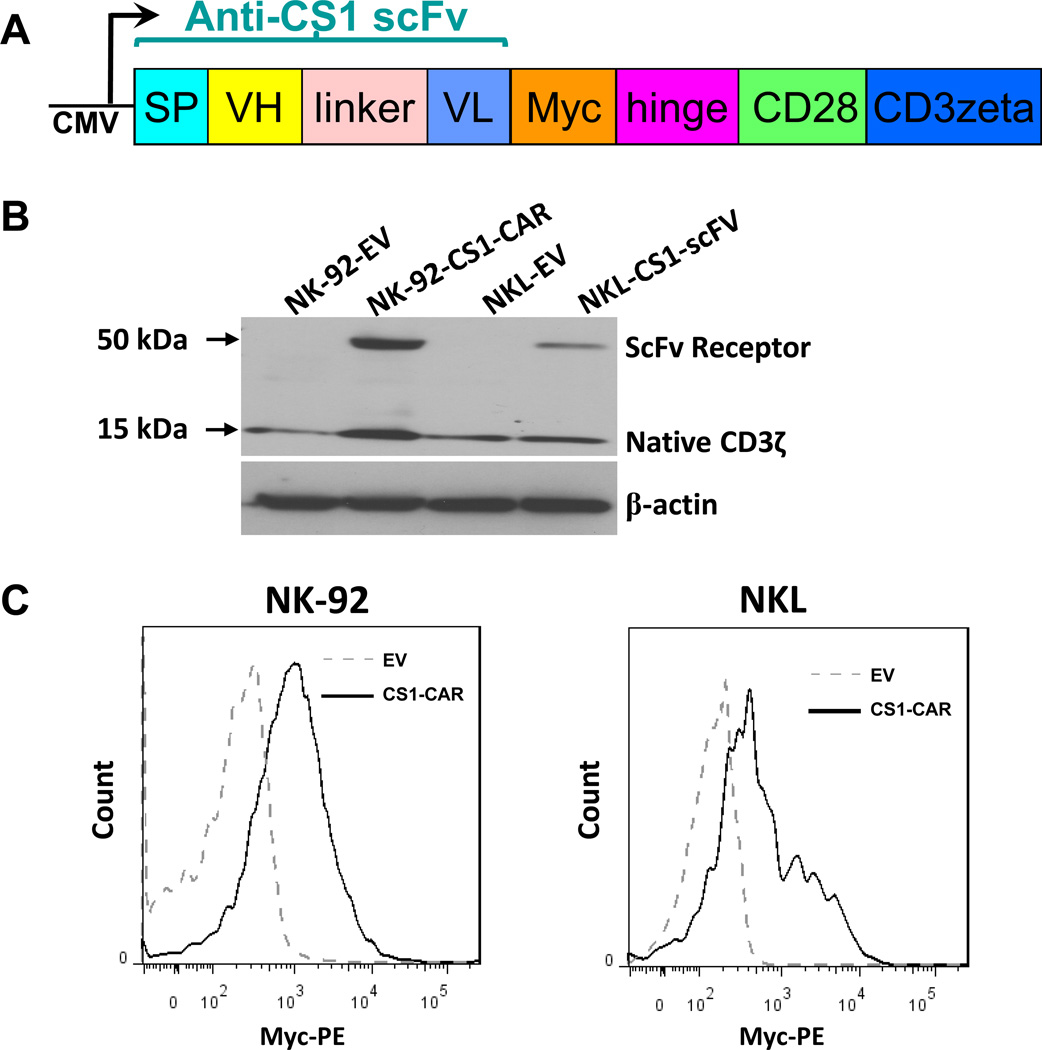
We generated a specific CS1-CAR construct with a PCDH lentiviral vector backbone, sequentially containing a signal peptide (SP), a heavy chain variable region (VH), a linker, a light chain variable region (VL), a Myc tag, a hinge, CD28 and CD3 ζ (Fig. 1A). NK-92 and NKL NK cell lines were transduced with the CAR construct and then sorted for expression of GFP, a marker expressed by the vector. Western blotting of the sorted cells demonstrated that CS1-CAR was successfully introduced and expressed, as evidenced by the expression of the chimeric CS1-scFv receptor containing CD3 ζ in both NK-92 and NKL cell lines transduced with the CAR construct rather than with the control vector (Fig. 1B). Moreover, a flow cytometric analysis after anti-Myc Ab surface staining indicated that CS1-CAR was expressed on the surface of both NK-92 and NKL cells transduced with the CS1-CAR construct (Fig. 1C).
Figure 1. Generation of a CS1-specific CAR and detecting its expression in CAR-transduced NK cells.
A) Schematic representation of the CS1-CAR lentiviral construct that we generated. B) Western blotting analysis of CS1-CAR expression using a CD3 ζ-specific Ab. Data shown are representative of three experiments with similar results. C) Expression of chimeric CS1 scFv on the surface of FACS-sorted NK-92 and NKL cells transduced with the CS1-CAR construct (NK-92-CS1-CAR and NKL-CS1-CAR) was analyzed by flow cytometry after cells were stained with an anti-myc antibody or IgG1 isotype control. Data shown are representative of three experiments with similar results.
CS1-CAR-modified NK cells more effectively eradicate CS1+ MM cells, but not CS1− cells, in vitro in comparison to mock-transduced NK cells
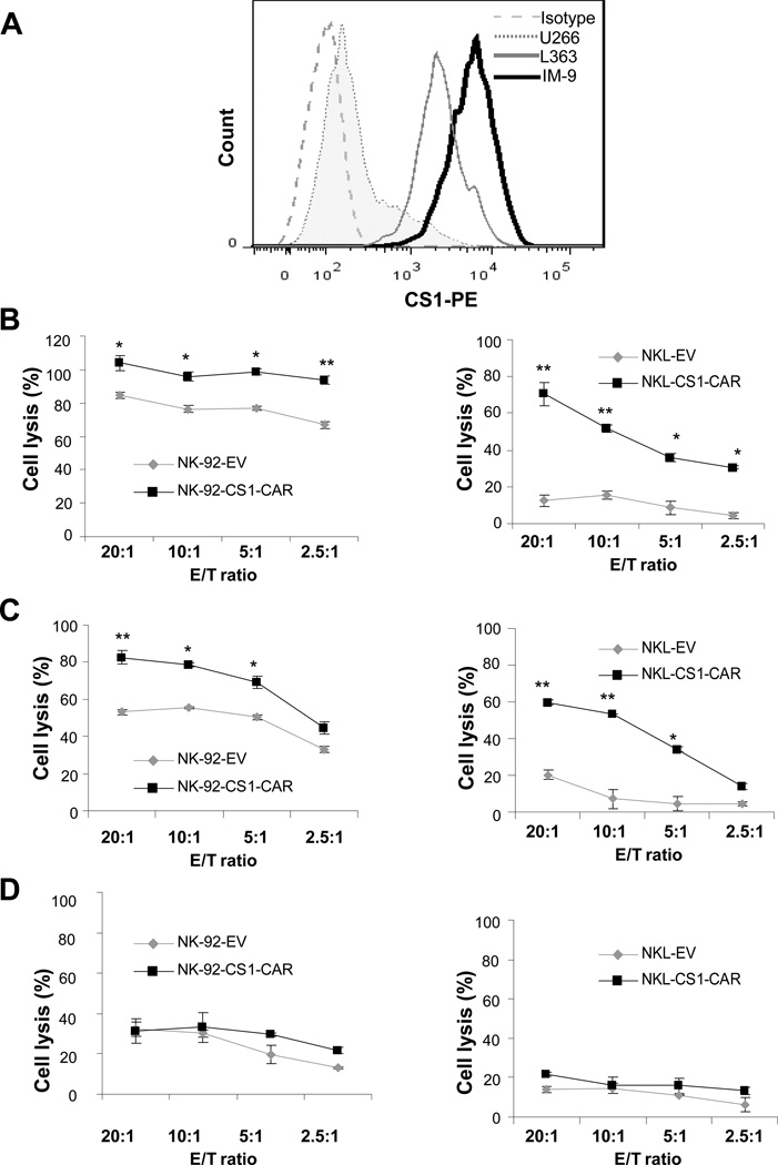
After generating the CS1-CAR NK cells, we determined whether they selectively kill CS1+ better than CS1− MM cells. For this purpose, we first confirmed that IM9 and L363 MM cells lines constitutively expressed CS1 protein on their surface, while constitutive expression of CS1 was negligible in U266 MM cells (Fig. 2A). Next, a 4 h chromium-51 release assay indicated that, compared with mock-transduced NK-92 cells, NK-92 cells transduced with CS1-CAR were significantly enhanced in their ability to kill CS1+ IM9 and L363 cells (Figs. 2B and 2C, left panels). Similar data were observed in experiments repeated using NKL cells transduced with CS1-CAR (Figs. 2B and 2C, right panels). However, both the CS1-CAR- and mock-transduced NK-92 or NKL cells were similar in their low levels of cytotoxicity against CS1− U266 myeloma cells (Fig. 2D). In addition, forced expression of CS1-CAR did not induce obvious apoptosis in NK-92 or NKL cells as determined by analyses of 7AAD/Annexin V-staining cells using flow cytometry (supplementary Fig. 1), suggesting that CS1-CAR expression did not cause cytotoxicity to the NK-92 or NKL cells themselves. Similarly, we also found that CS1-CAR expression in purified primary human NK cells augmented their cytotoxicity against CS1+ IM9 myeloma cells (data not shown).
Figure 2. CS1-CAR NK cells eradicate CS1+ but not CS1− MM cells.
A) Determination of CS1 expression on the surface of L363, IM9, and U266 MM cell lines by flow cytometry after cells were stained with anti-CS1 mAb or isotype-matched control antibody. B–D) Cytotoxic activity of mock-transduced or CS1-CAR-transduced NK-92 or NKL cells against IM9 (B), L363 (C) and U266 (D) cells using a standard chromium-51 release assay. NK-92-EV and NKL-EV indicate empty vector (EV) control-transduced NK-92 and NKL cells, respectively. NK-92-CS1-CAR and NKL-CS1-CAR indicate transduction of NK-92 and NKL cells, respectively, with a CS1-CAR construct. * and ** indicate P < 0.05 and P < 0.01, respectively.
CS1-CAR-modified NK cells secrete more IFN-γ than mock-transduced NK cells after exposure to CS1+ MM cells
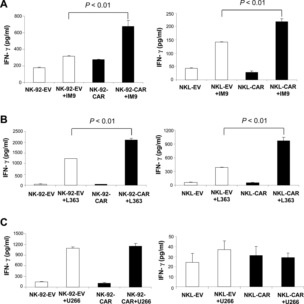
The signaling domain from the CD28 co-stimulatory molecule, which we included in our CAR construct, may enhance activation after recognition of the CS1 scFv with the CS1 antigen on the surface of MM cells. Therefore, the inclusion of this signaling domain may have the capacity to activate NK cells to not only have higher cytotoxicity, but also to produce more IFN-γ, the latter of which is also important for tumor surveillance and activation of CD8+ T cells and macrophages25–27. To test this, CS1-CAR-modified or control-engineered effector NK cells were either cultured alone or co-cultured with CS1+ myeloma cells including the IM9 and L363 MM cell lines. After 24 h, IFN-γ production was measured by ELISA. As shown in Fig. 3, both CS1-CAR-modified and mock-transduced NK-92 or NKL cells spontaneously produced low or negligible levels of IFN-γ when incubated alone. Co-culture with CS1+ MM tumor cells (IM9 or L363) induced IFN-γ in both CS1-CAR and mock-transduced NK-92 or NKL cell lines; however, significantly higher levels of IFN-γ were produced by CAR-modified NK-92 or NKL cells than by mock-transduced NK-92 (Fig. 3A and 3B, left panels) or NKL cells (Fig. 3A and 3B, right panels). When co-cultured with the CS1− MM cell line, U266, both mock-transduced and CS1-CAR-transduced NK-92 cells but not the transduced NKL cells produced higher levels of IFN-γ than corresponding cells that had not been co-cultured with U266 cells (Fig. 3C). This suggests that a unique interaction between NK cell receptors on NK-92 cells and their ligands on U266 cells may induce CS1-independent IFN-γ production by NK-92 cells. Moreover, CS1-CAR NK-92 cells and CS1-CAR NKL cells failed to produce more IFN-γ than mock-transduced NK-92 cells when they were co-cultured with U266 cells. (Fig. 3C). These results are in agreement with the aforementioned cytotoxicity data, and together indicate that modification with CS1-CAR can dramatically enhance NK cell effector functions, in terms of both cytotoxicity and IFN-γ production, in response to CS1+ but not CS1− myeloma cells.
Figure 3. Recognition of CS1+ MM cells induces a stronger response from CS1-CAR NK cells than from control NK cells.
A–C) Mock-transduced or CS1-CAR transduced NK-92 or NKL effector cells were co-cultured with an equal number of IM9 (A), L363 (B), or U266 (C) myeloma cells for 24 h. Supernatants were then harvested and measured for IFN-γ secretion using ELISA. NK-92-EV and NKL-EV indicate empty vector (EV) control-transduced NK-92 and NKL cells, respectively. NK-92-CS1-CAR and NKL-CS1-CAR indicate transduction of NK-92 and NKL cells, respectively, with a CS1-CAR construct.
Enforced CS1 expression in U266 cells enhances cytotoxicity and IFN-γ production of NK-92-CS1-CAR cells
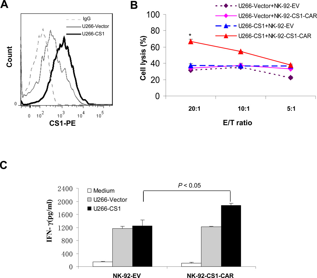
We next explored whether this enhanced activity of CS1-CAR NK cells relies on CS1 antigen expression on MM cells. Our aforementioned observation - that the introduction of CS1-CAR conferred NK-92 cells with increased cytotoxic activity and enhanced IFN-γ production in response to CS1+ myeloma cells, but not CS1− U266 myeloma cells - prompted us to investigate whether CS1 overexpression in U266 cells is sufficient to change the sensitivity of U266 cells to NK-92-CS1-CAR cells. For this purpose, we ectopically expressed CS1 in U266 cells by lentiviral infection. Flow cytometric analysis confirmed that CS1 protein was successfully expressed on the surface of the U266-CS1 cells (Fig. 4A). Chromium-51 release assay indicated that, when compared to mock-transduced NK-92 cells, there was a significant increase in the cytotoxic activity of CS1-CAR-transduced NK-92 cells towards U266 cells overexpressing CS1 (Fig 4B). Likewise, compared with parallel co-cultures instead containing mock-transduced NK-92 cells, NK-92-CS1-CAR cells co-cultured with U266 cells overexpressing CS1 secreted significantly higher levels of IFN-γ (Fig 4C). However, consistent with data in Fig. 2D and Fig. 3C, there was no difference in cytotoxicity and IFN-γ secretion between NK-92-CS1-CAR cells and mock-transduced NK-92 cells when they were incubated with U266 cells transduced with an empty vector control [Fig 4B and Fig 4C (gray)]. These results suggested that the increased recognition and killing of myeloma cells by NK-92-CS1-CAR cells occurs in a CS1-dependent manner.
Figure 4. Enhanced target recognition of NK-92-CS1-CAR cells depends on expression of CS1 on MM cells.
A) Flow cytometric staining for CS1 protein or IgG isotype control (dotted line) on the surface of U266 cells overexpressing CS1 (U266-CS1, solid heavy line) or an empty vector control (U266-Vector, solid light line). B) Cytotoxicity of mock- or CS1-CAR-transduced NK-92 cells (NK-92-EV and NK-92-CS1-CAR, respectively) against U266-Vector and U266-CS1 cells. U266-Vector or U266-CS1 cells were incubated with NK-92-CS1-CAR or NK-92-EV cells at different Effecor/Target (E/T) ratios for 4 h. Specific lysis was determined using a standard chromium-51 release assay. * indicates P < 0.05. C) NK-92-CS1-CAR or NK-92-EV cells were co-cultured with an equal number of U266-Vector or U266-CS1 myeloma cells for 24 h. Supernatants were then harvested for measurement of IFN-γ secretion using ELISA.
Phenotypic characterization of NK-92-CS1-CAR cells
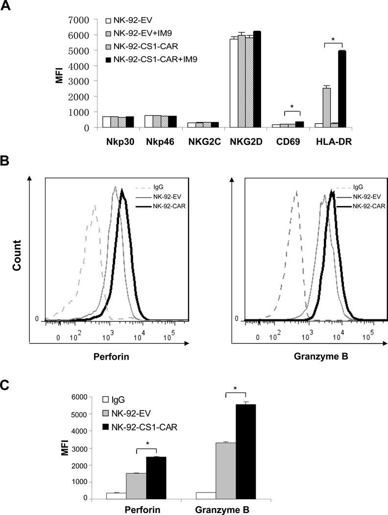
We next investigated whether the expression of a CS1-specific CAR could change NK cell phenotype. We employed flow cytometry to compare expression of antigens on the surface of CS1-CAR-transduced and mock-transduced NK-92 cells, following culture in the presence or absence of IM9 myeloma cells. As shown in Fig. 5A, we observed that there was no difference between CS1-CAR- and mock-transduced NK-92 cells, whether cultured in the presence or absence of IM9 cells, in the expression of NK cell receptors including NKp30, NKp46, NKG2C and NKG2D. We also assessed expression of the NK cell activation markers, CD6928 and HLA-DR29,30. We found that recognition of IM9 cells did not elicit CD69 expression on mock-transduced NK-92 cells, yet induced a moderate but significant increase in CD69 expression on the surface of CS1-CAR-transduced NK-92 cells (Fig 5A). Interestingly, co-incubation with IM9 cells caused a dramatic increase in the expression of HLA-DR in both CS1-CAR-transduced and mock-transduced NK-92 cells. In the absence of IM9 target cells, there was no obvious difference in HLA-DR expression between CS1-CAR-transduced and mock-transduced NK-92 cells; however, upon stimulation with IM9 cells, the expression of HLA-DR became significantly higher in NK-92-CS1-CAR cells than in mock-transduced NK-92 cells. Thus, the increase in the activation markers, especially HLA-DR, expressed on NK-92-CS1-CAR cells may have occurred in connection with the enhanced cytotoxicity and IFN-γ production by these cells when they are exposed to CS1+ MM cells. Using intracellular staining, we also observed that, when compared to mock-tranduced NK cells, NK-92-CS1-CAR cells had significantly higher levels of perforin and granzyme B (GZMB) expression, even in the absence of MM tumor cells (Figs. 5B and 5C). This is consistent with a previous report regarding the elevated expression of GZMB in CAR T cells31, and also consistent with the fact that perforin and granzyme B expression are generally correlated with cytotoxic activity of NK cells32.
Figure 5. Phenotypic characterization of CS1-CAR modified NK cells.
A) Mock- or CS1-CAR-transduced NK-92 cells (NK-92-EV and NK-92-CS1-CAR, respectively) were either cultured alone, or cultured with IM9 MM cells for 4 h. Surface expression of NKp30, NKp46, NKG2C, NKG2D, CD69 and HLA-DR was assessed by flow cytometry following staining with corresponding monoclonal antibodies (mAbs), and the mean fluorescence intensity (MFI) was recorded. * indicates P < 0.05. B) NK-92-EV and NK-92-CS1-CAR cells were permeabilized for intracellular staining with mAb specific for perforin or granzyme B, and analyzed by flow cytometry. The dotted line represents staining the NK-92-EV control cells with CS1 mAb, solid heavy line for NK-92-CS1-CAR cells with CS1 mAb, and the dashed line for the NK-92-EV control cells with isotype-matched control antibodies. C) Depicted MFI for histograms shown in (B).* indicates P < 0.05.
CS1-CAR-transduced NK-92 cells more effectively recognize and kill NK-resistant primary multiple myeloma cells ex vivo
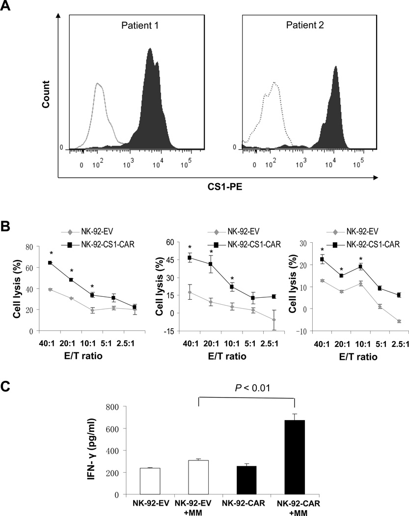
To make our findings more clinically relevant, we investigated whether CS1-CAR-modified NK-92 cells also harbored enhanced cytolytic activity and IFN-γ production when recognizing primary MM cells ex vivo. Flow cytometry was used to assess surface expression of CS1 on primary CD138+ magnetic bead-selected MM cells from MM patients (Fig. 6A). In accordance with the previous report, showing that CS1 protein was highly expressed on CD138-purified MM patient cells14,15, we observed that CS1 protein was indeed uniformly expressed on the surface of primary MM cells (Fig. 6A). By chromium-51 release assay, we found that primary myeloma cells freshly isolated from MM patients were highly resistant to NK-92 cell-mediated lysis even at E:T ratios as high as 40:1 and 20:1; however, this resistance could be partially overcome by NK-92 cell expression of CS1-CAR, which resulted in a dramatic increase in eradication of primary myeloma cells (Fig. 6B). In line with the cytotoxicity result, after 24 h co-culture with primary myeloma cells, CS1-CAR-transduced NK-92 cells also secreted significantly higher levels of IFN-γ than mock-transduced NK-92 cells (Fig. 6C).
Figure 6. CS1-CAR-transduced NK-92 cells enhance killing of primary human myeloma cells.
A) Flow cytometric staining for CS1 protein or IgG isotype control, demonstrating that CD138+ primary myeloma cells highly express CS1. The open and filled histograms represent staining with isotype-matched control antibodies and anti-CS1 antibodies, respectively. Data shown are representative of 2 out of 6 patient samples with similar results. B) Cytotoxic activity of mock- or CS1-CAR-transduced NK-92 cells (NK-92-EV and NK-92-CS1-CAR, respectively) against CD138+ primary myeloma cells from 3 of 6 patients with similar results using a standard chromium-51 release assay. E/T indicates effector cell/target cell ratio. * indicates P < 0.05. C) CD138+ primary myeloma cells were co-cultured with NK-92-EV or NK-92-CS1-CAR cells at an E/T ratio of 5:1 for 24 h. Supernatants were harvested for measurement of IFN-γ secretion using ELISA. Data shown are representative of 1 out of 3 patient samples with similar results.
CS1-CAR-transduced NK-92 cells inhibit MM tumor growth and prolong survival of tumor-bearing mice in an orthotopic xenograft MM model
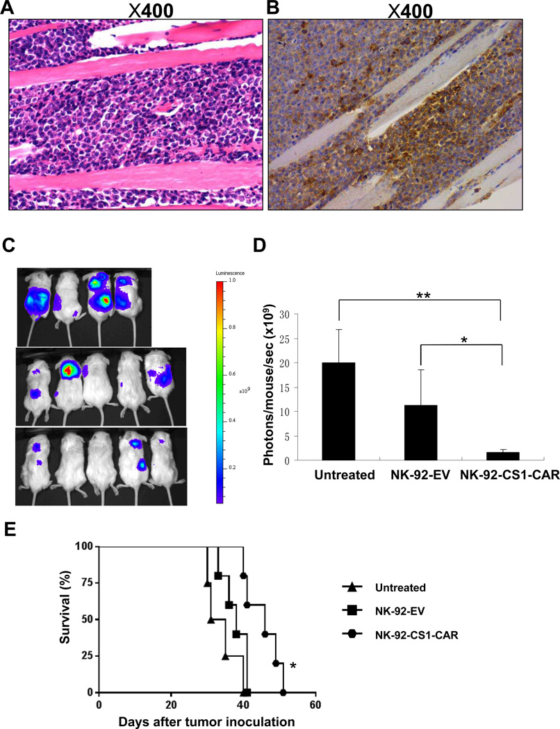
To further address the potential therapeutic application of NK-92-CS1-CAR cells, we further examined their antitumor activity in IM9-xenografted NSG mice. We generated an IM9 cell line expressing firefly luciferase (FFL) (IM9-Luc) by retrovirally transducing IM9 cells with virus expressing FFL, then performing GFP-based cell sorting. The expression of full-length FFL mRNA was confirmed by RT-PCR (supplementary Fig. 2A). Like typical myeloma cells, IM9-Luc cells expressed CD138 protein on their surface (supplementary Fig. 2B). In agreement with a previous report33, we observed that IM9-Luc-bearing NSG mice displayed disseminated disease, manifested by hind-limb paralysis and motor ataxia. Histological examination of spinal vertebrae in a mouse displaying hind-limb paralysis showed the presence of numerous tumor cells and osteolytic lesions in bone tissue (Fig. 7A). Immunohistochemical staining with human specific anti-CD138 antibody further confirmed the presence of tumor cells (Fig. 7B). Bioluminescence imaging was used to monitor the IM9-Luc tumor growth. As shown in Fig. 7C and 7D, and in agreement with the in vitro cytotoxicity data, comparing the mice who later received injections with mock-transduced control cells, IM9-Luc tumors were significantly suppressed in mice who instead later were administerd NK-92-CS1-CAR cells. Moreover, treatment with NK-92-CS1-CAR cells significantly prolonged the survival of mice bearing IM9-Luc tumors as compared to treatment with the mock-transduced NK-92 control cells (Fig. 7E). Of note, when NK-92-CS1-CAR cells or mock-transduced NK-92 cells were similarly administrated, but without i.v. injection of IM9-Luc cells, mice did not develop disseminated disease or die (data not shown).
Figure 7. CS1-CAR NK cells suppress in vivo growth of orthotopic human MM cells and prolong the survival of MM-bearing mice.
A) Massive infiltration of human IM9 cells, detected by Hematoxylin-Eosin (HE) staining, in the lumbar vertebrae bone lesions of one representative mouse displaying hind leg paralyses after intravenously injected with IM9 cells. B) Immunohistochemical staining of mouse lumbar vertebrae bone lesions with anti-human CD138 mAb. C) Dorsal bioluminescence imaging of mice bearing IM9 tumors. NSG mice were inoculated with 5 × 10⁵ luciferase-expressing IM9 cells via a tail vein injection (day 0). Seven days after inoculation, mice were treated with mock-transduced NK-92 cells (NK-92-EV), CS1-CAR tranduced NK-92 cells (NK-92-CS1-CAR) or PBS (a negative control) according to a schedule described in the Materials and Methods section. D) Quantification summary of units of photons per second per mouse from (B). * indicates P < 0.05; ** denotes P < 0.01. E) IM9-bearing mice treated with NK-92-CS1-CAR cells showed significantly increased survival compared to the mice treated with NK-92-EV cells (*, P < 0.05), as determined by Kaplan-Meier survival curves.
Discussion
Genetic manipulation of cytotoxic immune cells, including T cells and NK cells, to express a CAR that is specific for a tumor-associated antigen has emerged as a promising strategy for treating hematological malignancies8,10–12. Identification of suitable target antigens is a prerequisite for developing CAR-expressing immune cell therapies against malignancies, yet this has been the biggest hurdle for developing CAR T or NK cells13. Substantial progress has been made recently for the development of CAR immune cell therapy against B cell lineage malignancies, among which CD19-directed CAR T cells have been demonstrated to induce prolonged remission in advanced B-cell ALL and CLL8,10,11,34. Unfortunately, CD19-directed CAR T cells cannot be utilized to treat MM patients because over 95% of MM patients lack the expression of CD19 on their tumor cells35. Although many other surface antigens, such as CD40, CD56, CD138 and CD74 can also be expressed on MM cells, the clinical utility of each of these antigens is very limited given their lack of specificity for MM cells or variability between patients15. Both CD38 and B-cell maturation antigen (BCMA) were recently identified as promising immunotherapeutic targets in MM36–37; however, BCMA was found to be expressed by normal plasma cells and subsets of mature B cells as well38–40, and CD38 has widespread expression on hematopoietic and non-hematopoietic tissue41, despite the relative safety of naked CD38 antibodies in phase 1 trials42. Unlike these antigens, CS1 is highly and uniformly expressed on MM cells from all patients, and has a restricted pattern of expression in normal cells and tissues14. CS1 expression is maintained on MM cells of patients even after disease relapse14. In addition, mounting evidence has suggested that CS1-specific immunotherapy can target neoplastic cells without inducing major damage to normal cells, including immune cells like T and NK cells in MM patients43. On the basis of all these findings, it is tempting to speculate that CS1 may be an ideal antigen for targeting CAR NK cells against MM. In the present study, we have demonstrated for the first time that manipulating human NK cells to express a CS1-specific CAR can dramatically enhance their killing of CS1+ myeloma cell lines and primary myeloma cells, and can also augment their secretion of IFN-γ in vitro. More importantly, NK-92-CS1-CAR cells more efficiently eradicate human IM9 tumors established in NSG mice, resulting in improved overall survival of these IM9-bearing mice. All of this evidence strongly corroborates our hypothesis that CS1 represents an appropriate tumor antigen target for CAR NK cells against MM.
In contrast to first-generation CARs, which typically only bear the intracellular domain from CD3 ζ, second-generation CARs, which have already been widely used for generating and studying CAR-expressing T or NK cells, usually incorporate CD28 or 4-1BB (CD137) as a co-stimulatory signal44–46, and both of these co-stimulatory signals have been shown functional in CAR T cells8,10–12. In this study we showed that NK cells expressing a CS1-specific CAR containing CD28 and CD3 ζ signaling moieties display enhanced anti-MM activity, both in vitro and in vivo. Consistent with our study, others have demonstrated that primary NK cells grafted with a HER-2 specific CAR harboring CD28 and CD3 ζ signaling moieties specifically recognized and efficiently eradicated HER-2 positive carcinoma45. Regarding 4-1BB-CD3 ζ CAR in NK cells, it has been reported that primary NK cells grafted with a CD19-specific CAR harboring 4-1BB-CD3 ζ displays increased cytokine production and cytotoxic activity towards leukemic cells46. Therefore, it appears that the tumor antigen-specific CARs carrying either CD28-CD3 ζ or 4-1BB-CD3 ζ signaling moieties can efficiently redirect NK cells to specifically target and kill tumors cells expressing the corresponding tumor antigens, yet a direct comparison is needed to address which one is superior.
CAR T cells have been effectively used for treatment of refractory chronic lymphocytic leukemia and acute lymphoblastic leukemia8,10–12. One advantage of CAR NK cells as opposed to CAR T cells is that CAR T cells can induce a cytokine storm in the clinic7,10, while NK cells might be safer effector cells due to the lack of clonal expansion; on the other hand this might limit efficacy. Another advantage of CAR NK cells versus CAR T cells is that CAR NK cells may be used in the setting of allogeneic transplantation, enhancing an all too often weak graft-versus-tumor effect without inducing graft-versus-host disease20. We speculate that co-administration of CAR NK cells, either at the time of allograft or post-transplant, could enhance the graft-versus-tumor effect. Moreover, allogeneic NK cells should be more potent than autologous NK cells for lysis of MM cells due to mismatched killer-cell immunoglobulin-like receptors (KIR) effects21,47. It is noteworthy that normal CD34+ hematopoietic stem cells, which are often utilized at the time of autologous stem cell transplant, do not express CS1, suggesting an opportunity for CAR NK cell therapy at a time when there is minimal residual disease14.
It is generally believed that the initial control of malignant plasma cells by NK cells is often attenuated in the inexorable progression of MM5. This suggests that it is critical to infuse NK cells that are more potent, such as CAR NK cells. Of note, adoptive NK cell immunotherapy for MM has already been evaluated in several promising studies, which have fostered ongoing interest48,49. However, adoptive immunotherapy with primary NK cells is often complicated by the difficulties in expansion of these cells. This barrier might be overcome by the recent success of an effective NK cell expansion strategy, which utilizes 4-1BB ligand50,51. Another option for overcoming these limitations is to use established NK cell lines52,53. The large-scale expansion of NK cell lines under GMP conditions is easier, more cost effective, and makes these cells more readily available for use in clinical adoptive therapy54. The NK-92 cell line, one of the cell lines that we used in the present study, seems to be the best choice due to two reasons. First, NK-92 cells have been proven generally safe with mild and transient toxicities, and expansion of these cells is feasible55. Second, NK-92 cells have displayed appreciable in vitro and in vivo anti-cancer activity in a variety of malignancies including MM53,56. In fact, infusion of NK-92 cells is being used to treat hematological malignancies including MM (http://www.clinicaltrials.gov).
In the treatment of cancer such as MM, a single agent may not work as effectively as combination therapy. This could imply that other therapeutic approaches designed to synergize with CS1-CAR-directed NK cell therapy may be needed to achieve the best possible MM-killing outcome. Examples of these approaches include but are limited to: JAK inhibitors that can increase the susceptibility of MM cells to NK cell-mediated cytotoxicity57; the anti-CS1 monoclonal antibody, elotuzumab that can enhance the ADCC, natural cytoxcity, and IFN-γ production of NK cells16; the pan-KIR antibody, IPH2101, that can enhance NK cell activity through blocking the KIR inhibitory function58; and even circulating or exogenous microRNAs that can activate NK cells through Toll-like receptor signaling24.
In conclusion, CS1 is a promising target for the use of CAR NK cells for MM treatment. We have successfully generated a CAR that specifically recognizes CS1, which is uniformly expressed on the surface of MM cells. We also present evidence that NK cells armed with this CS1-specific CAR can more efficiently and specifically recognize and eradicate myeloma cells both in vitro and in vivo. Our study may help pave the way towards the clinical application of CS1-CAR-modified immune cells, either used alone or in combination with other approaches, for the treatment of MM.
Supplementary Material
ACKNOWLEDGMENTS
The project was supported in part by Multiple Myeloma Opportunities for Research and Education (MMORE), and grants from the National Cancer Institute (CA155521 to J.Y. and CA095426 and CA068458 to M.A.C.), a 2012 scientific research grant from the National Blood Foundation (J.Y.), an institutional research grant (IRG-67-003-47) from the American Cancer Society (J.Y.), and The Ohio State University Comprehensive Cancer Center Pelotonia grant (J.Y.).
Footnotes
CONFLICT OF INTEREST
The authors declare no conflict of interest.
AUTHORSHIP
Contributions: J.C. designed research, performed experiments, and wrote the paper; Y.D., S.H, Y.P., H.M., L.Y. performed experiments; D.M., G.K., X.H., S.M.D., X.Z., M.A.C. designed research; T.H. reviewed and edited the manuscript; J.Z. analyzed data; C.C.H. devised the concept, designed research and reviewed the manuscript; J.Y. devised the concept, designed research, supervised the study, and wrote the paper.
Supplementary information is available at Leukemia's website.
REFERENCES
- 1.Kyle RA, Rajkumar SV. Multiple myeloma. Blood. 2008;111:2962–2972. doi: 10.1182/blood-2007-10-078022. [DOI] [PMC free article] [PubMed] [Google Scholar]
- 2.Siegel R, Naishadham D, Jemal A. Cancer statistics, 2012. CA Cancer J Clin. 2012;62:10–29. doi: 10.3322/caac.20138. [DOI] [PubMed] [Google Scholar]
- 3.Palumbo A, Rajkumar SV. Treatment of newly diagnosed myeloma. Leukemia. 2009;23:449–456. doi: 10.1038/leu.2008.325. [DOI] [PMC free article] [PubMed] [Google Scholar]
- 4.Podar K, Chauhan D, Anderson KC. Bone marrow microenvironment and the identification of new targets for myeloma therapy. Leukemia. 2009;23:10–24. doi: 10.1038/leu.2008.259. [DOI] [PMC free article] [PubMed] [Google Scholar]
- 5.Godfrey J, Benson DM., Jr The role of natural killer cells in immunity against multiple myeloma. Leuk Lymphoma. 2012;53:1666–1676. doi: 10.3109/10428194.2012.676175. [DOI] [PubMed] [Google Scholar]
- 6.Narni-Mancinelli E, Vivier E, Kerdiles YM. The 'T-cell-ness' of NK cells: unexpected similarities between NK cells and T cells. Int Immunol. 2011;23:427–431. doi: 10.1093/intimm/dxr035. [DOI] [PubMed] [Google Scholar]
- 7.Morgan RA, Yang JC, Kitano M, Dudley ME, Laurencot CM, Rosenberg SA. Case report of a serious adverse event following the administration of T cells transduced with a chimeric antigen receptor recognizing ERBB2. Mol Ther. 2010;18:843–851. doi: 10.1038/mt.2010.24. [DOI] [PMC free article] [PubMed] [Google Scholar]
- 8.Porter DL, Levine BL, Kalos M, Bagg A, June CH. Chimeric antigen receptor-modified T cells in chronic lymphoid leukemia. N Engl J Med. 2011;365:725–733. doi: 10.1056/NEJMoa1103849. [DOI] [PMC free article] [PubMed] [Google Scholar]
- 9.Fauriat C, Mallet F, Olive D, Costello RT. Impaired activating receptor expression pattern in natural killer cells from patients with multiple myeloma. Leukemia. 2006;20:732–733. doi: 10.1038/sj.leu.2404096. [DOI] [PubMed] [Google Scholar]
- 10.Grupp SA, Kalos M, Barrett D, Aplenc R, Porter DL, Rheingold SR, et al. Chimeric antigen receptor-modified T cells for acute lymphoid leukemia. N Engl J Med. 2013;368:1509–1518. doi: 10.1056/NEJMoa1215134. [DOI] [PMC free article] [PubMed] [Google Scholar]
- 11.Brentjens RJDM, Riviere I, Park J, Wang X, Cowell LG, Bartido S, Stefanski J, Taylor C, Olszewska M, Borquez-Ojeda O, Qu J, Wasielewska T, He Q, Bernal Y, Rijo IV, Hedvat C, Kobos R, Curran K, Steinherz P, Jurcic J, Rosenblat T, Maslak P, Frattini M, Sadelain M. CD19-targeted T cells rapidly induce molecular remissions in adults with chemotherapy-refractory acute lymphoblastic leukemia. Sci Transl Med. 2013;5:177ra138. doi: 10.1126/scitranslmed.3005930. [DOI] [PMC free article] [PubMed] [Google Scholar]
- 12.Kalos M, Levine BL, Porter DL, Katz S, Grupp SA, Bagg A, et al. T cells with chimeric antigen receptors have potent antitumor effects and can establish memory in patients with advanced leukemia. Sci Transl Med. 2011;3:95ra73. doi: 10.1126/scitranslmed.3002842. [DOI] [PMC free article] [PubMed] [Google Scholar]
- 13.Maus MV, June CH. Zoom Zoom: racing CARs for multiple myeloma. Clin Cancer Res. 2013;19:1917–1919. doi: 10.1158/1078-0432.CCR-13-0168. [DOI] [PMC free article] [PubMed] [Google Scholar]
- 14.Hsi ED, Steinle R, Balasa B, Szmania S, Draksharapu A, Shum BP, et al. CS1, a potential new therapeutic antibody target for the treatment of multiple myeloma. Clin Cancer Res. 2008;14:2775–2784. doi: 10.1158/1078-0432.CCR-07-4246. [DOI] [PMC free article] [PubMed] [Google Scholar]
- 15.Tai YT, Dillon M, Song W, Leiba M, Li XF, Burger P, et al. Anti-CS1 humanized monoclonal antibody HuLuc63 inhibits myeloma cell adhesion and induces antibody-dependent cellular cytotoxicity in the bone marrow milieu. Blood. 2008;112:1329–1337. doi: 10.1182/blood-2007-08-107292. [DOI] [PMC free article] [PubMed] [Google Scholar]
- 16.Benson DM, Jr, Byrd JC. CS1-directed monoclonal antibody therapy for multiple myeloma. J Clin Oncol. 2012;30:2013–2015. doi: 10.1200/JCO.2011.40.4061. [DOI] [PubMed] [Google Scholar]
- 17.Tai YT, Soydan E, Song W, Fulciniti M, Kim K, Hong F, et al. CS1 promotes multiple myeloma cell adhesion, clonogenic growth, and tumorigenicity via c-maf-mediated interactions with bone marrow stromal cells. Blood. 2009;113:4309–4318. doi: 10.1182/blood-2008-10-183772. [DOI] [PMC free article] [PubMed] [Google Scholar] [Retracted]
- 18.van de Donk NW, Kamps S, Mutis T, Lokhorst HM. Monoclonal antibody-based therapy as a new treatment strategy in multiple myeloma. Leukemia. 2012;26:199–213. doi: 10.1038/leu.2011.214. [DOI] [PubMed] [Google Scholar]
- 19.Lonial S, Vij R, Harousseau JL, Facon T, Moreau P, Mazumder A, et al. Elotuzumab in combination with lenalidomide and low-dose dexamethasone in relapsed or refractory multiple myeloma. J Clin Oncol. 2012;30:1953–1959. doi: 10.1200/JCO.2011.37.2649. [DOI] [PubMed] [Google Scholar]
- 20.Olson JA, Leveson-Gower DB, Gill S, Baker J, Beilhack A, Negrin RS. NK cells mediate reduction of GVHD by inhibiting activated, alloreactive T cells while retaining GVT effects. Blood. 2010;115:4293–4301. doi: 10.1182/blood-2009-05-222190. [DOI] [PMC free article] [PubMed] [Google Scholar]
- 21.Ruggeri L, Capanni M, Urbani E, Perruccio K, Shlomchik WD, Tosti A, et al. Effectiveness of donor natural killer cell alloreactivity in mismatched hematopoietic transplants. Science. 2002;295:2097–2100. doi: 10.1126/science.1068440. [DOI] [PubMed] [Google Scholar]
- 22.Yu J, Wei M, Becknell B, Trotta R, Liu S, Boyd Z, et al. Pro- and antiinflammatory cytokine signaling: reciprocal antagonism regulates interferon-gamma production by human natural killer cells. Immunity. 2006;24:575–590. doi: 10.1016/j.immuni.2006.03.016. [DOI] [PubMed] [Google Scholar]
- 23.Yu J, Mao HC, Wei M, Hughes T, Zhang J, Park IK, et al. CD94 surface density identifies a functional intermediary between the CD56bright and CD56dim human NK-cell subsets. Blood. 2010;115:274–281. doi: 10.1182/blood-2009-04-215491. [DOI] [PMC free article] [PubMed] [Google Scholar]
- 24.He S, Chu J, Wu LC, Mao H, Peng Y, Alvarez-Breckenridge CA, et al. MicroRNAs activate natural killer cells through Toll-like receptor signaling. Blood. 2013;121:4663–4671. doi: 10.1182/blood-2012-07-441360. [DOI] [PMC free article] [PubMed] [Google Scholar]
- 25.Martin-Fontecha A, Thomsen LL, Brett S, Gerard C, Lipp M, Lanzavecchia A, et al. Induced recruitment of NK cells to lymph nodes provides IFN-gamma for T(H)1 priming. Nat Immunol. 2004;5:1260–1265. doi: 10.1038/ni1138. [DOI] [PubMed] [Google Scholar]
- 26.Tu SP, Quante M, Bhagat G, Takaishi S, Cui G, Yang XD, et al. IFN-gamma inhibits gastric carcinogenesis by inducing epithelial cell autophagy and T-cell apoptosis. Cancer Res. 2011;71:4247–4259. doi: 10.1158/0008-5472.CAN-10-4009. [DOI] [PMC free article] [PubMed] [Google Scholar]
- 27.Ma J, Chen T, Mandelin J, Ceponis A, Miller NE, Hukkanen M, et al. Regulation of macrophage activation. Cell Mol Life Sci. 2003;60:2334–2346. doi: 10.1007/s00018-003-3020-0. [DOI] [PMC free article] [PubMed] [Google Scholar]
- 28.Borrego F, Pena J, Solana R. Regulation of CD69 expression on human natural killer cells: differential involvement of protein kinase C and protein tyrosine kinases. Eur J Immunol. 1993;23:1039–1043. doi: 10.1002/eji.1830230509. [DOI] [PubMed] [Google Scholar]
- 29.Phillips JH, Le AM, Lanier LL. Natural killer cells activated in a human mixed lymphocyte response culture identified by expression of Leu-11 and class II histocompatibility antigens. J Exp Med. 1984;159:993–1008. doi: 10.1084/jem.159.4.993. [DOI] [PMC free article] [PubMed] [Google Scholar]
- 30.Spits H, Lanier LL. Natural killer or dendritic: what's in a name? Immunity. 2007;26:11–16. doi: 10.1016/j.immuni.2007.01.004. [DOI] [PubMed] [Google Scholar]
- 31.Koehler H, Kofler D, Hombach A, Abken H. CD28 costimulation overcomes transforming growth factor-beta-mediated repression of proliferation of redirected human CD4+ and CD8+ T cells in an antitumor cell attack. Cancer Res. 2007;67:2265–2273. doi: 10.1158/0008-5472.CAN-06-2098. [DOI] [PubMed] [Google Scholar]
- 32.Krzewski K, Coligan JE. Human NK cell lytic granules and regulation of their exocytosis. Front Immunol. 2012;3:335. doi: 10.3389/fimmu.2012.00335. [DOI] [PMC free article] [PubMed] [Google Scholar]
- 33.Francisco JA, Donaldson KL, Chace D, Siegall CB, Wahl AF. Agonistic properties and in vivo antitumor activity of the anti-CD40 antibody SGN-14. Cancer Res. 2000;60:3225–3231. [PubMed] [Google Scholar]
- 34.Kochenderfer JN, Rosenberg SA. Treating B-cell cancer with T cells expressing anti-CD19 chimeric antigen receptors. Nat Rev Clin Oncol. 2013;10:267–276. doi: 10.1038/nrclinonc.2013.46. [DOI] [PMC free article] [PubMed] [Google Scholar]
- 35.Mateo G, Montalban MA, Vidriales MB, Lahuerta JJ, Mateos MV, Gutierrez N, et al. Prognostic value of immunophenotyping in multiple myeloma: a study by the PETHEMA/GEM cooperative study groups on patients uniformly treated with high-dose therapy. J Clin Oncol. 2008;26:2737–2744. doi: 10.1200/JCO.2007.15.4120. [DOI] [PubMed] [Google Scholar]
- 36.Mihara K, Bhattacharyya J, Kitanaka A, Yanagihara K, Kubo T, Takei Y, et al. T-cell immunotherapy with a chimeric receptor against CD38 is effective in eliminating myeloma cells. Leukemia. 2012;26:365–367. doi: 10.1038/leu.2011.205. [DOI] [PubMed] [Google Scholar]
- 37.Carpenter RO, Evbuomwan MO, Pittaluga S, Rose JJ, Raffeld M, Yang S, et al. B-cell Maturation Antigen Is a Promising Target for Adoptive T-cell Therapy of Multiple Myeloma. Clin Cancer Res. 2013;19:2048–2060. doi: 10.1158/1078-0432.CCR-12-2422. [DOI] [PMC free article] [PubMed] [Google Scholar]
- 38.Laabi Y, Gras MP, Carbonnel F, Brouet JC, Berger R, Larsen CJ, et al. A new gene, BCM, on chromosome 16 is fused to the interleukin 2 gene by a t(4;16)(q26;p13) translocation in a malignant T cell lymphoma. EMBO J. 1992;11:3897–3904. doi: 10.1002/j.1460-2075.1992.tb05482.x. [DOI] [PMC free article] [PubMed] [Google Scholar]
- 39.Laabi Y, Gras MP, Brouet JC, Berger R, Larsen CJ, Tsapis A. The BCMA gene, preferentially expressed during B lymphoid maturation, is bidirectionally transcribed. Nucleic Acids Res. 1994;22:1147–1154. doi: 10.1093/nar/22.7.1147. [DOI] [PMC free article] [PubMed] [Google Scholar]
- 40.O'Connor BP, Raman VS, Erickson LD, Cook WJ, Weaver LK, Ahonen C, et al. BCMA is essential for the survival of long-lived bone marrow plasma cells. J Exp Med. 2004;199:91–98. doi: 10.1084/jem.20031330. [DOI] [PMC free article] [PubMed] [Google Scholar]
- 41.Malavasi F, Deaglio S, Funaro A, Ferrero E, Horenstein AL, Ortolan E, et al. Evolution and function of the ADP ribosyl cyclase/CD38 gene family in physiology and pathology. Physiol Rev. 2008;88:841–886. doi: 10.1152/physrev.00035.2007. [DOI] [PubMed] [Google Scholar]
- 42.van de Donk NW, Lokhorst HM. New developments in the management and treatment of newly diagnosed and relapsed/refractory multiple myeloma patients. Expert Opin Pharmacother. 2013 doi: 10.1517/14656566.2013.805746. [DOI] [PubMed] [Google Scholar]
- 43.Bae J, Song W, Smith R, Daley J, Tai YT, Anderson KC, et al. A novel immunogenic CS1-specific peptide inducing antigen-specific cytotoxic T lymphocytes targeting multiple myeloma. Br J Haematol. 2012;157:687–701. doi: 10.1111/j.1365-2141.2012.09111.x. [DOI] [PMC free article] [PubMed] [Google Scholar]
- 44.Jena B, Dotti G, Cooper LJ. Redirecting T-cell specificity by introducing a tumor-specific chimeric antigen receptor. Blood. 2010;116:1035–1044. doi: 10.1182/blood-2010-01-043737. [DOI] [PMC free article] [PubMed] [Google Scholar]
- 45.Kruschinski A, Moosmann A, Poschke I, Norell H, Chmielewski M, Seliger B, et al. Engineering antigen-specific primary human NK cells against HER-2 positive carcinomas. Proc Natl Acad Sci U S A. 2008;105:17481–17486. doi: 10.1073/pnas.0804788105. [DOI] [PMC free article] [PubMed] [Google Scholar]
- 46.Imai C, Iwamoto S, Campana D. Genetic modification of primary natural killer cells overcomes inhibitory signals and induces specific killing of leukemic cells. Blood. 2005;106:376–383. doi: 10.1182/blood-2004-12-4797. [DOI] [PMC free article] [PubMed] [Google Scholar]
- 47.Kroger N, Shaw B, Iacobelli S, Zabelina T, Peggs K, Shimoni A, et al. Comparison between antithymocyte globulin and alemtuzumab and the possible impact of KIR-ligand mismatch after dose-reduced conditioning and unrelated stem cell transplantation in patients with multiple myeloma. Br J Haematol. 2005;129:631–643. doi: 10.1111/j.1365-2141.2005.05513.x. [DOI] [PubMed] [Google Scholar]
- 48.Tricot G, Vesole DH, Jagannath S, Hilton J, Munshi N, Barlogie B. Graft-versus-myeloma effect: proof of principle. Blood. 1996;87:1196–1198. [PubMed] [Google Scholar]
- 49.Alici E, Konstantinidis KV, Sutlu T, Aints A, Gahrton G, Ljunggren HG, et al. Anti-myeloma activity of endogenous and adoptively transferred activated natural killer cells in experimental multiple myeloma model. Exp Hematol. 2007;35:1839–1846. doi: 10.1016/j.exphem.2007.08.006. [DOI] [PubMed] [Google Scholar]
- 50.Lapteva N, Durett AG, Sun J, Rollins LA, Huye LL, Fang J, et al. Large-scale ex vivo expansion and characterization of natural killer cells for clinical applications. Cytotherapy. 2012;14:1131–1143. doi: 10.3109/14653249.2012.700767. [DOI] [PMC free article] [PubMed] [Google Scholar]
- 51.Fujisaki H, Kakuda H, Shimasaki N, Imai C, Ma J, Lockey T, et al. Expansion of highly cytotoxic human natural killer cells for cancer cell therapy. Cancer Res. 2009;69:4010–4017. doi: 10.1158/0008-5472.CAN-08-3712. [DOI] [PMC free article] [PubMed] [Google Scholar]
- 52.Tonn TBS, Esser R, Schwabe D, Seifried E. Cellular immunotherapy of malignancies using the clonal natural killer cell line NK-92. J Hematother Stem Cell Res. 2001;10:535–544. doi: 10.1089/15258160152509145. [DOI] [PubMed] [Google Scholar]
- 53.Swift BE, Williams BA, Kosaka Y, Wang XH, Medin JA, Viswanathan S, et al. Natural killer cell lines preferentially kill clonogenic multiple myeloma cells and decrease myeloma engraftment in a bioluminescent xenograft mouse model. Haematologica. 2012;97:1020–1028. doi: 10.3324/haematol.2011.054254. [DOI] [PMC free article] [PubMed] [Google Scholar]
- 54.Cheng M, Chen Y, Xiao W, Sun R, Tian Z. NK cell-based immunotherapy for malignant diseases. Cell Mol Immunol. 2013;10:230–252. doi: 10.1038/cmi.2013.10. [DOI] [PMC free article] [PubMed] [Google Scholar]
- 55.Arai S, Meagher R, Swearingen M, Myint H, Rich E, Martinson J, et al. Infusion of the allogeneic cell line NK-92 in patients with advanced renal cell cancer or melanoma: a phase I trial. Cytotherapy. 2008;10:625–632. doi: 10.1080/14653240802301872. [DOI] [PubMed] [Google Scholar]
- 56.Maki G, Hayes GM, Naji A, Tyler T, Carosella ED, Rouas-Freiss N, et al. NK resistance of tumor cells from multiple myeloma and chronic lymphocytic leukemia patients: implication of HLA-G. Leukemia. 2008;22:998–1006. doi: 10.1038/leu.2008.15. [DOI] [PubMed] [Google Scholar]
- 57.Bellucci R, Nguyen HN, Martin A, Heinrichs S, Schinzel AC, Hahn WC, et al. Tyrosine kinase pathways modulate tumor susceptibility to natural killer cells. J Clin Invest. 2012;122:2369–2383. doi: 10.1172/JCI58457. [DOI] [PMC free article] [PubMed] [Google Scholar]
- 58.Benson DM, Jr, Hofmeister CC, Padmanabhan S, Suvannasankha A, Jagannath S, Abonour R, et al. A phase 1 trial of the anti-KIR antibody IPH2101 in patients with relapsed/refractory multiple myeloma. Blood. 2012;120:4324–4333. doi: 10.1182/blood-2012-06-438028. [DOI] [PMC free article] [PubMed] [Google Scholar]
Associated Data
This section collects any data citations, data availability statements, or supplementary materials included in this article.


