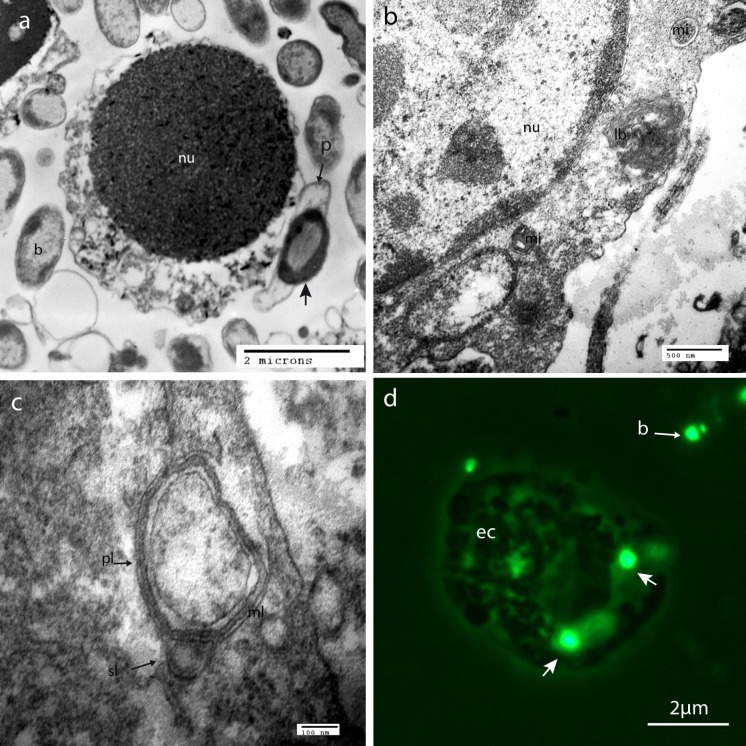
Fig. 4.
Phagocytosis by Haliotis tubeculata gill cells after 2 h of contact observed by TEM and fluorescent microscopy. a Internalization of ORM4 bacteria by hemocytes, b cytoplasm of an epithelial cell showing internalized bacteria, lysosomal body and condensed mitochondria, c detail of lysosome digesting bacteria, d fluorescent micrograph showing ORM4-GFP tagged bacteria in close association with gill cell cytoplasm. b: bacteria around cells, ec: epithelial cell, lb: lysosomal body, pl: primary lysosome, sl: secondary lysosome, nu: nucleus, m: mitochondria, ml: lysosomal multilamellar membrane, p: pseudopodia of the phagocytosis vesicle, arrows: internal bacteria

