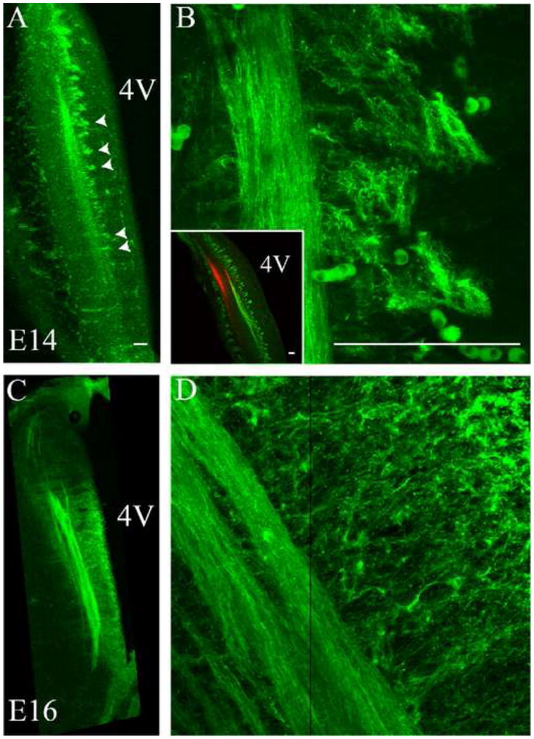
Figure 3. Neuropilin-2 is expressed in the ST and associated tuft-like structures.

Neuropilin-2 expressing fibers and associated tuft-like structures were observed in the ST at E14 and E16, not in the trigeminal tract. A. At E14, the Npn-2 expressing ST and tuft-like structures are seen medial to the ST (arrowheads), extending towards the 4th ventricle. B. Higher magnification shows the tuft-like structures in detail. The inset emphasizes the highly patterned nature of the tufts, parallel to the ST. The inset also can be directly compared to Figure 1A and illustrates that Npn-2 is within the ST only, not in the trigeminal tract, labeled here with DiI (red). C. At E16, Npn-2 continues to be expressed in the developing ST and associated tuft-like structures. D. Higher magnification images show a more mesh-like network of fibers and tuft-like structures extending towards the 4th ventricle. A vertical black line in panel D is the border where two images were aligned in a montage to form one image. 4V = 4th ventricle. Scale bars = 100 μm.
