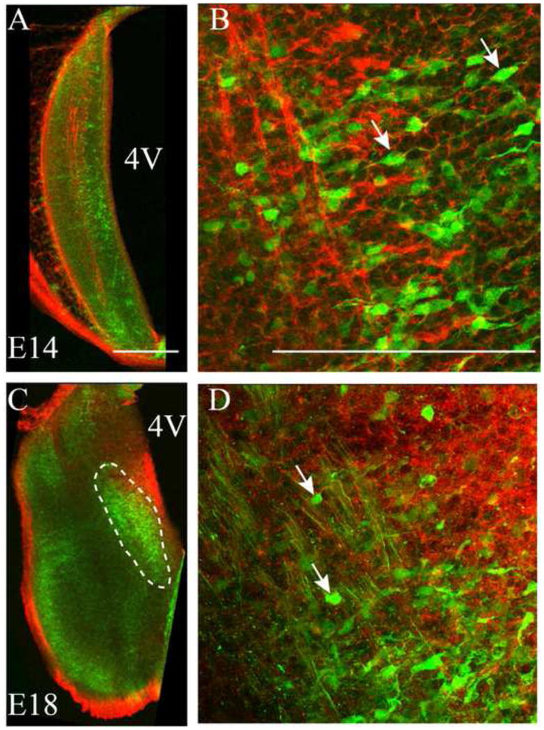
Figure 7. Calbindin-positive cells form a presumptive rNST.

A. At E14, calbindin-positive cells (green) are present along the medial length of the ST. B. At E14, a migratory appearance, characterized by bipolar morphology and elongated soma shapes, is apparent for several neurons (arrows point to examples). The ST is labeled with Npn-2 (red) to highlight the relative location of calbindin-positive cells. C. At E18, the medial rNST is obvious as a group of green, clustered, calbindin-positive neurons located on the medial side of the ST, indicated by dashed lines. D. At E18 it is apparent that most neurons have lost their migratory appearance and most neuron somata are ovoid in shape (arrows point to examples). 4V = 4th ventricle; scale bars =100 μm.
