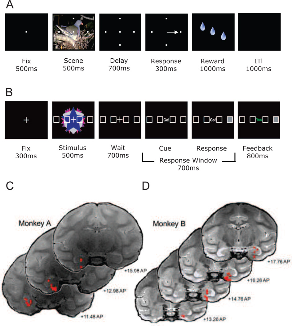
Figure 1. Schematic diagram depicting the within trial sequence of the conditional motor association tasks used for monkeys and humans.
(A) Monkeys initiated trials by fixating on a central spot for 500ms. Fixation was followed by a 500ms scene period, during which four targets were shown superimposed over a large complex visual scene that took up much of the computer monitor. The scene period was followed by a 700ms delay period during which the scene disappeared but the four targets remained. At the end of the delay period the monkeys were cued by the disappearance of the fixation spot, and had 300ms to make a saccade to one of the four targets. If correct, the monkeys received several drops of juice as a reward over approximately 1000ms. The reward period was followed by an inter-trial interval (ITI) period of 1000ms after which the next trial was started. If the monkeys made an error, the ITI period began immediately. (B) Human subjects initiated each trial by fixating on a centrally located “+” for 300ms.Fixation was followed by a 500ms stimulus period during which four targets placed in a horizontal array appeared superimposed across abstract kaleidoscopic images. The stimulus period was followed by a 700ms wait period during which the kaleidoscopic images were removed, but the targets remained. At the end of the delay period the subjects were cued by replacing the fixation cross with the cue (“Go!”), subsequent to which they had 700ms to press one of 4 button response keys that matched the horizontal array. Directly following their response the selected box was filled on the screen and feedback was presented for 800 ms: “yes” (shown in green) if they were correct, and “no” (shown in red) if they made an error. Panels C and D: Selected T2 weighted coronal MRI sections displaying the entorhinal and hippocampal locations (red circles) of the LFP recording sessions from monkeys A and B. In this illustration, the left side of the MRI image reflects the left hemisphere and the right side reflects the right hemisphere. Anterior-posterior locations of the slices are based on the surgical coordinates for centering the grid.

