Cartilage grafts have numerous uses in rhinoplasty; however, the use of intact grafts has several limitations and drawbacks. Intact cartilage grafts often leave contour abnormalities such as sharp edges or step-offs, especially in thin-skinned patients. This faceting of graft edges can become more noticeable over time.1,2 In contrast, morselized or crushed cartilage tends to be more pliable and easier to mold at the surgeon’s discretion and is thus used to soften transitions, conceal irregularities, and fill defects in circumstances in which structural support is not required.1–5
Some surgeons are hesitant to use crushed cartilage owing to its perceived clinical unpredictability. Considerable debate continues amid contradictory findings on the ultimate fate of grafts after morselization, and on whether the cartilage remains viable, resorbs, or is replaced by scar tissue.1–7 Previous studies use cell viability analysis methods that likely underestimate cell death and lack ability to visualize the distribution of nonviable cells. We examined the distribution of live and dead chondrocytes in crushed cartilage and correlate the degree of morselization with viability and mechanical stability.
Methods
In accordance with institutional review board guidelines, we obtained the leftover nasal septal cartilage from 6 healthy patents who had undergone nasal surgery at the University of California, Irvine, Medical Center. Cartilage specimens (approximately 15 × 10 mm), were stripped of perichondrium and placed into normal saline solution at ambient temperature. Each specimen was then cut into 5 identical pieces measuring 3 × 10 mm. Using a cartilage morselizer (Cottle cartilage crusher, model 523900; Karl Storz GmbH & Co, Tuttlingen, Germany), each individual piece was crushed to varying degrees based on a system by Cakmak and Buyuklu1 and Cakmak et al2,3 and categorized as follows: intact, slightly crushed, moderately crushed, significantly crushed, and severely crushed (Table).
Table.
Classification of Cartilage Crushinga
| Category | Description |
|---|---|
| Intact | No manipulation of cartilage |
| Slightly crushed | 1 Moderate-force hit to increase pliability without reducing cartilage structural integrity |
| Moderately crushed | 2 Moderate-force hits to increase pliability and also reduce structural integrity |
| Significantly crushed | 3–4 Moderate-force hits, enough to cause the graft to bend with gravity |
| Severely crushed | 5–6 Hits forceful enough to totally destroy the integrity of the cartilage and result in complete malleability of the graft |
Within 12 hours of explantation, viability analysis was performed using a Live/Dead assay (Molecular Probes Inc, Eugene, Oregon) in conjunction with confocal microscopy (Meta 510; Carl Zeiss LSM, Peabody, Massachusetts) as previously described.8 This is an established technique for analyzing chondrocyte viability and produces images in which live cells are colored green and dead cells are colored red.8–10
Three representative digital images were obtained for each specimen. Manual cell counts of live and dead cells were performed by 2 trained undergraduate students in a blinded fashion. Viability of cells was calculated by dividing the number of live cells by the number of live and dead cells for each image. The mean was computed from the aggregate score of the 3 images per specimen. The scores of the 2 counters were then averaged to determine the final chondrocyte viability.
Results
The degree of cartilage crushing produced using a Cottle morselizer varies widely and is surgeon dependent. Slightly crushing cartilage results in increased pliability without reducing cartilage structural integrity. Moderately crushing cartilage increases pliability and also reduces structural integrity. Significantly crushing cartilage causes the graft to bend with gravity. Severely crushing cartilage destroys the integrity of the cartilage and results in complete malleability of the graft (Figure 1).
Figure 1.
Varying degrees of cartilage crushing. From left to right: intact, slightly crushed, moderately crushed, significantly crushed, and severely crushed. The scale represents millimeters.
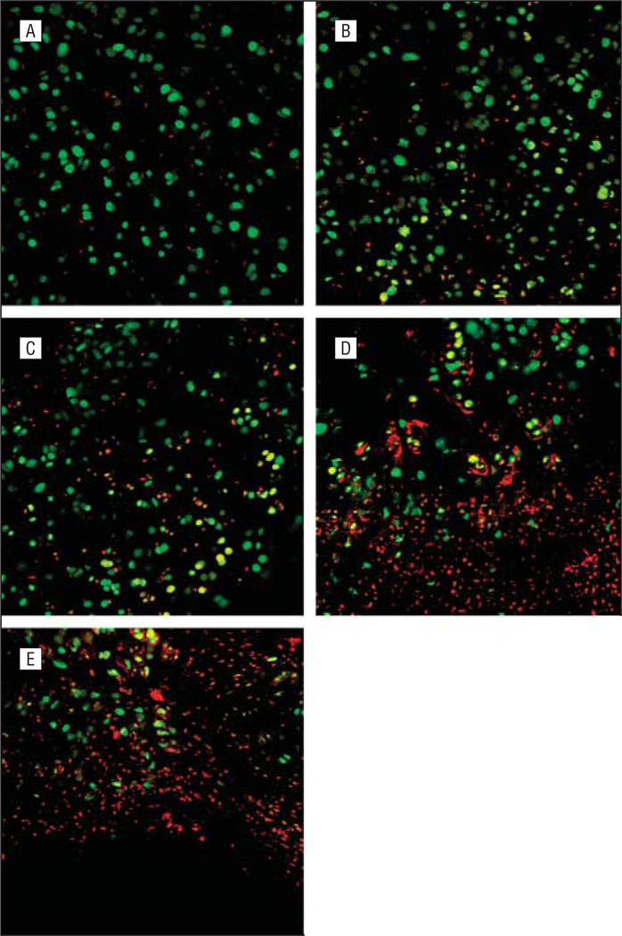
The immediate cell viability counts for intact, slightly crushed, moderately crushed, significantly crushed, and severely crushed cartilage after preparation were 74%, 67%, 55%, 39%, and 25%, respectively. Nonviable cells appeared to group in clusters, and these clusters increased with the severity of crushing. Intact specimens appear as uniform sheets of viable cells with seldom interspersed nonviable cells. Slightly and moderately crushed specimens tended to have uniform distribution of viable and nonviable cells with occasional clusters of nonviable cells. Significantly crushed specimens showed more frequent clusters of nonviable cells. Severely crushed specimens appeared as mostly nonviable cell clusters with few background viable cells (Figure 2).
Figure 2.
Images from Live/Dead assay (Molecular Probes Inc, Eugene, Oregon) using confocal microscopy (original magnification ×40). A, Intact; B, slightly crushed; C, moderately crushed; D, significantly crushed; and E, severely crushed.
Comment
There are varying opinions and contradictory conclusions from numerous studies regarding the immediate and long-term effect of morselization on cartilage graft viability. Because of the potential of resorption, many authors believe overcorrection is necessary with use of crushed cartilage, whereas others refute this point.1–7 This discrepancy may be due to differing methods used in chondrocyte viability analysis. There are numerous cartilage crushing products and devices on the market, and the degree of crushing produced by each is very surgeon dependent. Each alters the pliability and structural integrity of a cartilage graft via different mechanisms. This lack of uniformity in instruments results in difficulty in assessing outcomes of morselization. The grafting environment can also affect crushed graft viability, such that revision rhinoplasties with clinically significant scar tissue may lead to increased chondrocyte death owing to lack of robust perfusion in regions adjacent to the graft.
In contrast to previously published works, the present study has identified that some degree of chondrocyte injury occurs with even modest crushing. Cakmak and Buyuklu1 and Bujía6 digested intact and crushed cartilage and used the trypan blue dye exclusion test for analysis of chondrocyte viability. Digestion of tissue leads to partial loss of nonviable cells when performing cell counts because they are digested during the process. This results in a reduced number of measured nonviable cells, thus yielding an overestimate of the number of living or viable cells. Also, their method1,6 provides no information about spatial distribution of viable and nonviable cells in their specimens.
The viability assay used in our study provides the capability of examining the distribution of viable and nonviable cells in the morselized specimens. Nonviable cells appeared to group in clusters, and these clusters increased with the severity of crushing. Slightly and moderately crushed specimens tended to have uniform distribution of viable and non-viable cells. Based on our findings, significantly and severely crushed grafts may have heterogeneous regions of cell viability, and thus variable graft survival owing to focal areas of cell death, which may lead to unanticipated contour irregularities. By extension, slightly crushed grafts will have more uniform tissue survival and appearance.
This study was limited by its small sample size and inability to address long-term chondrocyte viability, which is technically challenging to perform with tissue explants. At our institution and in our local area, most surgeons do not perform submucous resection (SMR)in septoplasty surgery and are equally conservative in rhinoplasty operations. Large SMR specimens are also more difficult to obtain, and hence additional specimens for study are difficult to acquire.
Our results support the findings of Cakmak and Buyuklu1 and Cakmak et al2,3 that aggressive morselization reduces chondrocyte viability, although we believe their results likely underestimate the degree of chondrocyte injury. With the Cottle crusher method, there is a trade off between crushing cartilage to a clinically useful pliability and maintaining chondrocyte viability in the graft. Thus, the degree and intensity of morselization need to be thoughtfully considered. Other approaches besides the Cottle method may produce the same mechanical changes in cartilage with less graft injury. We advocate performing the least aggressive morselization of cartilage necessary to achieve the desired cosmetic outcome based on our results and experience.
In conclusion, crushed cartilage grafts are often used to soften transitions, conceal irregularities, and fill defects. Increasing the intensity of morselization using the Cottle method results in increased chondrocyte death. Nonviable cells appeared to group in clusters, and these clusters increase with the severity of crushing. Aggressive crushing of cartilage grafts should be avoided because it causes significant chondrocyte cell death and clinically unpredictable grafts. Slight to moderate crushing of cartilage likely results in the most functional and reliable graft.
Acknowledgments
Funding/Support: This study was supported by grants DC005572 and P41RR01192 from the National Institutes of Health (NIH).
Role of the Sponsor: The NIH played no role in the design and conduct of the study; collection, management, analysis, and interpretation of the data; and preparation, review, or approval of the manuscript.
Footnotes
Financial Disclosure: None reported.
References
- 1.Cakmak O, Buyuklu F. Crushed cartilage grafts for concealing irregularities in rhinoplasty. Arch Facial Plast Surg. 2007;9(5):352–357. doi: 10.1001/archfaci.9.5.352. [DOI] [PubMed] [Google Scholar]
- 2.Cakmak O, Buyuklu F, Yilmaz Z, Sahin FI, Tarhan E, Ozluoglu LN. Viability of cultured human nasal septum chondrocytes after crushing. Arch Facial Plast Surg. 2005;7(6):406–409. doi: 10.1001/archfaci.7.6.406. [DOI] [PubMed] [Google Scholar]
- 3.Cakmak O, Bircan S, Buyuklu F, Tuncer I, Dal T, Ozluoglu LN. Viability of crushed and diced cartilage grafts: a study in rabbits. Arch Facial Plast Surg. 2005;7(1):21–26. doi: 10.1001/archfaci.7.1.21. [DOI] [PubMed] [Google Scholar]
- 4.Stoksted P, Ladefoged C. Crushed cartilage in nasal reconstruction. J Laryngol Otol. 1986;100(8):897–906. doi: 10.1017/s0022215100100295. [DOI] [PubMed] [Google Scholar]
- 5.Hamra ST. Crushed cartilage grafts over alar dome reduction in open rhinoplasty. Plast Reconstr Surg. 1993;92(2):352–356. doi: 10.1097/00006534-199308000-00026. [DOI] [PubMed] [Google Scholar]
- 6.Bujía J. Determination of the viability of crushed cartilage grafts: clinical implications for wound healing in nasal surgery. Ann Plast Surg. 1994;32(3):261–265. [PubMed] [Google Scholar]
- 7.Rudderman RH, Guyuron B, Mendelsohn G. The fate of fresh and preserved, noncrushed and crushed autogenous cartilage in the rabbit model. Ann Plast Surg. 1994;32(3):250–254. doi: 10.1097/00000637-199403000-00004. [DOI] [PubMed] [Google Scholar]
- 8.Choi IS, Chae YS, Zemek A, Protsenko DE, Wong B. Viability of human septal cartilage after 1.45 micron diode laser irradiation. Lasers Surg Med. 2008;40(8):562–569. doi: 10.1002/lsm.20663. [DOI] [PMC free article] [PubMed] [Google Scholar]
- 9.Li C, Protsenko DE, Zemek A, Chae YS, Wong B. Analysis of Nd:YAG laser-mediated thermal damage in rabbit nasal septal cartilage. Lasers Surg Med. 2007;39(5):451–457. doi: 10.1002/lsm.20514. [DOI] [PubMed] [Google Scholar]
- 10.Karam AM, Protsenko DE, Li C, et al. Long-term viability and mechanical behavior following laser cartilage reshaping. Arch Facial Plast Surg. 2006;8(2):105–116. doi: 10.1001/archfaci.8.2.105. [DOI] [PubMed] [Google Scholar]


