Abstract
Some prospective studies showed that rabbit antithymocyte globulin was inferior to horse antithymocyte globulin as first-line therapy for patients with severe aplastic anemia. We retrospectively analyzed the clinical outcome of 455 children with severe aplastic anemia who received horse antithymocyte globulin (n=297) or rabbit antithymocyte globulin (n=158) combined with cyclosporine as first-line therapy between 1992 and 2010. The response rates were comparable between the horse and rabbit antithymocyte globulin groups at 3 months [46% (136/294) versus 42% (66/153), P=0.55] and 6 months [60% (178/292) versus 55% (87/143), P=1.0]. Using multivariate analysis, differences in antithymocyte globulin preparations were not associated with response rates. However, 2-year and 10-year overall survival rates in the horse antithymocyte globulin group were significantly better than those in the rabbit antithymocyte globulin group (2-year overall survival: 96% versus 87%, 10-year overall survival: 92% versus 84%, P=0.004). On the basis of multivariate analysis, use of rabbit antithymocyte globulin was a significant adverse factor for overall survival (hazard ratio = 3.56, 95% confidence interval, 1.53 – 8.28, P=0.003). Rabbit antithymocyte globulin caused more profound immunosuppression, which might be responsible for the higher incidence of severe infections. Considering that there are no studies showing the superiority of rabbit antithymocyte globulin over horse antithymocyte globulin, horse antithymocyte globulin should be recommended as a first-line therapy. However, our results justify the use of rabbit antithymocyte globulin as first-line therapy if horse antithymocyte globulin is not available.
Introduction
Idiopathic severe aplastic anemia is an uncommon disease characterized by pancytopenia and hypocellular bone marrow. Immunosuppressive therapy with antithymocyte globulin (ATG) and cyclosporine is the standard treatment for patients with severe aplastic anemia who do not have a human leukocyte antigen-matched related donor; it leads to a response rate of 60 to 70%.1,2 Historically, in Europe and the United States, horse ATG has been used for first-line therapy and rabbit ATG has been used for relapsed patients or non-responders to horse ATG.3,4 Because horse ATG (Lymphoglobulin, Genzyme, Cambridge, MA, USA) was withdrawn from the market and replaced by rabbit ATG (Thymoglobulin, Genzyme), rabbit ATG was prospectively evaluated as first-line immunosuppressive therapy in the United States and Europe.5,6 Both studies showed inferior results with a combination of rabbit ATG and cyclosporine compared with horse ATG and cyclosporine. However, the follow-up periods were relatively short in both studies; the median follow-up was 839 days (range, 2 to 1,852 days) in the study from the National Institutes of Health5 and 397 days (range, 6 to 805 days) in the study from the European Blood and Marrow Transplant Group.6
In Asian countries, both horse ATG and rabbit ATG have been used as first-line immunosuppressive treatment since the early 1990s, which provides an opportunity to compare long-term outcomes of patients who received horse ATG or rabbit ATG. We retrospectively evaluated the outcome of 297 patients with severe aplastic anemia who received horse ATG plus cyclosporine and 158 severe aplastic anemia who received rabbit ATG plus cyclosporine.
Methods
We analyzed data from 455 patients with severe aplastic anemia who received immunosuppressive therapy with horse ATG plus cyclosporine or rabbit ATG plus cyclosporine in Japan, China, and Korea between 1992 and 2010. The outcome of 205 patients who received horse ATG and that of 40 children who received rabbit ATG were previously reported.7,8 Patients with idiopathic severe aplastic anemia were included in the study if they were younger than 18 years and received immunosuppressive therapy within 6 months after diagnosis without specific prior treatments. The severity of the aplastic anemia was classified according to currently used criteria.9
Immunosuppressive therapy consisted of horse ATG (Lymphoglobulin, 15 mg/kg/day for 5 days) or rabbit ATG (Thymoglobulin, 2.3 to 5.0 mg/kg/day for 5 days), cyclosporine (6 mg/kg/day for at least 6 months), and methylprednisolone (2 mg/kg/day for 5 days) with subsequent halving of the dose every week until discontinuation on day 28. The dose of cyclosporine was adjusted to maintain whole blood concentrations between 150 and 250 ng/mL.
Hematologic response was evaluated at 3 and 6 months after the start of therapy. The definitions of complete response, partial response, and relapse have been previously published.1 Briefly, a complete response was defined for all patients as a neutrophil count more than 1.5×10⁹/L, a platelet count more than 100×109/L, and a hemoglobin level more than 11.0 g/dL. A partial response was defined as a neutrophil count more than 0.5×10⁹/L, a platelet count more than 20×109/L, a hemoglobin level more than 80 g/L and no requirement for blood transfusions. Second-line therapies for non-responders to the first immunosuppressive therapy depended on the policies of the individual hospitals.
The Mann-Whitney U test was used to compare continuous variables and Pearson χ2 test was used for categorical variables. Survival rates were calculated using two methods: one indicated when data on patients were censored at the time of stem cell transplantation (transplant-free survival) and the other indicated when data on patients were not censored at the time of transplantation (overall survival).
Survival rates were analyzed using the Kaplan-Meier method. Treatment groups were compared with the long-rank test. Cox proportional hazards models were used to assess which factors could predict response as well as risk factors for survival using both univariate and multivariate analyses. The estimated magnitude of the hazard ratio (HR) is shown along with the 95% confidence interval (95% CI). P values less than 0.05 are considered statistically significant.
This study was approved by ethics committees of the Catholic University of Korea, Nagoya University Graduate School of Medicine, and Chinese Academy of Medical Science and Pekin Union Medical College.
Results
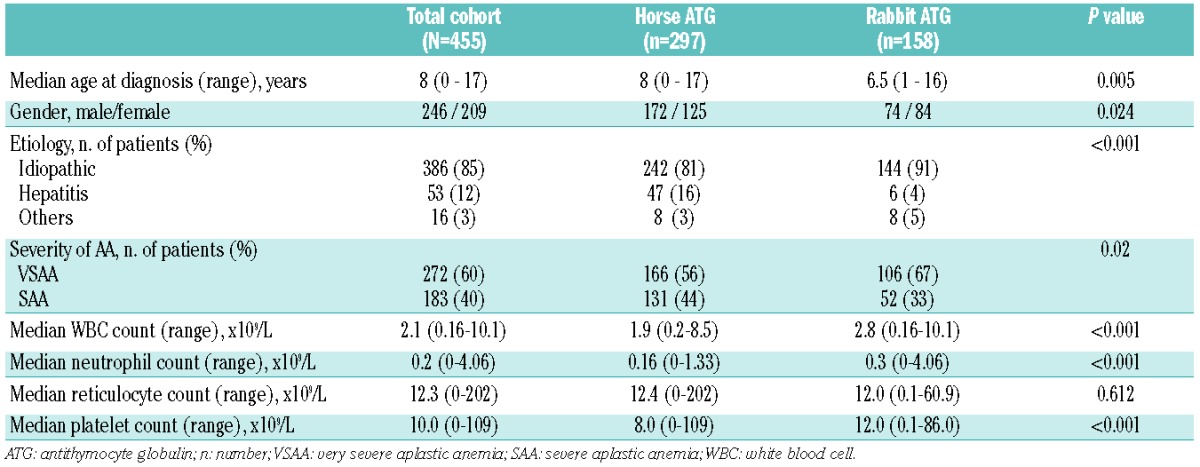
The patients’ characteristics are shown in Table 1. Overall, 455 patients fulfilled the eligibility criteria; 297 patients received horse ATG and 158 patients received rabbit ATG. The median follow-up periods were 82 months (range, 1 to 215 months) in the horse ATG group and 20 months (range, 1 to 182 months) in the rabbit ATG group. The median age at diagnosis was significantly older in the horse ATG group than in the rabbit ATG group. In addition, percentages of males, very severe aplastic anemia, and hepatitis-associated aplastic anemia were significantly higher in the horse ATG group than in the rabbit ATG group (Table 1).
Table 1.
Patients’ characteristics.
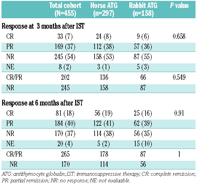
We compared hematologic responses between the horse ATG group and the rabbit ATG group (Table 2). After 3 months, in the horse ATG group, 24 (8%) patients had achieved a complete response and 112 (38%) had achieved a partial response, for an overall response rate of 46%. In the rabbit ATG group, 9 (6%) patients achieved a complete response and 57 (36%) achieved a partial response, for an overall response rate of 42%. After 6 months, in the horse ATG group, 178 of 292 evaluable patients achieved a response (61%) including 56 (19%) who achieved a complete response and 122 (42%) who achieved a partial response. In the rabbit ATG group, 87 of 143 evaluable patients achieved a response (55%) including 25 (16%) who achieved a complete response and 62 (39%) who achieved a partial response. The response rates between the horse ATG group and the rabbit ATG group were not significantly different at 3 months (P=0.55) or 6 months (P=1.0).
Table 2.
Response to horse ATG or rabbit ATG at 3 and 6 months after ATG treatment.
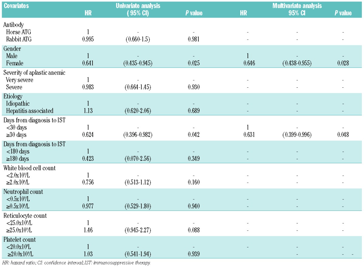
To determine predictors of response to immunosuppressive therapy at 6 months, we compared differences in pretreatment variables between responders and non-responders. The following variables were included in the analysis: etiology, interval between diagnosis and treatment, severity of disease, gender, type of ATG preparation, white blood cell count, reticulocyte count, and platelet count. In both univariate and multivariate analyses, differences in ATG preparations were not associated with response to immunosuppressive therapy. Male gender was a significant predictor of better response in multivariate analysis (Table 3).
Table 3.
Univariate and multivariate analyses for response to immunosuppressive therapy at 6 months after treatment.
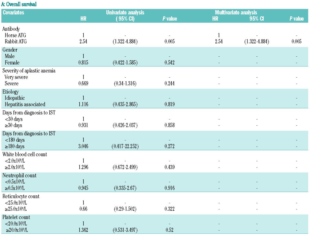
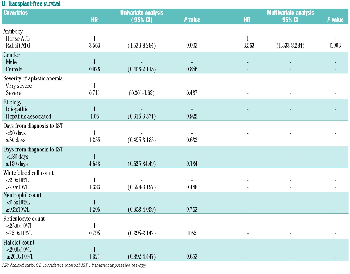
The overall survival rates at 2 and 10 years differed significantly between groups and were 96% (95% CI: 93–98%) and 92% (95% CI: 88–95%), respectively, in the horse ATG group compared with 87% (95% CI: 80–92%) and 84% (95% CI: 74–90%), respectively, in the rabbit ATG group (P=0.004). Because 81 of 297 (27%) patients in the horse ATG group and 21 of 158 (13%) patients in the rabbit ATG group underwent stem cell transplantation as salvage therapy, we calculated the overall survival when data were censored at the time of the transplant. The transplant-free survival rates at 2 and 10 years also differed significantly between groups and were 99% (95% CI: 95–99%) and 95% (95% CI: 90–98%), respectively, in the horse ATG group compared with 91% (95% CI: 84–95%) and 88% (95% CI: 80–94%), respectively, in the rabbit ATG group (P=0.002).
We assessed prognostic factors associated with overall survival and transplant-free survival in univariate and multivariate analyses. Rabbit ATG was the only significant adverse factor for both overall and transplant-free survival in the univariate and multivariate analyses (Table 4).
Table 4.
Prognostic factors for overall survival (A) and transplant-free survival (B) by univariate and multivariate analyses.
Clonal evolution to myelodysplastic syndrome/acute myelogenous leukemia appeared in 13 patients in the horse ATG group and in one patient in the rabbit ATG group. Cytogenetic analysis at the time of evolution showed the following abnormalities: monosomy 7 (n=7), trisomy 8 (n=2), trisomy 8 and del(7) (n=1), monosomy X (n=1), ins (1;¿)(q21;¿) (n=1), and t(3;3)(q21;q26) (n=1) in the horse ATG group. In the rabbit ATG group, only one patient developed myelodysplastic syndrome with a normal karyotype. Evolution to clinical paroxysmal nocturnal hemoglobinuria was rare, with only three patients developing this condition: two in the horse ATG group and one in the rabbit ATG group.
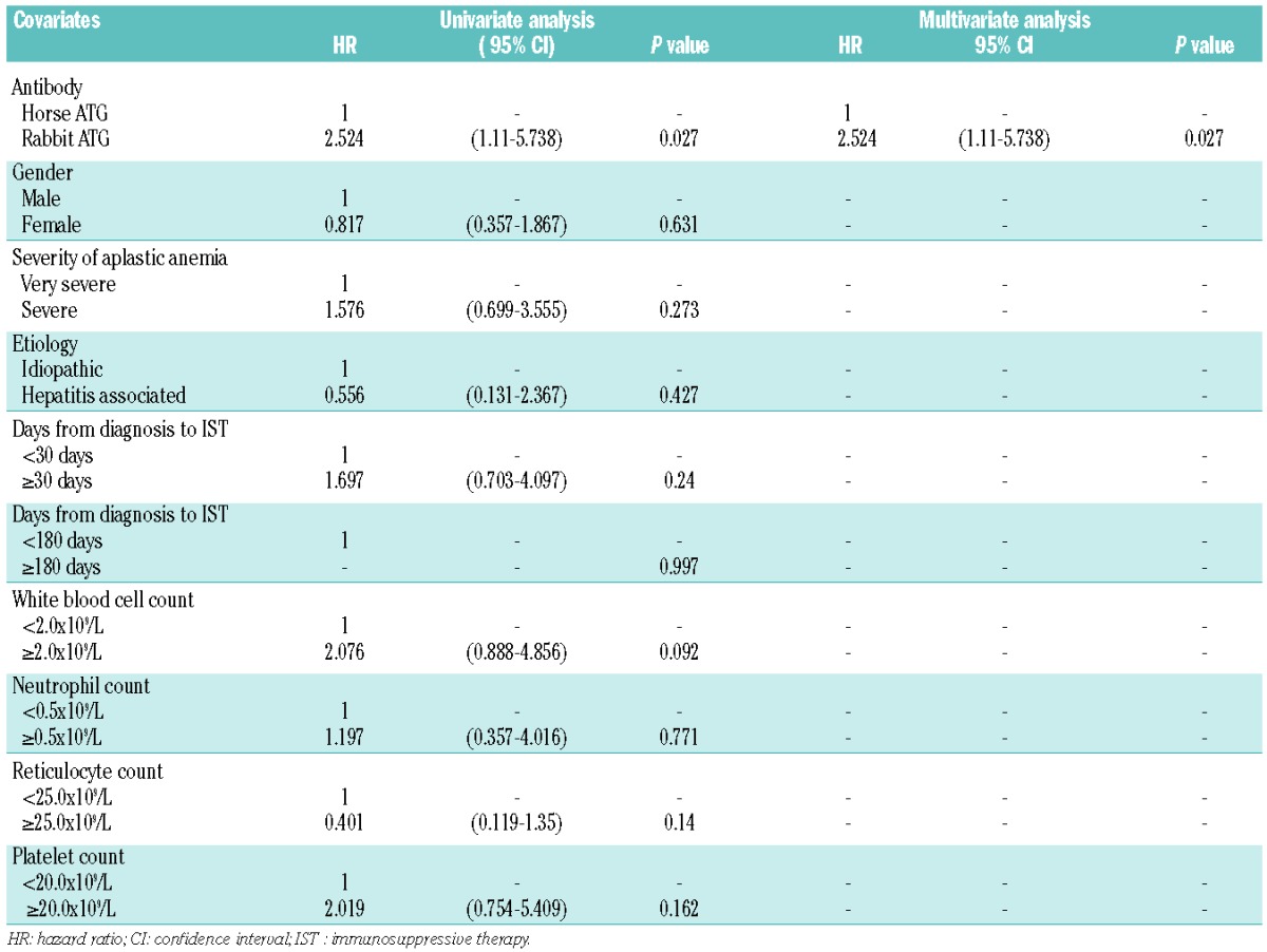
The cumulative incidence of clonal evolution at 2 years and 10 years, including both myelodysplastic syndrome/acute myelogenous leukemia and clinical paroxysmal nocturnal hemoglobinuria, was not significantly different between the two groups, 4% (95% CI: 2–7%) and 6% (95% CI: 4–10%) in the horse ATG group and 0% (95% CI: 0–0%) and 5% (95% CI: 1–19%) in the rabbit ATG group, respectively (P=0.2). However, we must be aware that the median follow-up period was much shorter in the rabbit ATG group than in the horse ATG group. On the other hand, the 2- and 10-year cumulative incidences of relapse were significantly higher in the rabbit ATG group (6%, 95% CI: 2–13% and 15%, 95% CI: 8–28%, respectively) than in the horse ATG group (3%, 95% CI:1–6% and 7%, 95% CI: 4–12%, respectively) (P=0.02). Both univariate and multivariate analyses showed that rabbit ATG was the only risk factor for relapse (Table 5).
Table 5.
Risk factors for relapse by univariate and multivariate analysis.
There were 19 deaths in the horse ATG group: the causes of death were transplant-related toxicity (n=7), myelodysplastic syndrome/acute myelogenous leukemia (n=3), infection within 3 months of immunosuppressive therapy (n=2), infection more than 3 months after immunosuppressive therapy (n=4), hemochromatosis (n=1), hemolysis (n=1), and an accident (n=1). Similarly, there were 18 deaths in the rabbit ATG group: these deaths were caused by infection within 3 months after immunosuppressive therapy (n=4), infection more than 3 months after immunosuppressive therapy (n=3), bleeding (n=6), transplant-related toxicity (n=4), and an accident (n=1).
Figure 1.
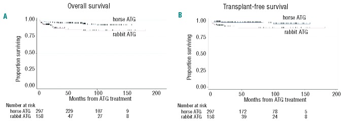
(A) Overall survival for patients treated with rabbit ATG and cyclosporine compared to horse ATG and cyclosporine. (B) Transplant-free survival for patients treated with rabbit ATG and cyclosporine compared to horse ATG and cyclosporine. Transplantation is considered as an event.
Figure 2.
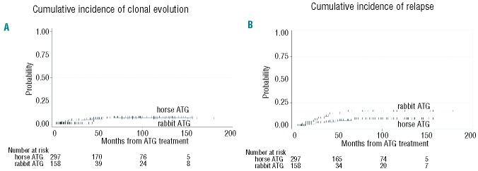
(A) Cumulative incidence of clonal evolution in patients treated with rabbit ATG and cyclosporine compared to horse ATG and cyclosporine. (B) Cumulative incidence of relapse in patients treated with rabbit ATG and cyclosporine compared to horse ATG and cyclosporine.
Discussion
Unlike the prospective National Institutes of Health study comparing horse ATG and rabbit ATG,5 our study showed a comparable response rate at 6 months between patients treated with horse ATG or rabbit ATG, that is, 60% versus 55%, respectively. Although our study has several limitations, including its retrospective design and significant differences in patients’ characteristics between groups, the large number of patients and long follow-up period may overcome these drawbacks. The prospective randomized study from the National Institutes of Health5 and a retrospective study from Brazil10 showed significantly lower response rates in the rabbit ATG group. The response rates at 6 months for patients treated with rabbit ATG were only 37% and 34%, respectively, which translated into inferior overall survival rates. However, several other studies showed comparable response rates between patients treated with horse ATG or rabbit ATG.11–15 In the European Blood and Marrow Transplant Group study, the best total response rate was not significantly different between groups, that is, 67% versus 60% for horse ATG and rabbit ATG, respectively.6 Of note, all three retrospective studies from China,11 Korea,12 and Japan13 showed comparable response rates between patients treated with horse ATG or rabbit ATG, which suggests that ethnicity may be responsible for differences in response rates between studies.
Another issue is the difference in dose of rabbit ATG used in different studies. In the studies from China,11 Korea,12 and Spain,15 the dose of rabbit ATG was 2.5 mg/kg/day for 5 days, which was lower than the standard dose of rabbit ATG in Europe and United States. In the study from Guagzhou, China, 46 children received rabbit ATG at a dose of 2.5 mg/kg/day for 5 days plus cyclosporine.11 The response rate was 65% at 6 months, which was comparable to results reported in previous studies with horse ATG.1,2
Regulatory T cells are key elements in suppressing immune responses.16 These cells are decreased in patients with severe aplastic anemia and an increase in numbers of regulatory T cells may be one of the mechanisms by which to restore hematopoiesis after ATG therapy.17 Rabbit ATG, but not horse ATG, induces an expansion of functional regulatory T cells in in vitro cultures.18 However, in the National Institutes of Health study, the number of regulatory T cells was much lower in the weeks after treatment with rabbit ATG because of a profound decrease of total CD4 lymphocytes in the rabbit ATG group compared with the horse ATG group.5 It would be interesting to compare the number of regulatory T cells between groups of patients receiving lower and higher doses of rabbit ATG.
Despite the comparable response rates, both overall survival and transplant-free survival rates were significantly inferior in the rabbit ATG group than the horse ATG group in our study. Multivariate analysis revealed that use of rabbit ATG was the only adverse factor for survival. The incidence of deaths due to infections and bleeding was higher in the rabbit ATG group than in the horse ATG group. In the report from Brazil, the excess of deaths observed in the rabbit ATG group occurred mainly within 60 days following administration of ATG and all deaths were caused by infections or bleeding.9 In the European Blood and Marrow Transplant Group study, 11 of 35 patients treated with rabbit ATG died of infections (n=9), bleeding (n=1), or transplant-related toxicity (n=1).6
Epstein-Barr virus remains latent in B cells after primary infection and is controlled by virus-specific T lymphocytes. Compromising cellular immune responses can put the patient at high risk of reactivation of this virus, which can lead to lymphoproliferative disorders.19 Scheinberg et al. reported on a series of 78 patients with aplastic anemia who received four different immunosuppressive therapies: the median peak of Epstein-Barr virus copy numbers was significantly higher and the median duration of polymerase chain reaction positivity significantly longer in the rabbit ATG group than in the other groups.20 Fortunately, progression to clinical disease was not observed after Epstein-Barr virus reactivation. The authors did not recommend routine monitoring of Epstein-Barr virus copy numbers. However, recently, a fatal case of Epstein-Barr virus-lymphoproliferative disorder was reported in a patient from Japan treated with rabbit ATG as first-line therapy.21 There is some concern that prolonged T-cell suppression and more active Epstein-Barr virus reactivation caused by immunosuppressive therapy with rabbit ATG might increase the incidence of Epstein-Barr virus-related lymphoproliferative disorder.
Based on these findings, intensive supportive care may decrease the mortality rate and improve the survival rate after rabbit ATG therapy. In fact, among 24 adult patients who received intensive supportive care with levofloxacin, fluconazole, valacyclovir, and granulocyte colony-stimulating factor in the MD Anderson Cancer Center, there were no early deaths in the first 100 days and the overall survival rate was favorable, being 70% at 3 years.22
Considering that there are no studies showing the superiority of rabbit ATG over horse ATG, horse ATG should be recommended as first-line immunosuppressive therapy. However, horse ATG is not available everywhere, especially in Asian countries. In these cases, first-line therapy with rabbit ATG may be justified. A prospective study is warranted to determine the optimal use of rabbit ATG.
Footnotes
Authorship and Disclosures
Information on authorship, contributions, and financial & other disclosures was provided by the authors and is available with the online version of this article at www.haematologica.org.
References
- 1.Kojima S, Hibi S, Kosaka Y, Yamamoto M, Tsuchida M, Mugishima H, et al. Immunosuppressive therapy using antithymocyte globulin, cyclosporine, and danazol with or without human granulocyte colony-stimulating factor in children with acquired aplastic anemia. Blood. 2000;96(6):2049–54 [PubMed] [Google Scholar]
- 2.Fuhrer M, Rampf U, Baumann I, Faldum A, Niemeyer C, Janka-Schaub G, et al. Immunosuppressive therapy for aplastic anemia in children: a more severe disease predicts better survival. Blood. 2005;106(6):2102–4 [DOI] [PubMed] [Google Scholar]
- 3.Di Bona E, Rodeghiero F, Bruno B, Gabbas A, Foa P, Locasciulli A, et al. Rabbit antithymocyte globulin (r-ATG) plus cyclosporine and granulocyte colony stimulating factor is an effective treatment for aplastic anaemia patients unresponsive to a first course of intensive immunosuppressive therapy. Gruppo Italiano Trapianto di Midollo Osseo (GITMO). Br J Haematol. 1999;107(2):330–4 [DOI] [PubMed] [Google Scholar]
- 4.Scheinberg P, Nunez O, Young NS. Retreatment with rabbit anti-thymocyte globulin and ciclosporin for patients with relapsed or refractory severe aplastic anaemia. Br J Haematol. 2006;133(6):622–7 [DOI] [PubMed] [Google Scholar]
- 5.Scheinberg P, Nunez O, Weinstein B, Scheinberg P, Biancotto A, Wu CO, et al. Horse versus rabbit antithymocyte globulin in acquired aplastic anemia. N Engl J Med. 2011;365(5):430–8 [DOI] [PMC free article] [PubMed] [Google Scholar]
- 6.Marsh JC, Bacigalupo A, Schrezenmeier H, Tichelli A, Risitano AM, Passweg JR, et al. Prospective study of rabbit antithymocyte globulin and cyclosporine for aplastic anemia from the EBMT Severe Aplastic Anaemia Working Party. Blood. 2012;119(23):5391–6 [DOI] [PubMed] [Google Scholar]
- 7.Kosaka Y, Yagasaki H, Sano K, Kobayashi R, Ayukawa H, Kaneko T, et al. Prospective multicenter trial comparing repeated immunosuppressive therapy with stem-cell transplantation from an alternative donor as second-line treatment for children with severe and very severe aplastic anemia. Blood. 2008;111(3):1054–9 [DOI] [PubMed] [Google Scholar]
- 8.Takahashi Y, Muramatsu H, Sakata N, Hyakuna N, Hamamoto K, Kobayashi R, et al. Rabbit antithymocyte globulin and cyclosporine as first-line therapy for children with acquired aplastic anemia. Blood. 2013;121(5):862–3 [DOI] [PubMed] [Google Scholar]
- 9.Camitta BM, Thomas ED, Nathan DG, Gale RP, Kopecky KJ, Rappeport JM, et al. A prospective study of androgens and bone marrow transplantation for treatment of severe aplastic anemia. Blood. 1979;53(3):504–14 [PubMed] [Google Scholar]
- 10.Atta EH, Dias DS, Marra VL, de Azevedo AM. Comparison between horse and rabbit antithymocyte globulin as first-line treatment for patients with severe aplastic anemia: a single-center retrospective study. Ann Hematol. 2010;89(9):851–9 [DOI] [PubMed] [Google Scholar]
- 11.Chen C, Xue HM, Xu HG, Li Y, Huang K, Zhou DH, et al. Rabbit-antithymocyte globulin combined with cyclosporin A as a first-line therapy: improved, effective, and safe for children with acquired severe aplastic anemia. J Cancer Res Clin Oncol. 2012;138(7):1105–11 [DOI] [PMC free article] [PubMed] [Google Scholar]
- 12.Jeong D-C, Chung NG, Lee JW, Jang P-S, Cho B, Kim H-K. Long-term outcome of immunosuppressive therapy with rabbit antithymocyte globulin (rATG) for childhood severe aplastic anemia for 15 Years. Blood. 2011;118(21):1346a [Google Scholar]
- 13.Yamazaki H, Saito C, Sugimori N, Hosokawa K, Kiyu Y, Mochizuki K, et al. Thymoglobuline is as effective as lymphoglobuline in Japanese patients with aplastic anemia possessing increased glycosylphosphatidylinositol-anchored protein (GPI-AP) deficient cells. Blood. 2011;118(21):1339a [Google Scholar]
- 14.Afable MG, 2nd, Shaik M, Sugimoto Y, Elson P, Clemente M, Makishima H, et al. Efficacy of rabbit anti-thymocyte globulin in severe aplastic anemia. Haematologica. 2011;96(9):1269–75 [DOI] [PMC free article] [PubMed] [Google Scholar]
- 15.Vallejo C, Montesinos P, Rosell A, Brunet S, Córdoba Raul, Pérez-Ceballos Elena, et al. Comparison between lymphoglobuline- and thymoglobuline-based immunosuppressive therapy as first-line treatment for patients with aplastic anemia. Blood. 2009;114(22):3194a [Google Scholar]
- 16.Sakaguchi S. Naturally arising Foxp3-expressing CD25+CD4+ regulatory T cells in immunological tolerance to self and non-self. Nat Immunol. 2005;6(4):345–52 [DOI] [PubMed] [Google Scholar]
- 17.Solomou EE, Rezvani K, Mielke S, Malide D, Keyvanfar K, Visconte V, et al. Deficient CD4+ CD25+ FOXP3+ T regulatory cells in acquired aplastic anemia. Blood. 2007;110(5):1603–6 [DOI] [PMC free article] [PubMed] [Google Scholar]
- 18.Feng X, Kajigaya S, Solomou EE, Keyvanfar K, Xu X, Raghavachari N, et al. Rabbit ATG but not horse ATG promotes expansion of functional CD4+CD25highFOXP3+ regulatory T cells in vitro. Blood. 2008;111(7):3675–83 [DOI] [PMC free article] [PubMed] [Google Scholar]
- 19.Long HM, Taylor GS, Rickinson AB. Immune defence against EBV and EBV-associated disease. Curr Opin Immunol. 2011;23(2):258–64 [DOI] [PubMed] [Google Scholar]
- 20.Scheinberg P, Fischer SH, Li L, Nunez O, Wu CO, Sloand EM, et al. Distinct EBV and CMV reactivation patterns following antibody-based immunosuppressive regimens in patients with severe aplastic anemia. Blood. 2007;109(8):3219–24 [DOI] [PMC free article] [PubMed] [Google Scholar]
- 21.Ohata K, Iwaki N, Kotani T, Kondo Y, Yamazaki H, Nakao S. An Epstein-Barr virus-associated leukemic lymphoma in a patient treated with rabbit antithymocyte globulin and cyclosporine for hepatitis-associated aplastic anemia. Acta Haematol. 2012;127(2):96–9 [DOI] [PubMed] [Google Scholar]
- 22.Kadia TM, Borthakur G, Garcia-Manero G, Faderl S, Jabbour E, Estrov Z, et al. Final results of the phase II study of rabbit anti-thymocyte globulin, ciclosporin, methylprednisone, and granulocyte colony-stimulating factor in patients with aplastic anaemia and myelodysplastic syndrome. Br J Haematol. 2012;157(3):312–20 [DOI] [PMC free article] [PubMed] [Google Scholar]


