Abstract
Background
Balancing the relative advantages and disadvantages of unicompartmental knee arthroplasties (UKAs) against those for TKAs can be challenging. Survivorship is one important end point; arthroplasty registers repeatedly report inferior midterm survival rates, but longer-term data are sparse. Comparing survival directly by using arthroplasty register survival reports also may be inadequate because of differences in indications, implant designs, and patient demographics in patients having UKAs and TKAs.
Questions/purposes
The aims of this study were to assess the survivorship of UKA in the context of one large, northern European registry, and to compare the rates of survivorship with those of cemented TKAs performed for primary knee osteoarthritis during the same 27-year period.
Methods
From the Finnish Arthroplasty Register, we obtained the data for 4713 patients undergoing UKAs for primary osteoarthritis (mean age, 63.5 years; minimum followup, 0 years; mean, 6.0 years; range, 0–24 years) who had surgical revision between 1985 and 2011. From this cohort, we calculated the Kaplan-Meier survivorship for revision performed for any reason and compared it with the survivorship of 83,511 patients (mean age, 69.5 years; minimum followup 0 years; mean, 6.4 years; range, 0–27 years) with TKAs treated for primary osteoarthritis during the same period. Data were adjusted for age and sex in a comparative analysis.
Results
Kaplan-Meier survivorship of UKAs was 89.4% at 5 years, 80.6% at 10 years, and 69.6% at 15 years; the corresponding rates for TKAs were 96.3%, 93.3%, and 88.7%, respectively. UKAs had inferior long-term survivorship compared with cemented TKAs, even after adjusting for the age and sex of the patients (hazard ratio 2.2, p < 0.001).
Conclusions
A UKA offers tempting advantages compared with a TKA; however, the revision frequency for UKAs in widespread use, as measured in a large, national registry, was poorer than that of TKAs. When choosing between a UKA and a TKA, patients should be informed of advantages of both procedures, but they also should be advised about the generally higher revision risk after UKA.
Level of Evidence
Level III, therapeutic study. See the Instructions for Authors for a complete description of levels of evidence.
Introduction
Unicompartmental knee arthroplasties (UKA) are widely used to treat isolated unicompartmental knee osteoarthritis (OA). Advantages of a UKA include lower postoperative morbidity, quicker return to activities, and more normal feeling of the knee in comparison to the advantages after a TKA [1, 8, 9], and some single-center studies report UKA results that are comparable to those of TKAs [12, 13, 15, 18, 22]. However, these studies are not supported by available implant register data from Finland, Norway, Sweden, Australia, New Zealand, and the United Kingdom [10, 23–27], which repeatedly report inferior midterm survivorship of UKAs compared with TKAs.
However, less is known regarding comparative survivorship between UKA and TKA for the longer term. In addition, comparing survival of UKAs and TKAs directly by using arthroplasty register survival reports may be inadequate because of differences in indications, implant designs, and patient demographics between patients having UKAs and TKAs which are not consistently considered in registry reports.
The purpose of this population-based study was to assess survivorship of UKAs performed for patients with primary knee OA in Finland during a 27-year period and to compare the survivorship rate of patients who underwent UKAs with that of a cohort of patients who received cemented TKAs during the same period.
Patients and Methods
Patients Undergoing UKA
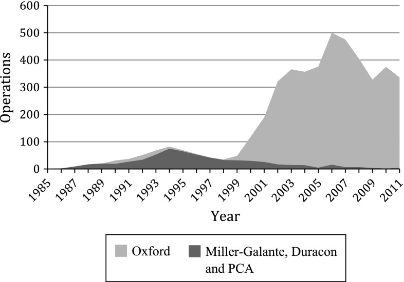
Records for patients who had UKAs from 1985 to 2011 for treatment of primary knee OA were extracted from the data in the Finnish Arthroplasty Register. We obtained information regarding 5211 UKAs with one of 11 different implant designs. After identifying all the UKAs, we excluded the designs that had been implanted in less than 100 knees during the study period (n = 208) or for which fewer than 20 knees were at risk after 10 years (n = 112). Similar exclusion criteria were used in previous register-based studies [2, 4]. In addition, we excluded UKAs which had been performed for reasons other than primary knee OA (n = 178). This resulted in retention of data for 4713 UKAs, which represented 90% of all UKAs (Table 1). The number of UKAs performed was relatively small until 1998 when the Oxford® Phase 3 (Biomet Inc, Warsaw, IN, USA) was introduced (Fig. 1).
Table 1.
Demographic data of the patients
| Variables | UKA | TKA | ||
|---|---|---|---|---|
| Males | Females | Males | Females | |
| Number of knees (%) | 1757 | 2956 (62.7) | 24,929 | 58,581 (70.1) |
| Age (years) | ||||
| Mean (SD) [range] | 62.9 (9.3) [33 to 94] | 62.9 (9.4) [33 to 91] | 68.2 (8.7) [29 to 106] | 70.0 (8.5) [23 to 106] |
| Followup (years) | ||||
| Mean (SD) [range] | 5.6 (3.9) [0 to 24] | 6.2 (4.2) [0 to 24] | 5.6 (4.2) [0 to 25] | 6.7 (4.8) [0 to 27] |
| Implant design, Number (%) | ||||
| Oxford® | 4084 (86.7) | 83,510 37 different designs* |
||
| Miller-Galante | 318 (6.7) | |||
| PCA® | 171 (3.6) | |||
| Duracon™ | 140 (3.0) | |||
UKA= unicompartmental knee arthroplasty; Oxford®, Biomet Inc, Warsaw, IN, USA; Miller-Galante, Zimmer, Warsaw, IN, USA; Porous Coated Anatomic (PCA®), Howmedica, Rutherford, NJ, USA; Duracon™, Stryker, Mahwah, NJ, USA; * cemented, cruciate retaining, or posterior stabilized.
Fig. 1.
The numbers of UKAs performed for primary knee OA during the study are shown. Oxford®, Biomet Inc, Warsaw, IN, USA; Duracon™, Stryker, Mahwah, NJ, USA; Miller-Galante, Zimmer, Warsaw, IN, USA; Porous Coated Anatomic (PCA®), Howmedica, Rutherford, NJ, USA.
Patients Undergoing TKA
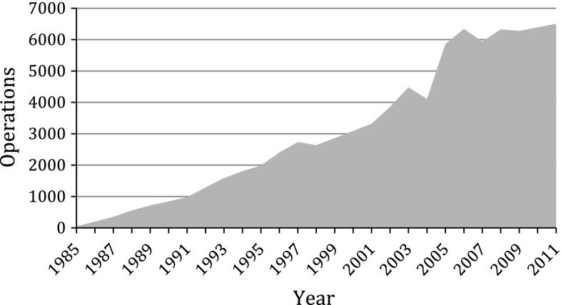
The TKA group included all patients who had undergone cemented TKAs for primary knee OA during the study period. Constrained models other than cruciate-retaining or posterior-stabilized prostheses were excluded. In total, we included 83,511 TKAs (Table 1). The number of TKAs performed has increased steadily during the study period (Fig. 2).
Fig. 2.
The numbers of cemented, cruciate-retaining, or posterior-stabilized TKA designs implanted for primary knee OA during the study period are shown.
To obtain TKA designs corresponding to four UKA designs included in the study, we extracted data for four TKAs during the study period that fulfilled the inclusion criteria; ie, more than 100 procedures performed with 20 or more at risk after 15 years and limited to those performed for primary knee OA. The data for these TKAs were extracted starting with the most commonly used implants then proceeding to the less frequently used designs.
Demographic Differences and Changing Demographics Among Patients
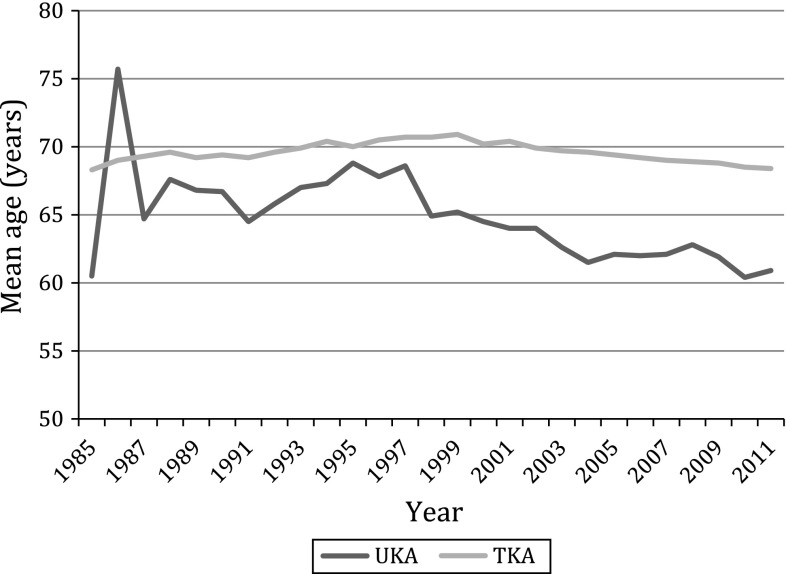
Patients undergoing UKAs were younger than patients undergoing TKAs, with mean ages of 63 years (range, 33–91 years) and 69 years (range, 23–96 years), respectively (p < 0.001). The mean age of the patients undergoing UKAs also decreased during the study period, being 69.1 years between 1985 and 1993, 66.4 years between 1994 and 2002, and 62.1 years between 2003 and 2011. The corresponding mean ages of patients undergoing TKAs were 69.5, 70.4, and 69.1 years, respectively (Fig. 3). The decline in mean age during the study period also was steeper in patients undergoing UKAs compared with patients undergoing TKAs (p < 0.001).
Fig. 3.
The mean ages of patients undergoing UKAs and TKAs are shown.
Statistics
In the Kaplan-Meier survivorship analysis, the survival end point was defined as revision of the knee for any reason. Revisions included changing, removing, or adding any component to the prosthesis. Information regarding patient deaths or emigrations was obtained from the Finnish Population Centre. In the Kaplan-Meier analysis, patients were censored at the date of death or emigration, but they were not excluded from the study. Univariate analysis for comparison of the two groups was performed using the log-rank test.
The Cox proportional hazard model was used to obtain the hazard ratio (HR) and the 95% CIs. Three different Cox proportional hazards models were created to compare: (1) UKAs and TKAs, (2) different UKA designs, and (3) different TKA designs. Age and sex were used as adjusting factors. Age was categorized (≤ 65 years and > 65 years), because the linearity assumption did not hold. Differences between groups were considered statistically significant if the p value was less than 0.05 in a two-tailed test. SPSS software (Version 19.0, IBM, Armonk, NY, USA) was used for the statistical analyses.
Results
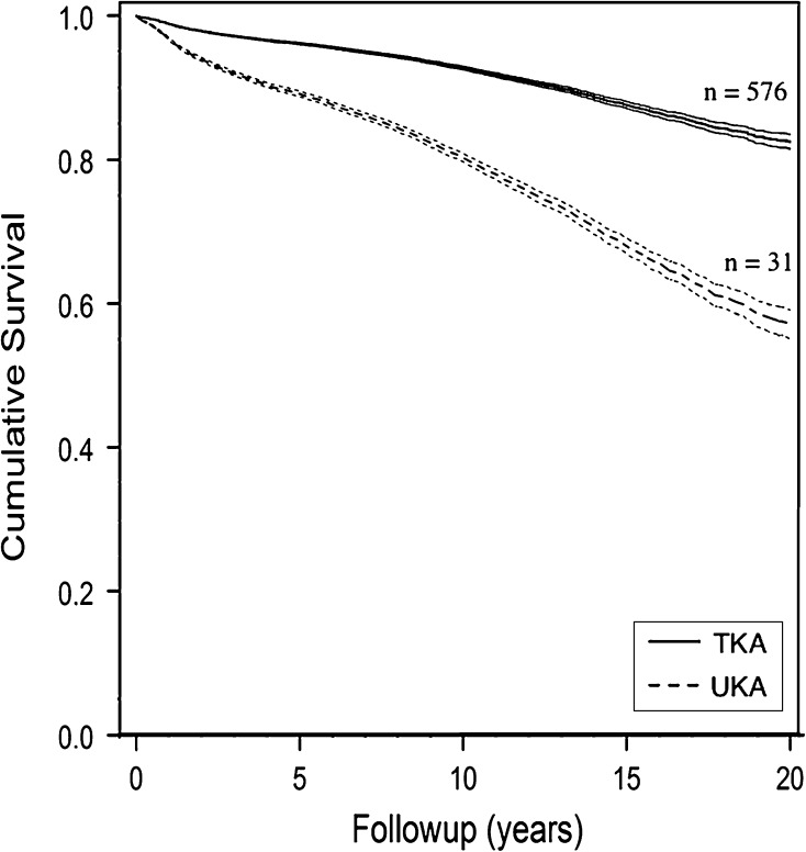
Overall, Kaplan-Meier survivorship of UKAs was 89.4% (95% CI, 88.4–90.2) at 5 years, 80.6% (95% CI, 79.4–81.7) at 10 years, and 69.6% (95% CI, 68.2–70.9) at 15 years. Survivorship of TKAs was 96.3% at 5 years (95% CI, 96.2–96.4), 93.3% at 10 years (95% CI, 93.1–93.4), and 88.7% at 15 years (95% CI, 88.5–88.9) (Fig. 4).
Fig. 4.
The overall Kaplan-Meier survivorship rates for UKAs and TKAs during the study period are shown. The end point was defined as any revision, including when either the whole implant or any one component was removed, exchanged, or implanted for any reason. Adjustments were made for age and sex.
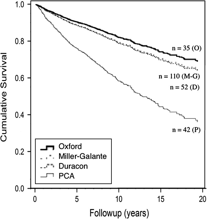
Aseptic loosening was the most common reason for revisions in patients undergoing UKAs and TKAs but was more common among patients undergoing UKAs (HR, 4.42; 95% CI, 3.7–4.8; p < 0.001 adjusted by sex and age) (Table 2). In patients undergoing UKAs, neither age (≤ 65 years or > 65 years) nor sex affected the revision rate. In patients undergoing TKAs, patients 65 years or younger had an increased revision rate compared with patients older than 65 years (HR, 2.1; 95% CI, 1.9–2.2; p < 0.001). UKAs had inferior survivorship compared with TKAs, even after adjusting for patient age and sex (HR, 2.2; 95% CI, 2.0–2.4; p < 0.001). Survivorship varied by UKA prosthesis design (Fig. 5), and the PCA® (Howmedica, Rutherford, NJ, USA) had shorter survivorship when compared with other designs (Table 3); Kaplan-Meier analysis performed on UKAs with PCA® prostheses removed (data not shown) did not change the overall conclusion of our study, because the PCA® accounted for a relatively small proportion of implants in the UKAs reported in the register. For comparison, we also obtained survival data for four different TKA prosthesis designs that corresponded to the UKA prosthesis designs (Table 4).
Table 2.
Reasons for revisions
| Reason for revision | UKA, number (%) | TKA, number (%) |
|---|---|---|
| Aseptic loosening | 310 (46.8) | 1178 (26.7) |
| Malalignment | 40 (6.0) | 395 (9.0) |
| Prosthesis fracture | 29 (4.4) | 144 (3.3) |
| Instability | 19 (2.9) | 101 (2.3) |
| Infection | 18 (2.7) | 528 (12.0) |
| Fracture | 14 (2.1) | 92 (2.1) |
| Patella complication | 1 (0.2) | 478 (10.8) |
| Other reason | 232 (35.0) | 1496 (33.9) |
| Total | 663 (100) | 4412 (100) |
Fig. 5.
The Kaplan-Meier survivorship rates for different UKA designs used during the study period are shown. The end point was defined as any revision, including when either the whole implant or any one component was removed, exchanged, or implanted for any reason. Adjustments were made for age and sex. O = Oxford® (Biomet Inc, Warsaw, IN, USA); M-G = Miller-Galante (Zimmer, Warsaw, IN, USA); D = Duracon™ (Stryker, Mahwah, NJ, USA); P = Porous Coated Anatomic (PCA®) (Howmedica, Rutherford, NJ, USA).
Table 3.
Survivorship of different UKA designs
| UKA design | Years | Number* | Mean followup years/(range) | At 5 years** (95% CI) | At 10 years** (95% CI) | At 15 years** (95% CI) | HR (95% CI) | p value |
|---|---|---|---|---|---|---|---|---|
| Oxford® | 1990–2011 | 439/4084 | 5.2 (0–21) | 90.4 (89.4,91.3) | 83.7 (82.5,84.8) | 76.9 (75.6,78.2) | 1.0 | – |
| Miller-Galante | 1990–2005 | 90/318 | 11.3 (1–21) | 89.1 (85.0,92.2) | 77.4 (72.4,81.8) | 66.7 (63.7,74.1) | 1.2 (0.9–1.5) | 0.19 |
| Duracon™ | 1993–2000 | 39/140 | 10.5 (0–19) | 84.8 (77.6,90.1) | 76.7 (68.7,83.3) | 70.7 (62.3,77.9) | 1.2 (0.9–1.7) | 0.29 |
| PCA® | 1985–1995 | 95/171 | 9.5 (1–24) | 75.7 (68.4,81.7) | 53.1 (45.3,60.7) | 38.7 (31.4,46.4) | 2.7 (2.2–3.5) | < 0.001 |
UKA= unicompartmental knee arthroplasty; (years); * number of revisions/number of total operations; **survivorship obtained via Kaplan-Meier analysis.hazard ratio (HR) according to Cox proportional hazards model (adjusted by sex and age); Oxford®, Biomet Inc, Warsaw, IN, USA; Miller-Galante, Zimmer, Warsaw, IN, USA; Duracon™, Stryker, Mahwah, NJ, USA; Porous Coated Anatomic (PCA®), Howmedica, Rutherford, NJ, USA.
Table 4.
Survivorship of different TKA prosthesis designs
| TKA design | Years | Number* | Mean followup years/(range) | At 5 years** (95% CI) | At 10 years** (95% CI) | At 15 years** (95% CI) | HR (95% CI) | p value |
|---|---|---|---|---|---|---|---|---|
| Duracon™ | 1992–2011 | 990/18199 | 8.5 (0–19) | 96.4 (96.1,96.7) | 94.4 (94.0,94.7) | 91.9 (91.5,92.3) | 1.0 | – |
| PFC®
Sigma® |
1989–2011 | 590/13292 | 6.2 (0–22) | 96.8 (96.4,97.1) | 94.8 (94.4,95.2) | 88.9 (88.3,89.4) | 1.1 (1.0–1.2) | 0.24 |
| Miller-Galante II | 1989–1998 | 111/1008 | 11.5 (0–20) | 95.5 (94.0,96.7) | 90.8 (88.8,92.4) | 86.9 (84.7,88.9) | 1.5 (1.2–1.8) | < 0.001 |
| PCA®/ modular | 1985–1996 | 143/935 | 12.4 (0–23) | 94.3 (92.5,95.6) | 89.2 (87.0,91.1) | 83.2 (80.7,85.6) | 1.8 (1.5–2.2) | < 0.001 |
* Number of revisions/number of total operations; **survivorship obtained via Kaplan-Meier analysis; HR = hazard ratio, according to Cox proportional hazards model (adjusted by sex and age); Duracon™, Stryker, Mahwah, NJ, USA; PFC® Sigma®, DePuy Orthopaedics Inc, Warsaw, IN, USA; Miller-Galante II, Zimmer, Warsaw, IN, USA; Porous Coated Anatomic (PCA®)/modular, Stryker, Mahwah, NJ, USA.
Discussion
In the arthroplasty register reports, overall survivorship of UKA is poorer compared with TKA [23–27]. However, direct comparison of UKA and TKA survival may be inadequate because of different implant designs, indications for surgery, durations of followup, and differences in patient demographics. We compared age- and sex-adjusted survival for patients who had UKAs and cemented TKAs performed for primary knee OA during a 27-year period. In our study, the overall long-term survivorship of UKAs performed for primary knee OA, even the best-performing UKA prosthesis design, was inferior to that for cemented TKAs. In addition we found that the age of patients having TKAs and UKAs decreased during the study period, but the decline in age of patients having UKAs was significantly steeper. Finally, the overall number of UKAs performed decreased during the last 5 years of the study.
The weaknesses of the current study are those common to all register-based studies. Detailed data regarding the patients’ medical histories or knee radiographs were not available. Additionally, we do not have any information regarding the preoperative and postoperative functional outcomes which would better reflect the results of the operations than implant survival alone [5, 28]. However, to overcome the limitations of single-surgeon or hospital studies, there is a need for register-based studies that reflect the results of the operations in widespread use outside the centers of excellence. Second, comparing UKA results with TKA results is complicated. Even if one could match patients by age and sex, which we could not do in this large registry setting, the characteristics of patients undergoing UKAs and those undergoing TKAs may be quite different. In general, it can be assumed that patients undergoing UKAs are more active and their expectations after surgery are higher. Such patients may be willing to accept a higher risk of failure to enjoy some of the perceived benefits of UKA; however, they need to be informed of this risk, and to not overstate the potential influence of age and sex (potential surrogates for activity level), we adjusted Cox models for age and sex. In addition, the Finnish register does not provide surgeon-specific data by volume; a post hoc analysis by hospital volume (greater than versus less than 20 UKAs per year) did not show a difference in survivorship by hospital volume (HR, 1.1, p = 0.26). Finally, the UKA group included an outlier design (PCA®) for which the survival was poor. The inclusion of this implant may bias the results; however, we repeated the Kaplan-Meier analysis with this implant excluded (data not shown), and because the numbers of PCA® prostheses in the registry was relatively small, the overall conclusions of our study remained substantially unchanged.
The reasons for the higher revision rate and decreasing number of UKAs are likely multifactorial. First, there are unique reasons for UKA revisions; in particular, the progression of arthritis to a contralateral compartment, which does not exist for TKAs and may partly explain the higher revision rates. Second, aseptic loosening is more frequent with patients undergoing UKAs compared with patients undergoing TKAs [3, 23–27]. The higher rate of loosening may be explained by the smaller contact area between the implant and bone compared with TKAs. Additionally, the UKA cementing technique is technically demanding, particularly if limited incisions are used and if the bone is sclerotic [7, 14]. We believe that aseptic loosening is a substantial reason for revision, but, especially for the Oxford® UKA prosthesis (Biomet Inc, Warsaw, IN, USA), radiolucent lines under the tibial tray may be misleading [6], but only in a relatively small number of patients. Even so, it appears that reliable fixation for UKAs remains an unsolved problem [20]. Third, patients undergoing UKAs are younger compared with patients undergoing TKAs, therefore, their expectations are high, and the results of UKAs in these patients may be disappointing [21]. Finally, there is increasing evidence that patients with mild or moderate OA who undergo knee arthroplasty, even those with severe symptoms, have a higher revision rate than patients with severe arthritis [16, 17, 19]. These patients may be overrepresented in the UKA group because the less invasive operation may be performed for patients with less severe arthritis.
Comparing the reasons for failure from the different arthroplasty register reports is difficult. Every register has individual classifications for revisions. For example, the most common reason for UKA revision in the Norwegian arthroplasty register was “pain,” which does not exist in the Finnish or United Kingdom registers [24, 26]. However, the register trends suggest that UKAs are revised more often because of aseptic loosening, pain, and progression of disease compared with TKAs. TKAs are revised more often because of infection [23–27].
A UKA offers tempting advantages compared with a TKA, including lower infection rates, preserving bone stock, minimizing invasiveness, and restoring knee kinematics without sacrificing ligaments [11]. However, in our registry and others [23–27], the survivorship of UKAs is poorer than that of TKAs. Aseptic loosening is a particular problem and tibial component fixation of UKAs appears to remain an unsolved problem. Additional research is needed to develop more reliable fixation of UKA implants. When choosing between a UKA and a TKA, patients should be informed of the advantages of both procedures, but they also should be advised of the generally higher revision risk after UKA.
Footnotes
Each author certifies that he or she, or a member of his or her immediate family, has no funding or commercial associations (eg, consultancies, stock ownership, equity interest, patent/licensing arrangements, etc) that might pose a conflict of interest in connection with the submitted article.
All ICMJE Conflict of Interest Forms for authors and Clinical Orthopaedics and Related Research editors and board members are on file with the publication and can be viewed on request.
This work was performed at the Department of Surgery, Oulu University Hospital, Oulu, Finland.
References
- 1.Brown NM, Sheth NP, Davis K, Berend ME, Lombardi AV, Berend KR, Della Valle CJ. Total knee arthroplasty has higher postoperative morbidity than unicompartmental knee arthroplasty: a multicenter analysis. J Arthroplasty. 2012;27(8 suppl):86–90. doi: 10.1016/j.arth.2012.03.022. [DOI] [PubMed] [Google Scholar]
- 2.Dorey FJ. Survivorship analysis of surgical treatment of the hip in young patients. Clin Orthop Relat Res. 2004;418:23–28. doi: 10.1097/00003086-200401000-00005. [DOI] [PubMed] [Google Scholar]
- 3.Epinette JA, Brunschweiler B, Mertl P, Mole D, Cazenave A, The French Society for the Hip and Knee Unicompartmental knee arthroplasty modes of failure: wear is not the main reason for failure: a multicentre study of 418 failed knees. Orthop Traumatol Surg Res. 2012;98(suppl):S124–S130. doi: 10.1016/j.otsr.2012.07.002. [DOI] [PubMed] [Google Scholar]
- 4.Eskelinen A, Remes V, Helenius I, Pulkkinen P, Nevalainen J, Paavolainen P. Uncemented total hip arthroplasty for primary osteoarthritis in young patients: a mid- to long-term follow-up study from the Finnish Arthroplasty Register. Acta Orthop. 2006;77:57–70. doi: 10.1080/17453670610045704. [DOI] [PubMed] [Google Scholar]
- 5.Goodfellow JW, O’Connor JJ, Murray DW. A critique of revision rate as an outcome measure: re-interpretation of knee joint registry data. J Bone Joint Surg Br. 2010;92:1628–1631. doi: 10.1302/0301-620X.92B12.25193. [DOI] [PubMed] [Google Scholar]
- 6.Gulati A, Chau R, Pandit HG, Gray H, Price AJ, Dodd CA, Murray DW. The incidence of physiological radiolucency following Oxford unicompartmental knee replacement and its relationship to outcome. J Bone Joint Surg Br. 2009;91:896–902. doi: 10.1302/0301-620X.91B7.21914. [DOI] [PubMed] [Google Scholar]
- 7.Hamilton WG, Collier MB, Tarabee E, McAuley JP, Engh CA, Jr, Engh GA. Incidence and reasons for reoperation after minimally invasive unicompartmental knee arthroplasty. J Arthroplasty. 2006;21(6 suppl 2):98–107. doi: 10.1016/j.arth.2006.05.010. [DOI] [PubMed] [Google Scholar]
- 8.Hopper GP, Leach WJ. Participation in sporting activities following knee replacement: total versus unicompartmental. Knee Surg Sports Traumatol Arthrosc. 2008;16:973–979. doi: 10.1007/s00167-008-0596-9. [DOI] [PubMed] [Google Scholar]
- 9.Jahromi I, Walton NP, Dobson PJ, Lewis PL, Campbell DG. Patient-perceived outcome measures following unicompartmental knee arthroplasty with mini-incision. Int Orthop. 2004;28:286–289. doi: 10.1007/s00264-004-0573-y. [DOI] [PMC free article] [PubMed] [Google Scholar]
- 10.Koskinen E, Paavolainen P, Eskelinen A, Pulkkinen P, Remes V. Unicondylar knee replacement for primary osteoarthritis: a prospective follow-up study of 1,819 patients from the Finnish Arthroplasty Register. Acta Orthop. 2007;78:128–135. doi: 10.1080/17453670610013538. [DOI] [PubMed] [Google Scholar]
- 11.Laurencin CT, Zelicof SB, Scott RD, Ewald FC. Unicompartmental versus total knee arthroplasty in the same patient: a comparative study. Clin Orthop Relat Res. 1991;273:151–156. [PubMed] [Google Scholar]
- 12.Lim HC, Bae JH, Song SH, Kim SJ. Oxford phase 3 unicompartmental knee replacement in Korean patients. J Bone Joint Surg Br. 2012;94:1071–1076. doi: 10.1302/0301-620X.94B8.29372. [DOI] [PubMed] [Google Scholar]
- 13.Macaulay W, Yoon RS. Fixed-bearing, medial unicondylar knee arthroplasty rapidly improves function and decreases pain: a prospective, single-surgeon outcomes study. J Knee Surg. 2008;21:279–284. doi: 10.1055/s-0030-1247832. [DOI] [PubMed] [Google Scholar]
- 14.Miskovsky C, Whiteside LA, White SE. The cemented unicondylar knee arthroplasty: an in vitro comparison of three cement techniques. Clin Orthop Relat Res. 1992;284:215–220. [PubMed] [Google Scholar]
- 15.Newman J, Pydisetty RV, Ackroyd C. Unicompartmental or total knee replacement: the 15-year results of a prospective randomised controlled trial. J Bone Joint Surg Br. 2009;91:52–57. doi: 10.1302/0301-620X.91B1.20899. [DOI] [PubMed] [Google Scholar]
- 16.Niinimaki TT, Murray DW, Partanen J, Pajala A, Leppilahti JI. Unicompartmental knee arthroplasties implanted for osteoarthritis with partial loss of joint space have high re-operation rates. Knee. 2011;18:432–435. doi: 10.1016/j.knee.2010.08.004. [DOI] [PubMed] [Google Scholar]
- 17.Pandit H, Gulati A, Jenkins C, Barker K, Price AJ, Dodd CA, Murray DW. Unicompartmental knee replacement for patients with partial thickness cartilage loss in the affected compartment. Knee. 2011;18:168–171. doi: 10.1016/j.knee.2010.05.003. [DOI] [PubMed] [Google Scholar]
- 18.Pandit H, Jenkins C, Barker K, Dodd CA, Murray DW. The Oxford medial unicompartmental knee replacement using a minimally-invasive approach. J Bone Joint Surg Br. 2006;88:54–60. doi: 10.1302/0301-620X.88B1.17114. [DOI] [PubMed] [Google Scholar]
- 19.Polkowski GG, 2nd, Ruh EL, Barrack TN, Nunley RM, Barrack RL. Is pain and dissatisfaction after TKA related to early-grade preoperative osteoarthritis? Clin Orthop Relat Res. 2013;471:162–168. doi: 10.1007/s11999-012-2465-6. [DOI] [PMC free article] [PubMed] [Google Scholar]
- 20.Schroer WC, Barnes CL, Diesfeld P, LeMarr A, Ingrassia R, Morton DJ, Reedy M. The Oxford Unicompartmental Knee fails at a high rate in a high-volume knee practise. Clin Orthop Relat Res. 2013;471:3533–3539. doi: 10.1007/s11999-013-3174-5. [DOI] [PMC free article] [PubMed] [Google Scholar]
- 21.Scott CE, Howie CR, MacDonald D, Biant LC. Predicting dissatisfaction following total knee replacement: a prospective study of 1217 patients. J Bone Joint Surg Br. 2010;92:1253–1258. doi: 10.1302/0301-620X.92B9.24394. [DOI] [PubMed] [Google Scholar]
- 22.Skowronski J, Jatskewych J, Dlugosz J, Skowronski R, Bielecki M. The Oxford II medial unicompartmental knee replacement: a minimum 10-year follow-up study. Ortop Traumatol Rehabil. 2005;7:620–625. [PubMed] [Google Scholar]
- 23.The Australian National Joint Replacement Registry. Annual Report 2012. Available at: https://aoanjrr.dmac.adelaide.edu.au/annual-reports-2012. Accessed August 12, 2013.
- 24.The National Joint Registry of England, Wales and Northern Ireland. 9th Annual Report 2012. Available at: http://www.njrcentre.org.uk/njrcentre/Portals/0/Documents/England/Reports/9th_annual_report/NJR%209th%20Annual%20Report%202012.pdf. Accessed September 22, 2013.
- 25.The New Zealand Joint Registry. Thirteen Year Report: January 1999 to December 2011. Available at: http://nzoa.org.nz/system/files/NJR%2013%20Year%20Report.pdf Accessed August 10, 2013.
- 26.The Norwegian Arthroplasty Register. Annual Report 2011. Available at: http://nrlweb.ihelse.net/eng/Report_2010.pdf. Accessed August 10, 2013.
- 27.The Swedish Knee Arthroplasty Register. Annual Report 2012. Available at: http://www.knee.nko.se/english/online/uploadedFiles/117_SKAR_2012_Engl_1.0.pdf. Accessed August 10, 2013.
- 28.Wylde V, Blom AW. The failure of survivorship. J Bone Joint Surg Br. 2011;93:569–570. doi: 10.1302/0301-620X.93B5.26687. [DOI] [PubMed] [Google Scholar]


