Abstract
Elevated levels of maternal androgens in avian eggs affect numerous traits, including oxidative stress. However, current studies disagree as to whether prenatal androgen exposure enhances or ameliorates oxidative stress. Here, we tested how prenatal testosterone exposure affects oxidative stress in female domestic chickens (Gallus gallus) during the known oxidative challenge of an acute stressor. Prior to incubation, eggs were either injected with an oil vehicle or 5 ng testosterone. At either 17 or 18 days post-hatch, several oxidative stress markers were assessed from blood taken before and after a 20 min acute stressor, as well as following a 25 min recovery from the stressor. We found that, regardless of yolk treatment, during both stress and recovery all individuals were in a state of oxidative stress, with elevated levels of oxidative damage markers accompanied by a reduced total antioxidant capacity. In addition, testosterone-exposed individuals exhibited poorer DNA damage repair efficiencies in comparison with control individuals. Our work suggests that while yolk androgens do not alter oxidative stress directly, they may impair mechanisms of oxidative damage repair.
Keywords: testosterone, oxidative stress, antioxidants, repair, maternal effects, stress
1. Introduction
Hormone-mediated maternal effects have an important influence on offspring phenotype. In birds, prenatal exposure to androgens can alter multiple aspects of physiology and behaviour [1,2], including vulnerability to oxidative stress [3,4]. Oxidative stress is a physiological state in which free radical production exceeds antioxidant and cellular repair defences, resulting in oxidative damage to macromolecules [5]. Experimental elevation of testosterone levels in adult male zebra finches (Taeniopygia guttata) increases oxidative damage and inhibits antioxidant activity during a free radical attack [6]. However, the effects of prenatal testosterone on baseline levels of oxidative stress in females are not consistent across studies, with decreasing lipid peroxidation and increasing total antioxidant capacity (TAC) in one study [3], and no differences in TAC in another [4].
Because oxidative damage negatively impacts reproduction [7,8] and survival [7,9,10], an individual's resistance to oxidative stress has fitness implications. One context in which the relationship between oxidative stress and fitness might be especially influential is in stressful early-life environments. Recently, we showed that the rising glucocorticoid levels in response to an acute stressor are accompanied by a shift into a state of oxidative stress, with an increase in plasma oxidative damage markers and a decrease in plasma antioxidants. We refer to this effect as glucocorticoid-induced oxidative stress (GiOS) [11].
Given that effects of prenatal testosterone on oxidative stress remain unclear, we explored for the first time the hypothesis that prenatal testosterone exposure alters vulnerability to GiOS and DNA damage repair efficiency in juvenile female chickens (Gallus gallus).
2. Material and methods
(a). Egg injections and housing
One hundred and seventy-two fresh, unincubated Bovan White eggs, laid by different hens (Centurion Poultry Inc., Milton, PA, USA), were injected with either 50 µl of a sesame oil vehicle (control) or 5 ng of testosterone suspended in 50 µl of oil (testosterone) directly into the yolk (for details, see the electronic supplemental material). This dose of testosterone elevates yolk androgen levels within a physiological range for eggs from this supplier (12.5 ± 4.2 ng egg−1). Injected eggs were incubated as previously described [11]. At one day post-hatch, 20 females (n = 10 per treatment), sexed morphologically (details in the electronic supplementary materials), were retained for use in this study. Chicks were housed in a Brower brooder with ad libitum food (Start & Grow Sunfresh Recipe, Purina Mills, MO, USA) and water, and weighed daily [11].
(b). Acute stress and recovery
On either post-hatch day 17 or 18, chicks underwent an acute stress using a standard bag restraint protocol (treatments were evenly distributed across sampling days). A total of 140 µl of blood was taken from the alar vein within 3 min of entering the housing room (baseline). After the baseline sample, birds were placed in a breathable cloth bag for 20 min, and then bled again (stress sample). Birds were returned to their brooder and a third sample was taken 25 min later (recovery sample). Samples were centrifuged at 3000 r.p.m. for 5 min. Plasma was removed and stored in aliquots at −80°C until plasma oxidative stress analyses were conducted. Erythrocytes were immediately analysed via single-cell gel electrophoresis (SCGE).
(c). Oxidative stress measures
We assessed reactive oxygen metabolites (ROMs) and total plasma antioxidant capacity (TAC), using the d-ROMs and OXY-adsorbent tests (Diacron International, Grosseto, Italy), respectively, in all blood samples, as described previously ([11]; see the electronic supplementary materials, for more details). All analyses were run in duplicate and the intra- and inter-assay coefficients of variation were 4.01 and 4.33% for ROMs, and 3.85 and 4.45% for TAC. We used the SCGE assay [10] with modifications (see the electronic supplementary materials) to measure three distinct aspects of DNA damage and repair: in vivo DNA damage levels, vulnerability of DNA to a H2O2 challenge and DNA repair efficiency. These three measurements were taken for all blood samples, by making three slides per sample (untreated, challenge and repair). In SCGE, DNA damage levels are quantified as the average per cent DNA found in the electrophoresis-generated tail (damaged DNA migrates into tails [10]). In vivo DNA damage levels were quantified on 50 cells of an untreated slide. Vulnerability of DNA to the H2O2 challenge was estimated for each individual as the difference in damage on the challenge slide (exposed to 200 µM H2O2 for 5 min prior to SCGE) and the untreated slide. Repair efficiency was estimated for each individual as the difference in damage on the repair slide (exposed to 200 µM H2O2 for 5 min followed by incubation at 37°C and 5% CO2 in RPMI complete media for 1 h) and the untreated slide. This difference was then divided by the vulnerability measure, yielding the per cent of damage acquired during the H2O2 challenge that had been repaired within 1 h.
Statistics. Statistical analyses were run using JMP software (v. 10.0.0, SAS Institute Inc. 2012, Cary, NC, USA). For all analyses, we performed generalized linear mixed models of restricted maximum likelihood (REML-GLMM). For every model, we checked for homogeneity of variances (Levene's test), and for normality of residuals (Kolmogorov–Smirnov test). In each model, individual was introduced as a random factor to control for variance among individuals. For growth analyses, treatment was included as a fixed effect, and exponential growth rates for mass between days 4 and 17 were estimated as previously described [11]. For oxidative stress analyses, ROMs, TAC and the three measures of DNA damage were each modelled over the acute stress (three time points). Oxidative stress models included the fixed effects of time, treatment and all interactions (time × treatment, time × individual, treatment × individual, time × treatment × individual). Non-significant interactions were sequentially removed from the models, starting from the higher order interactions, and the analyses were repeated until we obtained a model with only significant terms. Post hoc comparisons were carried out using Tukey HSD tests.
3. Results
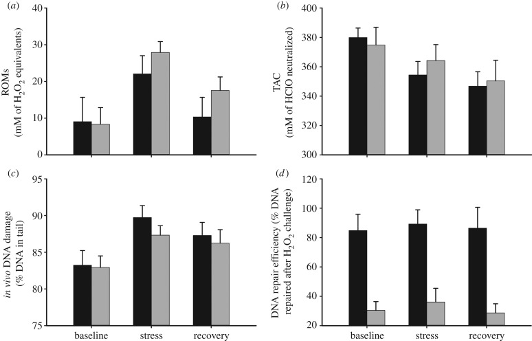
There was no effect of treatment on growth, ROMs or TAC (table 1). However, regardless of treatment, there was a significant change in ROMs (table 1 and figure 1a) and TAC (table 1 and figure 1b) over the acute stress and recovery period. Specifically, ROMs increased during the stress period and then decreased but did not return to baseline during the recovery period (figure 1a; Tukey HSD, p < 0.05). TAC decreased over the stress period and remained at a decreased level during the recovery period (figure 1b; Tukey HSD, p < 0.05).
Table 1.
Results of REML-GLMM on the response to prenatal testosterone exposure during growth and during GiOS. Individual was included as a random factor. For growth, N = 20 birds. For GiOS measures, N = 60 observations on 20 birds. Outcomes in italic refer to variables included in the final models (the rest were subsequently excluded from the models because they were not significant).
| variable | treatment (control and testosterone) | time (baseline, stress and recovery) | treatment × time |
|---|---|---|---|
| growth | F1,18 = 1.09, p = 0.32 | ||
| ROM | F1,13 = 0.65, p = 0.43 | F2,23 = 23.47, p < 0.0001 | F2,20 = 0.22, p = 0.80 |
| TAC | F1,18 = 0.02, p = 0.88 | F2,38 = 10.42, p = 0.0003 | F2,36 = 0.74, p = 0.48 |
| in vivo DNA | F1,18 = 0.55, p = 0.47 | F2,38 = 11.71, p = 0.0001 | F2,36 = 0.45, p = 0.64 |
| vulnerability to H2O2 challenge | F1,18 = 1.34, p = 0.28 | F2,36 = 1.55, p = 0.24 | F2,36 = 0.18, p = 0.84 |
| DNA repair efficiency | F1,18 = 26.12, p < 0.0001 | F2,38 = 0.31, p = 0.74 | F2,36 = 0.05, p = 0.95 |
Figure 1.
The effect of elevated embryonic testosterone on (a) ROMs, (b) TAC, (c) in vivo levels of DNA damage and (d) DNA repair efficiency after H2O2 challenge during an acute stress response (baseline: 0 min; stress: after 20 min stressor; recovery: 25 min post-stressor) in juvenile chickens (all groups, n = 10; mean ± s.e.m.). Black bars denote control and grey bars denote testosterone.
In vivo levels of DNA damage did not differ by treatment, but did change over the acute stress and recovery period (table 1 and figure 1c), increasing over the acute stress period and then remaining stable during the recovery period (figure 1c; Tukey HSD, p < 0.05). The H2O2 challenge effectively increased DNA damage above in vivo levels in all samples, but did not differ by treatment or time period (table 1). However, birds exposed to prenatal testosterone had impaired DNA repair efficiency at all time points in comparison with the control birds (table 1 and figure 1d).
4. Discussion
Based on our results, prenatal testosterone exposure has a potentially costly impact on juvenile female chickens’ ability to repair DNA oxidative damage. In accordance with prior work in our laboratory [11], during an acute stress response, birds experienced GiOS with increased levels of plasma oxidative damage and decreased antioxidant capacity. Importantly, however, individuals exposed to elevated prenatal testosterone had reduced DNA repair efficiency in comparison with controls. Thus, in response to oxidative challenges, offspring prenatally exposed to high levels of testosterone potentially accumulate DNA damage at a faster rate. Consistent with previous research, this effect of yolk testosterone on female oxidative status was not apparent in baseline samples [4], but was revealed by the imposition of an oxidative challenge.
While few studies have investigated the mechanisms underlying effects of maternal testosterone on offspring phenotype, proposed mechanisms include alterations in hormone secretion, androgen sensitivity of the offspring or, more generally, gene expression [2]. The effect of prenatal testosterone exposure on DNA repair may be caused by inhibition of DNA repair enzyme gene expression. DNA repair pathways are established in ovo [12], concurrent with embryonic exposure to yolk steroids [13]. In human cells, androgens inhibit expression of genes coding for enzymes involved in one of the major oxidative DNA damage repair pathways—long patch nucleotide excision repair [14–16]. Based on our findings, future work investigating the effects of testosterone on the expression and activity of DNA repair enzymes is warranted.
The costs of the decreased repair efficiency associated with exposure to elevated yolk testosterone are not yet clear. Despite consistent differences in repair capabilities, we saw no treatment-driven differences in oxidative damage accumulation before or after a GiOS challenge. This may be caused by the gradual rate of repair activity [17] and related to the fact that, even following the recovery period, all individuals were still experiencing GiOS. Alternatively, costs of this difference in repair capacity might only be incurred following multiple oxidative stress challenges that could increase long-term oxidative damage accumulation in cells. In terms of fitness, excessive oxidative damage has been linked to reduced survival [7,9,10] and reproductive success [7,8]. Accordingly, we propose that poor repair efficiency as a consequence of increased prenatal androgen exposure may be particularly costly to fitness in stressful early life environments. However, in assessing effects of yolk androgens on fitness, it is important to consider two things: (i) the apparent costs of androgens on oxidative damage repair shown here will be moderated by other benefits and (ii) some of the fitness consequences of prenatal androgens are likely to be context-dependent, and may not manifest until animals face challenges.
Acknowledgements
We thank C. Garrehy, K. Sweeny, V. Fasanello, C. Fischer, C. Rhone, N. Metcalfe and an anonymous reviewer.
All procedures were approved by the Bucknell University Institutional Animal Care and Use Committee.
Data accessibility
Data deposited in Dryad, doi:10.5061/dryad.7q5k2 [18].
Funding statement
Support came from a Bucknell University Swanson Research Fellowship to M.F.H. and Bucknell Kalman and Russo support to B.N.W. and L.A.T.
References
- 1.Groothuis TGG, Wendt M, von Engelhardt N, Carere C, Eising C. 2005. Maternal hormones as a tool to adjust offspring phenotype in avian species. Neurosci. Biobehav. Rev. 29, 329–352 (doi:10.1016/j.neubiorev.2004.12.002) [DOI] [PubMed] [Google Scholar]
- 2.Gil D. 2008. Hormones in avian eggs: physiology, ecology, and behavior. Adv. Study Behav. 38, 337–398 [Google Scholar]
- 3.Noguera JC, Alonso-Alvarez C, Kim SY, Morales J, Velando A. 2011. Yolk testosterone reduces oxidative damages during postnatal development. Biol. Lett. 7, 93–95 (doi:10.1098/rsbl.2010.0421) [DOI] [PMC free article] [PubMed] [Google Scholar]
- 4.Tobler M, Sandell MI, Chiriac S, Hasselquist D. 2013. Effects of prenatal testosterone exposure on antioxidant status and bill color in adult zebra finches. Physiol. Biochem. Zool. 86, 52–54 (doi:10.1086/670194) [DOI] [PubMed] [Google Scholar]
- 5.Monaghan P, Metcalfe NB, Torres R. 2009. Oxidative stress as a mediator of life history trade-offs: mechanisms, measurements, and interpretation. Ecol. Lett. 12, 75–92 (doi:10.1111/j.1461-0248.2008.01258.x) [DOI] [PubMed] [Google Scholar]
- 6.Alonso-Alvarez C, Bertrand S, Faivre B, Chastel O, Sorci G. 2007. Testosterone and oxidative stress: the oxidation handicap hypothesis. Proc. R. Soc. B 274, 819–825 (doi:10.1098/rspb.2006.3764) [DOI] [PMC free article] [PubMed] [Google Scholar]
- 7.Bize P, Devevey G, Monaghan P, Doligez B, Christe P. 2008. Fecundity and survival in relation to resistance to oxidative stress in a free-living bird. Ecology 89, 2584–2593 (doi:10.1890/07-1135.1) [DOI] [PubMed] [Google Scholar]
- 8.Noguera JC, Kim SY, Velando A. 2012. Pre-fledgling oxidative damage predicts recruitment in a long-lived bird. Biol. Lett. 8, 61–63 (doi:10.1098/rsbl.2011.0756) [DOI] [PMC free article] [PubMed] [Google Scholar]
- 9.Beckman KB, Ames BN. 1998. The free radical theory of aging matures. Physiol. Rev. 78, 547–581 [DOI] [PubMed] [Google Scholar]
- 10.Freeman-Gallant CR, Amidon J, Berdy B, Wein S, Taff CC, Haussmann MF. 2011. Oxidative damage to DNA related to survivorship and carotenoid-based sexual signal ornamentation in the common yellowthroat. Biol. Lett. 7, 429–432 (doi:10.1098/rsbl.2010.1186) [DOI] [PMC free article] [PubMed] [Google Scholar]
- 11.Haussmann MF, Longenecker AL, Marchetto NM, Juliano SA, Bowden RM. 2012. Embryonic exposure to corticosterone modifies the juvenile stress response, oxidative stress, and telomere length. Proc. R. Soc. B 279, 1447–1456 (doi:10.1098/rspb.2011.1913) [DOI] [PMC free article] [PubMed] [Google Scholar]
- 12.Pachkowski BF, Guyton KZ, Sonawane B. 2011. DNA repair during in utero development: a review of the current state of knowledge, research needs, and potential application in risk assessment. Mutat. Res. 728, 35–46 (doi:10.1016/j.mrrev.2011.05.003) [DOI] [PubMed] [Google Scholar]
- 13.von Engelhardt N, Henriksen R, Groothuis TGG. 2009. Steroids in chicken egg yolk: metabolism and uptake during early embryonic development. Gen. Comp. Endocr. 163, 175–183 (doi:10.1016/j.ygcen.2009.04.004) [DOI] [PubMed] [Google Scholar]
- 14.Dianov G, Bischoff C, Piotrowski J, Bohr VA. 1998. Repair pathways for processing of 8-Oxoguanine in DNA by mammalian cell extracts. J. Biol. Chem. 273, 33 811–33 816 (doi:10.1074/jbc.273.50.33811) [DOI] [PubMed] [Google Scholar]
- 15.Xu LL, Su YP, Labiche R, Segawa T, Shanmugam N, McLeod DG, Moul JW, Srivastava S. 2001. Quantitative expression profile of androgen-regulated genes in prostate cancer cells and identification of prostate-specific genes. Int. J. Cancer 92, 322–328 (doi:10.1002/ijc.1196) [DOI] [PubMed] [Google Scholar]
- 16.Mikkonen L, Pihlajamaa P, Sahu B, Zhang FP, Jänne OA. 2010. Androgen receptor and androgen-dependent gene expression in lung. Mol. Cell. Endocriol. 317, 14–24 (doi:10.1016/j.mce.2009.12.022) [DOI] [PubMed] [Google Scholar]
- 17.Gastaldo J, Viau M, Bouchot M, Joubert A, Charvet A, Foray N. 2008. Induction and repair rate of DNA damage: a unified model for describing effects of external and internal irradiation and contamination with heavy metals. J. Theor. Biol. 251, 68–81 (doi:10.1016/j.jtbi.2007.10.034) [DOI] [PubMed] [Google Scholar]
- 18.Haussmann MF, Treidel LA, Whitley BN, Benowitz-Fredericks ZM. 2013. Data from: Prenatal exposure to testosterone impairs oxidative damage repair efficiency in the domestic chicken (Gallus gallus). Dryad Digital Repository. (doi:10.5061/dryad.7q5k2) [DOI] [PMC free article] [PubMed] [Google Scholar]
Associated Data
This section collects any data citations, data availability statements, or supplementary materials included in this article.
Data Availability Statement
Data deposited in Dryad, doi:10.5061/dryad.7q5k2 [18].


