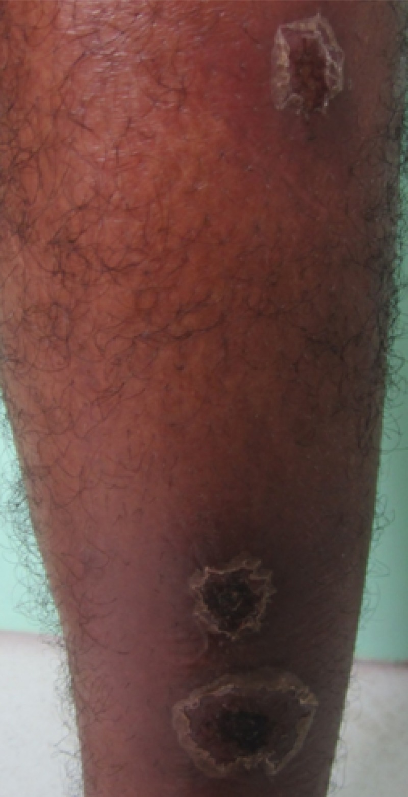
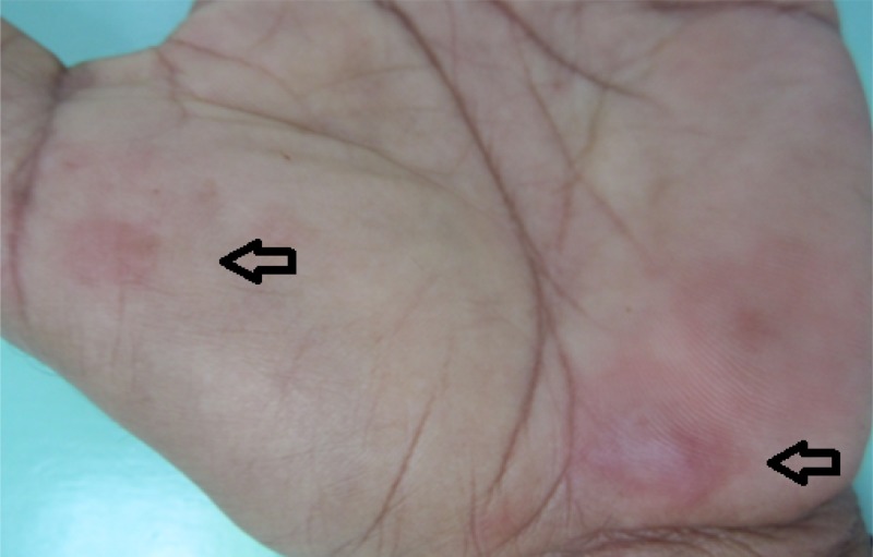
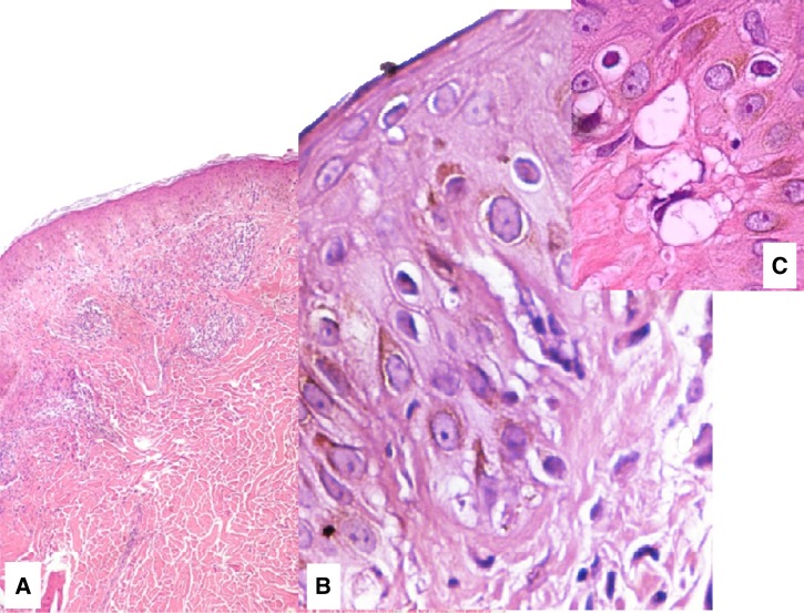
A 45-year-old otherwise healthy male from an endemic region for Leishmania braziliensis infection in Bahia, Brazil, presented with three erosive hemorrhagic infiltrated plaques on the left shin accompanied with lymphadenopathy in the groin since one month (Figure 1 ). A Leishmania skin test performed on the left forearm was strongly positive (20 × 18 mm).1 Two days later, the patient felt sick and feverish. Painful erythematous target lesions developed on the palms and scapula together with conjunctivitis (Figure 2 ). Histopathology confirmed erythema exsudativum multiforme (EEM) (Figure 3). Both EEM and cutaneous leishmaniasis were successfully treated with a 5-day course of prednisone 20 mg, and a 20-day course of intravenous pentavalent antimony, respectively.
Figure 1.

Erosive hemorraghic infiltrated plaques on the left shin, suspective for cutaneous leishmaniasis.
Figure 2.
Target lesions (arrows) on the palm of the left hand.
Figure 3.
Hematoxylin and eosin (H&E) stain showing dermatitis in the upper dermis and spongiosis at the dermal-epidermal junction (A) ×40. Vacuolization of epidermal basal cells (B) ×400, and the presence of necrotic keratinocytes (C) ×1,000.
This case supports the hypothesis that an exacerbated host immune response against Leishmania antigens may be associated with tissue damage and several clinical manifestations including EEM2,3; this case should alert the clinicians that Leishmania skin test is not totally risk free and may trigger hypersensitivity reactions.
ACKNOWLEDGMENTS
We acknowledge Dr. Sérgio Arruda for the histopathology.
Disclaimer: The authors have no conflict of interest to declare.
Footnotes
Authors' addresses: Bas S. Wind, Department of Dermatology, Academic Medical Center, Amsterdam, The Netherlands, E-mail: b.s.wind@amc.uva.nl. Luiz H. Guimarães and Paulo R. L. Machado, Serviço de Imunologia, Hospital Universitário Professor Edgard Santos, Canela, Salvador, and Universidade Federal da Bahia, Salvador, Bahia, Brazil, E-mails: imuno@ufba.br and prlmachado@uol.com.br.
References
- 1.Reed SG, Badaró R, Masur H, Carvalho EM, Lorenco R, Lisboa A, Teixeira R, Johnson WD, Jr, Jones TC. Selection of a skin test antigen for American visceral leishmaniasis. Am J Trop Med Hyg. 1986;35:79–85. doi: 10.4269/ajtmh.1986.35.79. [DOI] [PubMed] [Google Scholar]
- 2.Machado P, Araújo C, Da Silva AT, Almeida RP, D'Oliveira Jr A, Bittencourt A, Carvalho EM. Failure of early treatment of cutaneous leishmaniasis in preventing the development of an ulcer. Clin Infect Dis. 2002;34:E69–E73. doi: 10.1086/340526. [DOI] [PubMed] [Google Scholar]
- 3.Machado PR, Carvalho AM, Machado GU, Dantas ML, Arruda S. Development of cutaneous leishmaniasis after Leishmania skin test. Case Rep Med. 2011;2011(631079) doi: 10.1155/2011/631079. [DOI] [PMC free article] [PubMed] [Google Scholar]


