

Abstract
Orexin receptors are involved in many processes including energy homeostasis, wake/sleep cycle, metabolism, and reward. Development of potent and selective ligands is an essential step for defining the mechanism(s) underlying such critical processes. The goal of this study was to further investigate the structure–activity relationships of these peptides and to identify the truncated form of the orexin peptides active at OX1. Truncation studies have led to OXA (17–33) as the shortest active peptide known to date with a 23-fold selectivity for OX1 over OX2. Alanine, d-amino acid, and proline scans have highlighted the particular importance of Tyr17, Leu20, Asn25, and His26 for agonist properties of OXA(17–33). The conformation of the C-terminus might also be a defining factor in agonist activity and selectivity of the orexin peptides for the OX1 receptor.
Keywords: Orexin, peptide, structure−activity relationship
Orexin A and B (or hypocretin-1 and -2) are two neuropeptides discovered in 1998.1,2 Both peptides bind to two G protein-coupled receptors (GPCRs), orexin 1 (OX1) and orexin 2 (OX2). OX1 selectively binds orexin A, whereas OX2 is activated by both peptides nonselectively.2 The orexin system has been shown to play an important role in energy homeostasis,3 wake/sleep cycle,4 metabolism,5 modulating nonsleep energy consumption,6 and reward processing.7 Therefore, these receptors have been implicated as potential targets for treatment of a variety of conditions such as insomnia, narcolepsy, obesity, and drug addiction.8−10 Additionally, OX1 and OX2 receptors may modulate different physiological responses as suggested by their different distribution patterns in brain tissues.2,11,12 Although a list of subtype selective or dual orexin antagonists have been reported,13−15 progress on agonist development has been limited, and no small molecule agonists have been reported to date. Orexin A and B have been widely used for research purposes, but they display minimal subtype selectivity. Hence, identification of peptide analogues with increased selectivity is needed to further elucidate the respective functions of these receptors.
Orexin A consists of 33 amino acids with C-terminal amidation, an N-terminal pyrolgutamyl residue, and two intrachain disulfide bonds [Cys6-Cys12 and Cys7-Cys14]. Orexin B is a 28 amino acid linear peptide with an amidated C-terminus. While orexin A has a completely conserved sequence among several mammalian species, human orexin B has variations at two amino acid residues compared to the rodent sequence.1 High homology is present at the C-termini of orexin A and B, whereas the N-termini are more divergent in structure (11% homology),16 suggesting that the C-terminal region might be important for activity.
Figure 1.
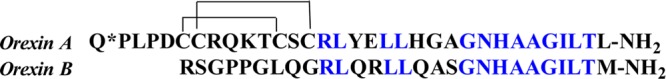
Orexin A and B peptide sequence. Q* corresponds to the pyroglutamoyl group.
Structure–activity relationship (SAR) studies on orexin A and B have been performed by several groups. The importance of the C-terminus for agonism has been confirmed.16−19 Several key amino acids in human orexin A and B required for binding to OX1 and OX2 have also been identified. Truncation studies have identified OXA (15–33) and OXB (10–28) as the shortest active peptides at OX1 and OX2 receptors, respectively.17,19 The two-point substituted analogue of orexin B, [Ala11, d-Leu15]orexin B (SB-668875-DM), was reported to have 400-fold selectivity for OX2 over OX120,21 although the selectivity was much weaker when studied by another group.22 Truncation of orexin B followed by one-point mutation studies resulted in orexin B (6–28) analogues with good selectivity for OX2 (>1000 fold).17 Among reported orexin A analogues, orexin A (2–33) is the only one that shows modest selectivity for OX1 (3-fold).17 We report herein our work on further studying the SAR of the orexin peptides in an effort to understand the structural requirements for peptides with minimal sequences that are selective for the OX1 receptor.
All target peptides were synthesized following conventional solid-phase technique using Fmoc/tBu strategy on an Apptec Focus XC peptide synthesizer (Supporting Information). Activity of the target compounds at the human OX1 and OX2 receptors was determined utilizing CHO-RD-HGA16 cells (Molecular Devices) engineered to stably express either the human OX1 or the human OX2 receptor. Test compound EC50 values were determined by running 8-point half-log concentration response curves.
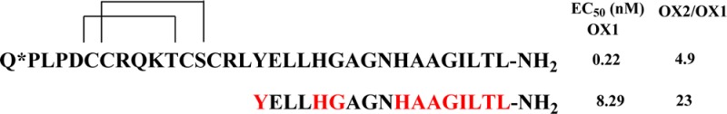
As the first step of the SAR studies, truncated analogues of both orexin A and B were prepared and tested in our assays (Table 1). Since the C terminus has been shown to be crucial for activity,16−19 truncation was carried out at the N terminus. The truncation of orexin A resulted in several analogues having agonist activity at the OX1 receptor (Table 1). OXA (19–33) displayed modest potency (EC50 = 1380 nM) at the OX1 receptor. Two peptides OXA (15–33) with EC50 of 64 nM and OXA (17–33) with EC50 = 8.29 nM displayed the highest potency among all analogues. In contrast to previous reports,19 orexin A (15–33) showed modest selectivity for the OX1 receptor (∼15-fold) in our hands. OXA (17–33) had slightly higher selectivity for OX1 (∼23-fold), which represents the shortest analogue with OX1 selectivity to date. Although the reason for this discrepancy is unclear, it has been proposed that orexin agonist discrimination is dependent on the expression system.23 Indeed, the EC50 of orexin A and B varies across several assay systems.17,19,20 Truncation on orexin B resulted in OXB (10–28) that showed activity at both receptors, confirming the previous notion that at least 19 amino acids are required for orexins to retain activity at orexin receptors.17,19
Table 1. Biological Activity of Orexin A and B and Their Truncates.
| peptides | EC50 (nM)a OX1 | EC50 (nM)a OX2 | OX2/OX1 |
|---|---|---|---|
| orexin A | 0.22 ± 0.06 | 1.07 ± 0.3 | 4.9 |
| orexin A (15–33) | 64.4 ± 6.9 | 976b | 15 |
| orexin A (17–33) | 8.29 ± 1.1 | 187b | 23 |
| orexin A(19–33) | 1380 ± 510 | 947 ± 300 | 0.7 |
| orexin A(21–33) | <50%c | ||
| orexin B (10–28) | 144 ± 35 | 99.3 ± 27 | 0.7 |
| orexin B (12–28) | <50%c | 1040 ± 300 |
Values are the mean ± SEM of at least three independent experiments in duplicate.
Values are the mean of two independent experiments in duplicate.
Percentage of <50% means activity at 10 μM was less than 50% of the control orexin A Emax.
Since orexin A is capped with a pyroglutamoyl group at the N-terminus and acylation may be important for activity, as is observed with Ghrelin,24 the corresponding N-acyl analogues were also synthesized (Supporting Information). No significant effect on agonist properties of truncated peptides was detected with the presence of the N-terminus “cap”.
OXA (17–33) was chosen for further SAR studies because of its good potency and OX1 preference. An alanine scan was first performed to determine the contributions of the side chains to the overall activity (Table 2). Substitution of the first six N-terminal amino acids (17–22) partially retained the agonist activity at the OX1 receptor, except for [Ala20] (EC50 > 3 μM), whose analogue with the acetyl cap had no OX1 activity at up to 10 μM. [Ala18] substitution showed the least effect on activity (EC50 79 nM). Alanine substitution at the 24–28 positions resulted in a more significant drop in activity, whereas changes in the last 5 amino acids (C-terminal) led to a complete loss in activity, confirming that modifications on the C-terminal region are not tolerated. A similar trend is present at the OX2 receptor. The preference for the OX1 receptor was generally maintained with substitution at 17–22 positions but was abolished with substitution at the 24–26 positions. Acylation on the N-terminus displayed minimal effects on agonist activity in this series of peptides (see Supporting Information).
Table 2. Biological Activity of Alanine Substituted OXA (17–33).
| peptides | EC50 (nM)a OX1 | EC50 (nM)b OX2 | OX2/OX1 |
|---|---|---|---|
| orexin A (17–33) | 8.29 ± 1.1 | 187 | 23 |
| [Ala17]OXA (17–33) | 1910 ± 620 | 4820 | 2.5 |
| [Ala18]OXA (17–33) | 78.9 ± 22 | 439 | 5.6 |
| [Ala19]OXA (17–33) | 212 ± 57 | 1890 ± 610a | 8.9 |
| [Ala20]OXA (17–33) | 3230 ± 1080 | 7850 | 2.4 |
| [Ala21]OXA (17–33) | 493 ± 49 | 1910 | 3.9 |
| [Ala22]OXA (17–33) | 422 ± 55 | 1440 | 3.4 |
| [Ala24]OXA (17–33) | 1040 ± 300 | 1360 | 1.3 |
| [Ala25]OXA (17–33) | 3070 ± 1180 | 2320 | 0.8 |
| [Ala26]OXA (17–33) | 3060 ± 940 | 1580 | 0.5 |
| [Ala29]OXA (17–33) | <50%c | <50%c | |
| [Ala30]OXA (17–33) | inactive at 10 μMb | inactive at 10 μM | |
| [Ala31]OXA (17–33) | inactive at 10 μMb | inactive at 10 μM | |
| [Ala32]OXA (17–33) | <50%b,c | <50%c | |
| [Ala33]OXA (17–33) | <50%b,c | inactive at 10 μM |
Values are the mean ± SEM of at least three independent experiments in duplicate.
Values are the mean of at least two independent experiments in duplicate.
Perecentage of <50% means activity at 10 μM was less than 50% of the control orexin A Emax.
The effect of side-chain chirality on the activity was examined by a d-amino acid scan on OXA (17–33) (Table 3). Similarly, changes on the C-terminal side of the peptide (28–33) led to a complete loss of activity. Changes in the remaining positions (17–27) revealed that [d-Glu18], [d-Leu19], and [d-Leu20] analogues retained some of the activity, whereas all other analogues had noticeable decreases in agonist potency. Interestingly, substitution to d-amino acids at positions 19–27 seemed to reverse the binding preference to favor OX2. For example, [d-Leu20] orexin (17–33) analogue had no change in activity at OX2 and a 64-fold drop in activity at OX1; [d-His26] OXA (17–33) had 9-fold reduction in activity at OX2 and almost 700-fold drop in activity at OX1. These results suggest a more pronounced impact of chain orientation on orexin receptor binding in this region. [Tyr17] and [Glu18] retained slight selectivity for OX1, although agonist activity of both analogues significantly decreased.
Table 3. Biological Activity of d-Amino Acid Substituted OXA (17–33).
| peptides | EC50 (nM)a OX1 | EC50 (nM)b OX2 | OX2/OX1 |
|---|---|---|---|
| [d-Tyr17]OXA (17–33) | 1360 ± 540b | 3220 | 2.4 |
| [d-Glu18]OXA (17–33) | 739 ± 190b | 2140 | 2.9 |
| [d-Leu19]OXA (17–33) | 526 ± 100 | 263 | 0.5 |
| [d-Leu20]OXA (17–33) | 535 ± 110 | 156 | 0.3 |
| [d-His21]OXA (17–33) | 1560 ± 310 | 610 | 0.4 |
| [d-Ala23]OXA (17–33) | 3070 ± 970 | 1920 | 0.6 |
| [d-Asn25]OXA (17–33) | inactive at 10 μMb | <50%c | |
| [d-His26]OXA (17–33) | 5700 ± 3420 | 1760 | 0.3 |
| [d-Ala27]OXA (17–33) | 1900 ± 530 | 1250 | 0.7 |
| [d-Ala28]OXA (17–33) | inactive at 10 μM | <50%c | |
| [d-Ile30]OXA (17–33) | inactive at 10 μMb | inactive at 10 μM | |
| [d-Leu31]OXA (17–33) | inactive at 10 μMb | <50%c | |
| [d-Thr32]OXA (17–33) | inactive at 10 μMb | inactive at 10 μM | |
| [d-Leu33]OXA (17–33) | inactive at 10 μMb | inactive at 10 μM |
Values are the mean of at least three independent experiments in duplicate.
Values are the mean of two independent experiments in duplicate.
Percentage of <50% means activity at 10 μM was less than 50% of the control orexin A Emax.
The effect of more dramatic changes on secondary structure of peptides was investigated using proline scans of OXA (17–33). Given that modification in the C-terminus resulted in total loss of activity in previous scans, only the first half from the N-terminus (17–22) was investigated. The analogues resulting from proline substitution at 17–19 position retained some of the original activity, with [Pro18] OXA (17–33) being the least affected by change (Table 4). A more significant drop in activity was observed for analogues at 20–22 positions. Selectivity of OX1 was slightly maintained or abolished in this series.
Table 4. Biological Activity of Proline Substituted OXA (17–33).
| peptides | EC50 (nM)a OX1 | EC50 (nM)b OX2 | OX2/OX1 |
|---|---|---|---|
| [Pro17]OXA (17–33) | 1370 ± 140 | 3250 | 2.4 |
| [Pro18]OXA (17–33) | 143 ± 16 | 729 | 5.1 |
| [Pro19]OXA (17–33) | 865 ± 350 | 1010 | 1.2 |
| [Pro20]OXA (17–33) | 1130 ± 450 | 1610 | 1.4 |
| [Pro21]OXA (17–33) | 1200 ± 360 | 1990 | 1.7 |
| [Pro22]OXA (17–33) | 2480 ± 1125 | 2180 | 0.9 |
Values are the mean ± SEM of at least three independent experiments in duplicate.
Values are the mean of two independent experiments in duplicate.
NMR and computational studies performed on orexin A (in aqueous25 and in membrane mimetic micellar solutions26) identified a highly conserved hydrophobic region on the C-terminus. This region contains two α-helices that are connected by a short linker.16,25 Similarly, solution structure of orexin B (in aqueous27 and in micellar solution28) also shows two α-helices at similar positions. These α-helices seem to differ in their length, the position of the loops, and their relative orientation between orexin A and B. The observed selectivity of OX1 toward orexin A was suggested to be due to specific recognition of the overall peptide conformation, especially the hydrophobic side chain in the C-terminal region of the receptor.16 In our study, two residues, His21 and Gly,22 were shown to be of moderate importance in the alanine scan; however, their proline analogues had more pronounced reduction in potency, especially at OX1. These two residues and Asn25 are bordering the two helices and the linker, according to Kim et al., who suggested the helices to be Cys14-His21 and Asn25-Leu31,25 and Takai et al., who proposed Leu16-Ala23 and Asn25-Thr32.16 Considering their close proximity to the linker our results may indicate that these three residues are important for orientation of the two α-helices.16 Since OX2 has lesser recognition requirements, the effect of these mutations on activity of corresponding peptides was less pronounced.
In the proposed orexin A structure, the residues that were deemed important for potency in truncated peptide OXA (15–33) (Leu16, Leu19, Leu20, His26, Gly29, Ile30, Leu31, Thr32, and Leu33) seemed to exist on one side of orexin A, forming a hydrophobic surface.16,25 This surface might also be important in binding to the cell membrane.26 Residues that were less important (Arg15, Tyr17, Glu18, His21, Gly22, Gly24, and Asn25) formed a hydrophilic surface on orexin A, which were thought to play a minimal role in potency. In our study, alanine and proline scans of OXA (17–33) have confirmed the importance of the hydrophobic surface residues Leu20 and His26, together with C-terminal residues 29–33. Leu18 showed no significant contribution to OXA (17–33) binding to OX1, agreeing with an earlier report on other truncated orexin A analogues.16 Interestingly, hydrophilic surface residues Tyr17 and Asn25 were also shown to be important for potency in our study.
It was previously suggested that OX1 had more strict recognition requirements than OX2 in binding endogenous ligands and their analogues.17 The potency of our truncated orexin A analogues showed more dramatic change in response at OX1 for all performed mutations. Both OXA (15–33) and OXA (17–33) showed some selectivity for OX1 in our hands (15- and 23-fold, respectively). Although mutations at Tyr17, one of the key amino acid residues, led to dramatically reduced potency at both receptors, the preference for OX1 was preserved. Interestingly, d-amino acid substitution at Leu20, His21, and His26 had not only abolished preference to OX1, but seemed to reverse it. Together, these results confirm that the binding pocket on OX1 might have more specific interactions with ligands than that on OX2.
In summary, truncated OXA (17–33) showed preferential agonist activity at OX1 in our study and is the shortest peptide with such selectivity. Given its shorter sequence and higher selectivity toward OX1, OXA (17–33) may serve as an improved biological probe of the OX1 receptor function. During single amino acid mutagenesis, OXA (17–33) seems to maintain structural requirements similar to those reported for orexin A and OXA (15–33). Our data suggest that Tyr17, Leu20, Asn25, and His26 play a key role in interaction of OXA (17–33) with OX1, whereas His21 and Gly22 might be important in the orientation of its two α-helices. Also in the majority of the active analogues studied to date, inversion of configuration at positions 19–27 has resulted in dramatic drops in potency at OX1, whereas potency at OX2 changed moderately. Finally, our results on OXA (17–33) suggest that its spatial conformation might be similar to that of human orexin A peptide and is important for binding preference of OX1.
Acknowledgments
This work was support by National Institute of Health/National Institute on Drug Abuse grant DA032837 and Research Triangle Institute. We are grateful for the help from Drs. Scott Runyon and Yanyan “Jasmine” Zhang in peptide synthesis. We thank Tiffany Langston, Allyson Smith, Keith Warner, and Dr. Elaine Gay for technical assistance.
Glossary
ABBREVIATIONS
- OX1
orexin 1 receptor
- OX2
orexin 2 receptor
- OXA
orexin A
- GPCR
G protein-coupled receptor
- SAR
structure–activity relationship
- F-moc
Fluorenylmethyloxycarbonyl
- tBu
t-butyl
- HPLC
high performance liquid chromatography
Supporting Information Available
Peptide synthesis, calcium assays, HPLC analysis of target peptides, and agonist activity of capped peptides. This material is available free of charge via the Internet at http://pubs.acs.org.
Author Contributions
The manuscript was written through contributions of all authors. All authors have given approval to the final version of the manuscript.
The authors declare no competing financial interest.
Funding Statement
National Institutes of Health, United States
Supplementary Material
References
- de Lecea L.; Kilduff T. S.; Peyron C.; Gao X.; Foye P. E.; Danielson P. E.; Fukuhara C.; Battenberg E. L.; Gautvik V. T.; Bartlett F. S. II; Frankel W. N.; van den Pol A. N.; Bloom F. E.; Gautvik K. M.; Sutcliffe J. G. The hypocretins: hypothalamus-specific peptides with neuroexcitatory activity. Proc. Natl. Acad. Sci. U.S.A. 1998, 95, 322–327. [DOI] [PMC free article] [PubMed] [Google Scholar]
- Sakurai T.; Amemiya A.; Ishii M.; Matsuzaki I.; Chemelli R. M.; Tanaka H.; Williams S. C.; Richardson J. A.; Kozlowski G. P.; Wilson S.; Arch J. R.; Buckingham R. E.; Haynes A. C.; Carr S. A.; Annan R. S.; McNulty D. E.; Liu W. S.; Terrett J. A.; Elshourbagy N. A.; Bergsma D. J.; Yanagisawa M. Orexins and orexin receptors: a family of hypothalamic neuropeptides and G protein-coupled receptors that regulate feeding behavior. Cell 1998, 92, 573–585. [DOI] [PubMed] [Google Scholar]
- Sakurai T. Roles of orexins and orexin receptors in central regulation of feeding behavior and energy homeostasis. CNS Neurol. Disord.: Drug Targets 2006, 5, 313–325. [DOI] [PubMed] [Google Scholar]
- Chemelli R. M.; Willie J. T.; Sinton C. M.; Elmquist J. K.; Scammell T.; Lee C.; Richardson J. A.; Williams S. C.; Xiong Y.; Kisanuki Y.; Fitch T. E.; Nakazato M.; Hammer R. E.; Saper C. B.; Yanagisawa M. Narcolepsy in orexin knockout mice: molecular genetics of sleep regulation. Cell 1999, 98, 437–451. [DOI] [PubMed] [Google Scholar]
- Lubkin M.; Stricker-Krongrad A. Independent feeding and metabolic actions of orexins in mice. Biochem. Biophys. Res. Commun. 1998, 253, 241–245. [DOI] [PubMed] [Google Scholar]
- Teske J. A.; Billington C. J.; Kotz C. M. Hypocretin/orexin and energy expenditure. Acta Physiol. 2010, 198, 303–312. [DOI] [PubMed] [Google Scholar]
- Sharf R.; Sarhan M.; Dileone R. J. Role of orexin/hypocretin in dependence and addiction. Brain Res. 2010, 1314, 130–138. [DOI] [PMC free article] [PubMed] [Google Scholar]
- Kukkonen J. P. Physiology of the orexinergic/hypocretinergic system: a revisit in 2012. Am. J. Physiol., Cell Physiol. 2013, 304, C2–32. [DOI] [PubMed] [Google Scholar]
- Scammell T. E.; Winrow C. J. Orexin receptors: pharmacology and therapeutic opportunities. Annu. Rev. Pharmacol. Toxicol. 2011, 51, 243–266. [DOI] [PMC free article] [PubMed] [Google Scholar]
- Gotter A. L.; Roecker A. J.; Hargreaves R.; Coleman P. J.; Winrow C. J.; Renger J. J. Orexin receptors as therapeutic drug targets. Prog. Brain Res. 2012, 198, 163–188. [DOI] [PubMed] [Google Scholar]
- Trivedi P.; Yu H.; MacNeil D. J.; Van der Ploeg L. H.; Guan X. M. Distribution of orexin receptor mRNA in the rat brain. FEBS Lett. 1998, 438, 71–75. [DOI] [PubMed] [Google Scholar]
- Marcus J. N.; Aschkenasi C. J.; Lee C. E.; Chemelli R. M.; Saper C. B.; Yanagisawa M.; Elmquist J. K. Differential expression of orexin receptors 1 and 2 in the rat brain. J. Comp. Neurol. 2001, 435, 6–25. [DOI] [PubMed] [Google Scholar]
- Roecker A. J.; Coleman P. J. Orexin receptor antagonists: medicinal chemistry and therapeutic potential. Curr. Top. Med. Chem. 2008, 8, 977–987. [DOI] [PubMed] [Google Scholar]
- Gatfield J.; Brisbare-Roch C.; Jenck F.; Boss C. Orexin receptor antagonists: a new concept in CNS disorders?. ChemMedChem 2010, 5, 1197–1214. [DOI] [PubMed] [Google Scholar]
- Boss C.; Brisbare-Roch C.; Jenck F. Biomedical application of orexin/hypocretin receptor ligands in neuroscience. J. Med. Chem. 2009, 52, 891–903. [DOI] [PubMed] [Google Scholar]
- Takai T.; Takaya T.; Nakano M.; Akutsu H.; Nakagawa A.; Aimoto S.; Nagai K.; Ikegami T. Orexin-A is composed of a highly conserved C-terminal and a specific, hydrophilic N-terminal region, revealing the structural basis of specific recognition by the orexin-1 receptor. J. Pept. Sci. 2006, 12, 443–454. [DOI] [PubMed] [Google Scholar]
- Lang M.; Soll R. M.; Durrenberger F.; Dautzenberg F. M.; Beck-Sickinger A. G. Structure–activity studies of orexin a and orexin B at the human orexin 1 and orexin 2 receptors led to orexin 2 receptor selective and orexin 1 receptor preferring ligands. J. Med. Chem. 2004, 47, 1153–1160. [DOI] [PubMed] [Google Scholar]
- Ammoun S.; Holmqvist T.; Shariatmadari R.; Oonk H. B.; Detheux M.; Parmentier M.; Akerman K. E.; Kukkonen J. P. Distinct recognition of OX1 and OX2 receptors by orexin peptides. J. Pharmacol. Exp. Ther. 2003, 305, 507–514. [DOI] [PubMed] [Google Scholar]
- Darker J. G.; Porter R. A.; Eggleston D. S.; Smart D.; Brough S. J.; Sabido-David C.; Jerman J. C. Structure–activity analysis of truncated orexin-A analogues at the orexin-1 receptor. Bioorg. Med. Chem. Lett. 2001, 11, 737–740. [DOI] [PubMed] [Google Scholar]
- Asahi S.; Egashira S.; Matsuda M.; Iwaasa H.; Kanatani A.; Ohkubo M.; Ihara M.; Morishima H. Development of an orexin-2 receptor selective agonist, [Ala(11), D-Leu(15)]orexin-B. Bioorg. Med. Chem. Lett. 2003, 13, 111–113. [DOI] [PubMed] [Google Scholar]
- Soffin E. M.; Gill C. H.; Brough S. J.; Jerman J. C.; Davies C. H. Pharmacological characterisation of the orexin receptor subtype mediating postsynaptic excitation in the rat dorsal raphe nucleus. Neuropharmacology 2004, 46, 1168–1176. [DOI] [PubMed] [Google Scholar]
- Putula J.; Turunen P. M.; Johansson L.; Nasman J.; Ra R.; Korhonen L.; Kukkonen J. P. Orexin/hypocretin receptor chimaeras reveal structural features important for orexin peptide distinction. FEBS Lett. 2011, 585, 1368–1374. [DOI] [PubMed] [Google Scholar]
- Putula J.; Turunen P. M.; Jantti M. H.; Ekholm M. E.; Kukkonen J. P. Agonist ligand discrimination by the two orexin receptors depends on the expression system. Neurosci. Lett. 2011, 494, 57–60. [DOI] [PubMed] [Google Scholar]
- Hosoda H.; Kojima M.; Matsuo H.; Kangawa K. Ghrelin and des-acyl ghrelin: two major forms of rat ghrelin peptide in gastrointestinal tissue. Biochem. Biophys. Res. Commun. 2000, 279, 909–913. [DOI] [PubMed] [Google Scholar]
- Kim H. Y.; Hong E.; Kim J. I.; Lee W. Solution structure of human orexin-A: regulator of appetite and wakefulness. J. Biochem. Mol. Biol. 2004, 37, 565–573. [DOI] [PubMed] [Google Scholar]
- Miskolzie M.; Kotovych G. The NMR-derived conformation of orexin-A: an orphan G-protein coupled receptor agonist involved in appetite regulation and sleep. J. Biomol. Struct. Dyn. 2003, 21, 201–210. [DOI] [PubMed] [Google Scholar]
- Lee J. H.; Bang E.; Chae K. J.; Kim J. Y.; Lee D. W.; Lee W. Solution structure of a new hypothalamic neuropeptide, human hypocretin-2/orexin-B. Eur. J. Biochem. 1999, 266, 831–839. [DOI] [PubMed] [Google Scholar]
- Miskolzie M.; Lucyk S.; Kotovych G. NMR conformational studies of micelle-bound orexin-B: a neuropeptide involved in the sleep/awake cycle and feeding regulation. J. Biomol. Struct. Dyn. 2003, 21, 341–351. [DOI] [PubMed] [Google Scholar]
Associated Data
This section collects any data citations, data availability statements, or supplementary materials included in this article.