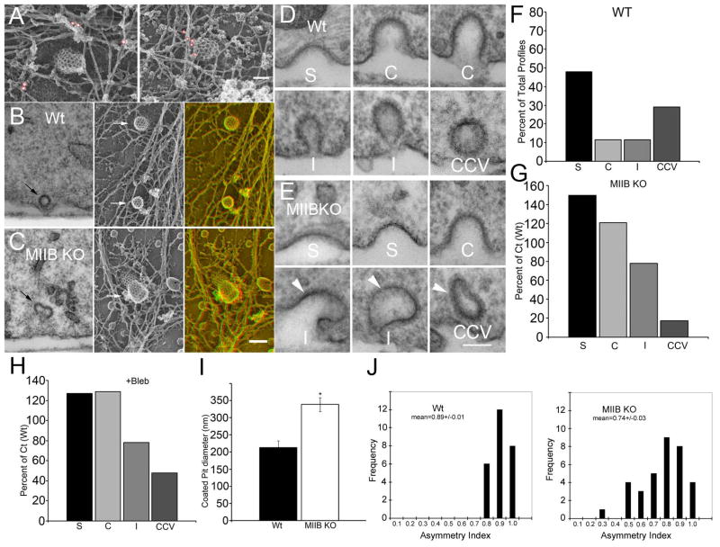
Figure 5.
MIIB is closely associated with coated pits in the Wt and clathrin-coated pit (CCP) structure is abnormal in MIIB KO cells. (A) Immunogold labeling for MIIB in Wt cells. Particles are circled in red for better visibility. Gold particles were identified in anaglyphs. From two separate preparations: on left, 18 nm gold, on right, 12 nm gold. Bar=90 nm. (B) Comparison of highly-invaginated pits in thin section (left) and in rotary shadowed replicas (right arrows indicate coated pit). Far right panel is an anaglyph. (C) The same comparison in MIIB KO cells. Note that the coated pits are distorted. Bar=150 nm. (D) CCP profiles in thin sections from Wt cells. S= shallow, C=curved, I=Invaginated, CCV=clathrin coated vesicle. (E) CCP profiles in MIIB KO cells. Arrowheads indicate distorted pits. Bar=100 nm. (F) Distribution of CCP profiles in Wt cells as a percentage of total (N=142 from 20 cells) observed. (G) Distribution of CCP profiles (N=132 from 15 cells) in MIIB KO cells as compared to Wt. The frequency of shallow and curved coated pits is increased, whereas the frequency of CCVs is greatly decreased. (H) Blebbistatin treatment results in a greater frequency of shallow and curved coated pits compared to untreated fibroblasts. The frequency of CCV’s is decreased. Untreated Ct, N=138 from 18 cells, bleb treated, N=144 from 16 cells. (I) Shallow coated pits measured in rotary shadowed replicas from unroofed fibroblasts are larger in diameter in MIIB KO cells. (T-test, P<0.001, N=20 each). The same result was obtained from measurements made from thin sections, except that the diameters for shallow coated pits in both Wt and MIIB KO cells were approximately 30% less than in replicas. (J) Histograms showing the distribution of coated pit symmetry (width in x divided by width in y) measured in rotary shadowed preparations. Coated pits are more asymmetrical in MIIB KO cells.

