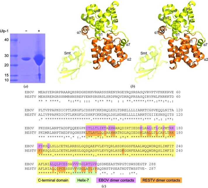
The crystal structure of the Reston ebolavirus VP30 C-terminal domain shows a rotated interface in comparison to the previous structure of the Zaire ebolavirus VP30 C-terminal domain.
Keywords: ebolaviruses, Reston ebolavirus, VP30 C-terminal domain
Abstract
The ebolaviruses can cause severe hemorrhagic fever. Essential to the ebolavirus life cycle is the protein VP30, which serves as a transcriptional cofactor. Here, the crystal structure of the C-terminal, NP-binding domain of VP30 from Reston ebolavirus is presented. Reston VP30 and Ebola VP30 both form homodimers, but the dimeric interfaces are rotated relative to each other, suggesting subtle inherent differences or flexibility in the dimeric interface.
1. Introduction
Ebolaviruses can cause hemorrhagic fever in humans, with a fatality rate as high as 90% (Burke et al., 1978 ▶). Four of the five ebolaviruses, including Ebola virus (EBOV; formerly Zaire ebolavirus), are found in Africa. However, Reston ebolavirus (RESTV) is uniquely Asian in origin. RESTV has been identified in bats and primates (Miranda et al., 1999 ▶; Rollin et al., 1999 ▶; Taniguchi et al., 2011 ▶), as well as swine (Barrette et al., 2009 ▶; Sayama et al., 2012 ▶). However, in humans RESTV appears to be nonpathogenic and transmits poorly for reasons that are not fully understood (Miranda et al., 1999 ▶).
Four proteins are essential for viral transcription: nucleoprotein (NP), VP30, VP35 and the RNA-dependent RNA polymerase (L) (Mühlberger et al., 1999 ▶). VP30 appears unique to the filoviruses and is essential for rescuing recombinant ebolavirus (Enterlein et al., 2006 ▶; Theriault et al., 2004 ▶). VP30 allows the polymerase to read beyond a cis-RNA element in the NP mRNA 5′ untranslated region (Weik et al., 2002 ▶) and to re-initiate transcription at gene junctions (Martínez et al., 2008 ▶). Furthermore, VP30 phosphorylation modulates the composition and function of the RNA synthesis machinery (Biedenkopf et al., 2013 ▶; Martinez et al., 2011 ▶).
Both VP30 N- and C-terminal domains have been associated with oligomerization and nucleocapsid-association functions (Hartlieb et al., 2003 ▶, 2007 ▶). A basic cluster in the C-terminal domain (CTD) contributes to the association of VP30 with NP and is essential for transcription activation (Hartlieb et al., 2007 ▶). Previous crystallographic studies demonstrated that the EBOV VP30 CTD forms a dimer by donation of the C-terminal helix 7 to the neighboring monomer (Hartlieb et al., 2007 ▶). To date, the EBOV VP30 CTD is the only filovirus VP30 structure available. Here, we present the crystal structure of the VP30 CTD from Reston ebolavirus.
2. Materials and methods
2.1. Cloning, expression and protein purification
RESTV (strain Reston-89) VP30 CTD, residues 142–266, was cloned using polymerase incomplete primer extension (PIPE; Klock & Lesley, 2009 ▶) into a modified Escherichia coli pET28 vector system engineered for N-terminal hexahistidine-Smt tags (clone ID EbreA.17250.a.D11). Expression was performed in E. coli BL21 (DE3) cells in TB medium (Teknova) at 37°C with 50 µg ml−1 kanamycin. Cells were induced at an OD600 of 0.7 with 1 mM IPTG and incubated overnight at 25°C.
Cells were resuspended in lysis buffer [25 mM Tris–HCl pH 8.0, 200 mM NaCl, 50 mM arginine, 1 mM tris(2-carboxyethyl)phosphine (TCEP), 10 mM imidazole, 0.5% glycerol, 125 U Benzonase, 0.71 mg ml−1 lysozyme and one EDTA-free complete protease inhibitor tablet (Roche)] and lysed via sonication. The lysate was clarified by centrifugation and filtration. The protein was eluted from HiTrap Ni2+ Chelating columns (GE Healthcare) with an imidazole step gradient. Pooled fractions were incubated for 18 h with Ulp-specific protease 1 (Ulp-1), intended to cleave the Smt domain from VP30. However, SDS–PAGE analysis showed that the His-Smt tag was unable to be removed (Fig. 1 ▶ a). The protein was dialyzed against 25 mM Tris pH 8.0, 200 mM NaCl, 1 mM TCEP, 1% glycerol for 12 h and was concentrated to 18 mg ml−1 for crystallization. Macromolecule-production information is given in Table 1 ▶.
Figure 1.
Overall Reston ebolavirus VP30 C-terminal domain structure. (a) Attempted cleavage of Smt-VP30 CTD with Ulp-1 protease. Lane 1, protein marker (labeled in kDa); lane 2, undigested protein; lane 3, Ulp-1 digestion. (b) The VP30 monomers forming the dimer are shown in orange and yellow. The ordered Smt domain is attached to the yellow VP30. The image is shown in wall-eyed stereo. (c) Sequence alignment of VP30 highlighting amino acids participating in the dimer interface. Residues in the dimer interfaces where amino acids are within 4 Å of the other chain in the corresponding structures are highlighted in purple (EBOV) and orange (RESTV).
Table 1. Macromolecule production.
| Source organism | Reston ebolavirus strain Reston-89 |
| DNA source | Synthetic |
| Forward primer | AAC AAA TCG GTG GAC TCA CTC TGG CAG TGT TAC TGC AGA T |
| Reverse primer | GGC CGC AAG CTT TTA CGT GCT GTT ATC CTG AGC AGG GTA C |
| Cloning vector | pET-28a |
| Expression vector | pET-28a |
| Expression host | E. coli BL21 (DE3) |
| Complete amino-acid sequence of construct produced | MGHHHHHHSGEVKPEVKPETHINLKVSDGSSEIFFKIKKTTPLRRLMEAFAKRQGKEMDSLRFLYDGIRIQADQTPEDLDMEDNDIIEAHREQIGGLTLAVLLQIAEHWATRDLRQIEDSKLRALLTLCAVLTRKFSKSQLGLLCETHLRHEGLGQDQADSVLEVYQRLHSDKGGNFEAALWQQWDRQSLIMFISAFLNIALQIPCESSSVVVSGLATLYPAQDNST |
2.2. Crystallization
Crystals grew by vapor diffusion in sitting-drop trays in 10% PEG 6000, 100 mM HEPES pH 7.0 in 2–3 weeks using 0.4 µl protein solution and 0.4 µl reservoir solution at 16°C. The rod-shaped crystals were cryoprotected with 20% ethylene glycol prior to flash-cooling in liquid nitrogen. Details are given in Table 2 ▶.
Table 2. Crystallization.
| Method | Vapor diffusion, sitting drop |
| Plate type | 96-well Compact Jr plates (Emerald Bio) |
| Temperature (°C) | 16 |
| Protein concentration (mg ml−1) | 18 |
| Buffer composition of protein solution | 25 mM Tris pH 8.0, 200 mM NaCl, 1 mM TCEP, 1% glycerol |
| Composition of reservoir solution | 10% PEG 6000, 100 mM HEPES pH 7.0 |
| Volume and ratio of drop | 0.8 µl, 1:1 |
| Volume of reservoir (µl) | 100 |
2.3. Data collection and processing
Data were collected on beamline 5.0.1 at the Advanced Light Source with the detector set at a distance of 300 mm, with 0.5° oscillations and 5 s exposures for a total of 300 frames The data were reduced using XDS (Kabsch, 2010 ▶) and XSCALE. Data-collection and processing statistics are given in Table 3 ▶.
Table 3. Data-collection and processing statistics.
Values in parentheses are for the highest shell.
| Data collection and processing | |
| Diffraction source | ALS 5.0.1 |
| Wavelength (Å) | 0.97740 |
| Temperature (°C) | −173 |
| Detector | ADSC Quantum 210 CCD |
| Crystal-to-detector distance (mm) | 300 |
| Rotation range per image (°) | 0.5 |
| Total rotation range (°) | 120 |
| Exposure time per image (s) | 5 |
| Space group | P212121 |
| Unit-cell parameters (Å, °) | a = 49.3, b = 93.7, c = 111.2, α = β = γ = 90 |
| Mosaicity (°) | 0.13 |
| Resolution (Å) | 50–2.25 (2.31–2.25) |
| Total reflections | 120225 (8910) |
| Unique reflections | 25173 (1831) |
| Completeness (%) | 99.7 (100.0) |
| Multiplicity | 4.8 (4.9) |
| 〈I/σ(I)〉 | 15.2 (3.2) |
| R p.i.m. | 0.048 (0.35) |
| R merge | 0.08 (0.51) |
| Overall B factor from Wilson plot (Å2) | 32.8 |
| Refinement | |
| Resolution range (Å) | 50–2.25 (2.31–2.25) |
| Completeness (%) | 99.7 (100) |
| No. of reflections, working set | 23841 (1577) |
| No. of reflections, test set | 1280 (95) |
| Final R cryst (%) | 0.19 (0.24) |
| Final R free(%) | 0.23 (0.32) |
| Cruickshank DPI (Å) | 0.20 |
| No. of non-H atoms | |
| Protein | 2554 |
| Ligand | 8 |
| Water | 150 |
| Total | 2712 |
| R.m.s. deviations | |
| Bonds (Å) | 0.011 |
| Angles (°) | 1.33 |
| Mean B factors (Å2) | |
| Overall | 32.6 |
| Protein | 32.4 |
| Ligand | 44.0 |
| Water | 34.9 |
| Model statistics | |
| Ramachandran plot | |
| Favored regions (%) | 98.8 |
| Additionally allowed (%) | 0.9 |
| MolProbity score | 1.23 [100th percentile] |
| Clashscore | 1.95 [100th percentile] |
| PDB code | 3v7o |
2.4. Structure solution, refinement and analysis
The structure was determined by molecular replacement using Phaser (McCoy et al., 2007 ▶) with the structure of EBOV VP30 CTD in monomeric form (Hartlieb et al., 2007 ▶; PDB entry 2i8b) as a search model. The structure was refined with REFMAC (Murshudov et al., 2011 ▶) and model building was performed with Coot (Emsley & Cowtan, 2004 ▶). The final structure was validated with MolProbity (Chen et al., 2010 ▶). Protein interfaces were analyzed using PISA (Krissinel & Henrick, 2007 ▶) and the shape-correlation statistic SC (Lawrence & Colman, 1993 ▶).
3. Results and discussion
In the crystal structure, there are two copies of the RESTV VP30 CTD in the asymmetric unit (Fig. 1 ▶ b). The VP30 domains retain their Smt fusion domains from purification (Fig. 1 ▶ a). One of the Smt domains helps facilitate crystal-packing interactions, whereas the second Smt domain is disordered. The two copies of RESTV VP30 in the asymmetric unit are nearly identical in structure and superimpose with an r.m.s.d. of 0.76 Å.
The fold of RESTV VP30 CTD is helical and closely resembles that of EBOV VP30, with an r.m.s.d. of 1.15–2.14 Å separating the Cα backbones of the EBOV and RESTV structures. Like EBOV VP30 (Hartlieb et al., 2007 ▶), RESTV VP30 forms a dimer by packing of its extended C-terminal helix 7 into a conserved hydrophobic face on the neighboring monomer (Fig. 1 ▶ c). Dimerization in both EBOV and RESTV VP30 is also facilitated by similar hydrophobic interactions between the globular domains and a conserved hydrogen bond between the side chain of Lys180 in helix 2 and the main chain of Cys251 in the linker between helices 6 and 7.
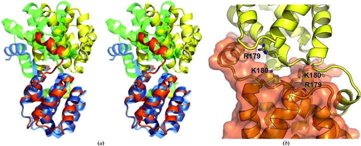
Superposition of the RESTV and EBOV VP30 CTD structures shows that the RESTV VP30 CTD assembly is significantly rotated about the dimer interface compared with EBOV (Fig. 2 ▶ a). The domain rotation lowers the buried surface area on each monomer (RESTV, 1630 Å2; EBOV, 1900 Å2) but maintains a similar surface complementarity (RESTV, SC = 0.69; EBOV, SC = 0.68), suggesting that the RESTV VP30 CTD dimer is a relevant conformation (Lawrence & Colman, 1993 ▶), and conservation of residues within the dimer interface suggests this conformation could also exist for EBOV.
Figure 2.
Reston VP30 CTD dimer. (a) Superimposition of the EBOV and RESTV VP30 C-terminal domain structures using a single domain shows a rotation about the dimer interface. The RESTV VP30 molecules are colored orange and yellow, while the EBOV VP30 molecules are colored blue and green. For clarity, the Smt is not illustrated. (b) The altered orientation of the RESTV VP30 CTD buries the basic residues Arg179 and Lys180 in the dimer interface.
It is difficult to discern the impact of the crystal-packing interactions on the EBOV or RESTV VP30 CTD conformations, as neither conformation is compatible with the packing of the other ebolavirus species. Additionally, the Smt domain in the RESTV VP30 CTD structure contributes to the crystal packing as well as making contacts with the opposite chain in the asymmetric unit (buries 545 Å2). However, there is no apparent structural reason for the Smt domain to constrain the VP30 conformation and the Smt domain and VP30 CTD are not expected to form a stable complex in solution (Krissinel & Henrick, 2007 ▶).
The observed rotation in the RESTV VP30 dimer buries Arg179 and Lys180 (Fig. 2 ▶ b), which are instead solvent-exposed in the EBOV structure. These residues are important in VP30 function and their mutation is detrimental to VP30 oligomerization, NP binding and transcription initiation (Hartlieb et al., 2007 ▶). The occlusion of Arg179 and Lys180 in the RESTV dimer interface suggests that mutations at these positions could disrupt VP30 dimerization.
EBOV and RESTV VP30 have 84% sequence identity within the CTD and conserve both the overall structure and hydrophobic interfaces (Fig. 1 ▶ c). While we are unable to rule out the influence of the Smt on the conformation of the RESTV dimer, the observation of two dimer conformations in the available structures suggests that there may be inherent differences between the African and Asian viral species. Alternatively, the VP30 CTD could adopt multiple conformations in solution: structural changes in VP30 may reflect its role in modulation of RNA synthesis or another role in the ebolavirus lifecycle.
Supplementary Material
PDB reference: Reston ebolavirus VP30 C-terminal domain, 3v7o
Acknowledgments
The authors thank the entire SSGCID team. This research was funded by the National Institute of Allergy and Infectious Diseases, National Institute of Health, Department of Health and Human Services under Federal Contract numbers HHSN272201200025C and HHSN272200700057C (PJM) and an Investigators in the Pathogenesis of Infectious Disease Award from the Burroughs Wellcome Fund (EOS). The assistance of the beamline 5.0.1 scientists at the Advanced Light Source in Berkeley, California is appreciated. The Advanced Light Source is supported by the Director, Office of Science, Office of Basic Energy Sciences, of the US Department of Energy under Contract No. DE-AC02-05CH11231. We thank Nina Madsen, Brianna Armour and Shellie Dieterich for technical assistance. This is manuscript 24094 from The Scripps Research Institute. The authors report no conflicts of interest.
References
- Barrette, R. W. et al. (2009). Science, 325, 204–206.
- Biedenkopf, N., Hartlieb, B., Hoenen, T. & Becker, S. (2013). J. Biol. Chem. 288, 11165–11174. [DOI] [PMC free article] [PubMed]
- Burke, J. et al. (1978). Bull. World Health Organ. 56, 271–293.
- Chen, V. B., Arendall, W. B., Headd, J. J., Keedy, D. A., Immormino, R. M., Kapral, G. J., Murray, L. W., Richardson, J. S. & Richardson, D. C. (2010). Acta Cryst. D66, 12–21. [DOI] [PMC free article] [PubMed]
- Emsley, P. & Cowtan, K. (2004). Acta Cryst. D60, 2126–2132. [DOI] [PubMed]
- Enterlein, S., Volchkov, V., Weik, M., Kolesnikova, L., Volchkova, V., Klenk, H. D. & Mühlberger, E. (2006). J. Virol. 80, 1038–1043. [DOI] [PMC free article] [PubMed]
- Hartlieb, B., Modrof, J., Mühlberger, E., Klenk, H. D. & Becker, S. (2003). J. Biol. Chem. 278, 41830–41836. [DOI] [PubMed]
- Hartlieb, B., Muziol, T., Weissenhorn, W. & Becker, S. (2007). Proc. Natl Acad. Sci. USA, 104, 624–629. [DOI] [PMC free article] [PubMed]
- Kabsch, W. (2010). Acta Cryst. D66, 125–132. [DOI] [PMC free article] [PubMed]
- Klock, H. E. & Lesley, S. A. (2009). Methods Mol. Biol. 498, 91–103. [DOI] [PubMed]
- Krissinel, E. & Henrick, K. (2007). J. Mol. Biol. 372, 774–797. [DOI] [PubMed]
- Lawrence, M. C. & Colman, P. M. (1993). J. Mol. Biol. 234, 946–950. [DOI] [PubMed]
- Martínez, M. J., Biedenkopf, N., Volchkova, V., Hartlieb, B., Alazard-Dany, N., Reynard, O., Becker, S. & Volchkov, V. (2008). J. Virol. 82, 12569–12573. [DOI] [PMC free article] [PubMed]
- Martinez, M. J., Volchkova, V. A., Raoul, H., Alazard-Dany, N., Reynard, O. & Volchkov, V. E. (2011). J. Infect. Dis. 204, S934–S940. [DOI] [PubMed]
- McCoy, A. J., Grosse-Kunstleve, R. W., Adams, P. D., Winn, M. D., Storoni, L. C. & Read, R. J. (2007). J. Appl. Cryst. 40, 658–674. [DOI] [PMC free article] [PubMed]
- Miranda, M. E., Ksiazek, T. G., Retuya, T. J., Khan, A. S., Sanchez, A., Fulhorst, C. F., Rollin, P. E., Calaor, A. B., Manalo, D. L., Roces, M. C., Dayrit, M. M. & Peters, C. J. (1999). J. Infect. Dis. 179, S115–S119. [DOI] [PubMed]
- Mühlberger, E., Weik, M., Volchkov, V. E., Klenk, H. D. & Becker, S. (1999). J. Virol. 73, 2333–2342. [DOI] [PMC free article] [PubMed]
- Murshudov, G. N., Skubák, P., Lebedev, A. A., Pannu, N. S., Steiner, R. A., Nicholls, R. A., Winn, M. D., Long, F. & Vagin, A. A. (2011). Acta Cryst. D67, 355–367. [DOI] [PMC free article] [PubMed]
- Rollin, P. E. et al. (1999). J. Infect. Dis. 179, S108–S114. [DOI] [PubMed]
- Sayama, Y. et al. (2012). BMC Vet. Res. 8, 82. [DOI] [PMC free article] [PubMed]
- Taniguchi, S. et al. (2011). Emerg. Infect. Dis. 17, 1559–1560. [DOI] [PMC free article] [PubMed]
- Theriault, S., Groseth, A., Neumann, G., Kawaoka, Y. & Feldmann, H. (2004). Virus Res. 106, 43–50. [DOI] [PubMed]
- Weik, M., Modrof, J., Klenk, H. D., Becker, S. & Mühlberger, E. (2002). J. Virol. 76, 8532–8539. [DOI] [PMC free article] [PubMed]
Associated Data
This section collects any data citations, data availability statements, or supplementary materials included in this article.
Supplementary Materials
PDB reference: Reston ebolavirus VP30 C-terminal domain, 3v7o


