

Abstract
Objective
The purpose of this case report is to describe the use of dry needling based on myofascial meridians for management of plantar fasciitis.
Clinical features
A 53-year-old man presented with bilateral chronic foot pain for more than 2 years. After 2 months of conventional treatment (ultrasound, plantar fascia and Achilles tendon stretching, and intrinsic foot strengthening), symptoms eventually improved; however, symptoms returned after prolonged standing or walking. Almost all previous treatment methods were localized in the site of pain that targeted only the plantar fascia. Initial examination of this individual revealed that multiple tender points were found along the insertion of Achilles tendon, medial gastrocnemius, biceps femoris, semimembranosus, and ischial tuberosity.
Intervention and outcome
Dry needling of the trigger points was applied. After 4 treatments over 2 weeks, the patient felt a 60% to 70% reduction in pain. His pressure pain threshold was increased, and pain was alleviated. The patient returned to full daily activities. The rapid relief of this patient’s pain after 2 weeks of dry needling to additional locations along the superficial back line suggests that a more global view on management was beneficial to this patient.
Conclusion
Dry needling based on myofascial meridians improved the symptoms for a patient with recurrent plantar fasciitis.
Key indexing terms: Plantar fasciitis, Meridians, Myofascial pain syndrome
Introduction
The plantar surface of the foot is often a source of pain.1 Plantar fasciitis (PF) or plantar heel pain is one of the most common conditions in runners or in individuals whose work requires long-term standing as well as sedentary populations.2,3 It also makes up 8% of all injuries to people participating in sport activities.4 The evaluation and management of PF continue to be a source of debate.5
Irving et al6 indicated that patients with chronic heel pain have inability to undertake physical activities, less energy to do daily tasks, and lower quality of life. There are many factors such as trauma; inflammation; heel spur; and degenerative, metabolic, nutritional, and psychological disorders that have been introduced for PF.7,8 However, myofascial pain syndrome or myofascial meridians involvement is recently believed to be an explanation for the pathological process of PF.9 Myofascial trigger point (MTrP) is a hyperirritable spot in skeletal muscle and is associated with a hypersensitive palpable nodule in a taut band and may result in characteristic referred pain, tenderness, motor dysfunction, and autonomic phenomena.10
Additionally, the lower parts of superficial back line (SBL) contains plantar fascia, Achilles tendon, gastrocnemius, hamstrings, sacrotuberous ligament, and erector spinae. Strain or tenderness in the abovementioned anatomical trains’ line might be considered in treating PF.9
Numerous rehabilitative interventions have been described for the treatment of PF that includes the following: orthotic devices,11 taping,12 injection,13 modalities,14 stretching and strengthening exercises,15 manual therapy,16 and dry needling (DN).17
Dry needling is a popular treatment technique in myofascial pain syndrome that involves inserting a fine filament needle into the trigger points of soft tissues.7 There is limited evidence supporting DN for PF.18 Moreover, there is a lack of studies specifically related to PF and DN based on myofascial meridians.
Case Report
A 53-year-old man with no known injury presented with bilateral foot pain. At first, he experienced a sharp, stabbing pain associated with feeling of fluid exit beneath the metatarsal heads during prayer stretch approximately 2½ years ago. His pain was worse in the morning when he first stepped out of bed. His pain (as measured using a 10-point numeric pain scale) was 8 for the right side and 6 for the left one. The patient spent most of his day sitting in front of the computer on a stool or swivel chair with a faulty position (Fig 1). Standing for more than 20 minutes exacerbated his pain. He was diagnosed with PF.
Fig 1.

The patient’s sitting position on a stool. (Color version of figure is available online.)
After 2 months of conventional treatment (ultrasound, plantar fascia and Achilles tendon stretching, and intrinsic foot strengthening), symptoms eventually became slightly better. However, his symptoms returned after prolonged standing or walking, after getting up from a seated position, and at the beginning of an activity and worsened by the end of the day and led him to go for further considerations. The next program included extracorporeal shockwave therapy, low-level laser therapy, ice, insoles and shoe modifications, and self-management for 2 months, which did not help relieve the condition.
The patient had a recurrence of PF that began 2 months prior to presentation to this clinic. The symptoms had disturbed patient’s daily activities and affected his full-time job. The situation became worse after a month, and he could not walk more than 10 minutes without having symptoms.
Upon physical examination, the patient experienced tenderness during palpation of the calcaneal attachment of the plantar fascia and the transverse arch below the first and fifth metatarsal heads. There was also moderate tenderness along the lower parts of the SBL. The SBL is a line of fascia that starts at the plantar surface and connects along the posterior of the body, ending in the frontal area of the head. Tender points were located 10 cm above insertion of Achilles tendon, 10 cm above previous point on medial gastrocnemius, about 5 cm above femoral condyles on biceps femoris and semimembranosus, and ischial tuberosity. Hip, knee, and ankle ranges of motion were all normal bilaterally. There were no contraindications for DN in this case.
Pressure algometry was used as an outcome measure because it successfully evaluates the immediate effects of treatment and long-term outcomes.19 A therapist who was experience in Active MTrP examination followed the identification procedures outlined by Simons and Travel.20 Consequently, the pressure pain threshold of the MTrPs was measured with an algometer (Force Gauge, FG-5005, RS232; Lutron Electronic, Phoenix, AZ) (Fig 2). The average of 3 rapid succession pressure pain threshold measurements was taken because Active MTrPs can be inactivated by sustained pressure.20
Fig 2.

Pressure algometery to measure the pressure pain threshold of a tender point. (Color version of figure is available online.)
Treatment was applied in the following manner. The patient lay prone on a plinth in a relaxed position. Once the MTrPs were identified and pressure pain threshold performed, DN began. The DN procedure was performed using a standard needle (Wuxi Jiajian Medical Instrument, Wuxi, Jiangsu, China) with 30 to 75 mm of length. The diameter of the needles was 0.30 mm. The active TrPs of the abovementioned tracks and stations were punctured (Fig 3).
Fig 3.
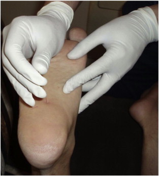
Dry needling in the insertion of the plantar fascia at the medial tuberosity of the calcaneus. (Color version of figure is available online.)
The areas of tenderness throughout the lower part of SBL (Fig 4) were punctured simultaneously. Following insertion, the needle was withdrawn partially and pressed in repeatedly to produce an appropriate response such as a local twitch, a dull ache, heaviness, distension, pressure, and/or a reproduction of the patient’s symptoms. If an appropriate response was not elicited, the needle was removed; and the patient was reexamined.2 If the patient was sensitive to the insertion of the needle, the manipulation was decreased. The needle was left in position for about 3 minutes. Dry needling was performed twice weekly for 2 weeks, and both sides of the lower extremity were treated. During the DN treatment, calf muscle stretching and postural corrections of sitting and standing positions were taught to the patient. The patient was asked to walk, and he felt an immediate reportable reduction of pain with a feeling of lightness in both sides. After 2 weeks of DN, there were a 60% to 70% reduction in pain (3 for the right side and 2 for the left side), dramatic increase of the pressure pain threshold, and complete relief of pain (Table 1). He reported a sense of well-being and was able to fully return to his job. No adverse effects of DN such as bruising and severe pain were observed. The patient consented to the publication of this case report.
Fig 4.

Markers along the lower part of the SBL and locations of needles according to Myers.9 (Color version of figure is available online.)
Table 1.
Mean of Pressure Pain Threshold (PPT) Measurements for Active MTrPs Before and After DN
| Pretreatment PPT (kg/cm2) |
Posttreatment PPT (kg/cm2) |
|||
|---|---|---|---|---|
| Myofascial Locations | Right | Left | Right | Left |
| Medial tuberosity of the calcaneus | 16.5 | 19.3 | 37.1 | 39.9 |
| Plantar surface of toe phalanges (beneath 5th metatarsal head) | 18.6 | 22.1 | 29.6 | 37.0 |
| Plantar surface of toe phalanges (beneath 1st metatarsal head) | 11.4 | 18.9 | 19.9 | 29.8 |
| Achilles tendon | 14.8 | 19.9 | 22.1 | 27.5 |
| Medial gastrocnemius | 19.9 | 28.4 | 35.5 | 46.1 |
| Biceps femoris | 18.5 | 25.9 | 28.9 | 39.9 |
| Semimembranosus | 14.6 | 18.9 | 23.2 | 27.5 |
| Ischial tuberosity | 8.6 | 9.9 | 14.6 | 15.9 |
Discussion
To the authors’ knowledge, this is the first case study for the evaluation of DN effects of myofascial meridians in the treatment of PF. The focus of this report was to describe how treating a myofascial meridian could be effective in resolving PF. Based on the success described in this case report, it may be of benefit for clinicians to consider myofascial continuity in their evaluations and treatment plans.
Plantar fascia is one of the ligamentous bands that maintain the longitudinal arch of the foot.5 It is a thin band of fibrous tissue that connects the inferior aspect of the calcaneus to the metatarsophalangeal joints and the basis of the proximal phalanges of the toes.4 Also, it is the first portion of the SBL distally. The postural function of the SBL is to protect the body in full upright extension and prevent the tendency into flexion. However, daily postural function requires a higher proportion of slow-twitch, endurance muscle fibers in the muscular portions of this myofascial band.9 A tense plantar fascia affects the other tracks of SBL that should be considered in the treatment of PF.9,11
Plantar fasciitis has been found to have a negative effect on foot-specific health-related quality of life.6 Regardless of numerous treatment protocols for PF,11-17 there is no strong evidence that supports the effectiveness of any specific treatment.21
The patient had received various treatment approaches before attempting DN. Almost all treatment methods were localized in the site of pain that targeted only the plantar fascia. The rapid relief of patient’s complaints after 2 weeks of DN may indicate the more global view on PF management.
Concepts of chains,22 trains,9 slings,23 and contractile fields24 have recently been developed in exercise and rehabilitative programs. According to Paoletti,22 this is critical to maintain motility or inherent micromovements of the system. Panayi25 showed the contribution of the deep longitudinal sling in hamstring strain. Also, Hungerford et al26 indicated that this sling may also become facilitated as a result of sacroiliac joint pain. According to fascial manipulation theory, Pedrelli et al27 proposed that patellar tendinopathy is often due to incorrect activation of knee extensor muscles. Therefore, treatment protocols should focus on the muscles of the extensor compartment and not just on the site of pain in patellar tendon.
Myofascial meridians are lines that transmit strain and maintain structural integrity of the body.9 They clearly have some overlap with the meridians of acupuncture but are not equivalent.9,28 Dorsher28 quantitatively explored an 89% overlap in the distribution of the anatomically derived myofascial meridians and the acupuncture principal meridian. Zhang et al29 showed the efficacy and specificity of acupuncture treatment for PF. They suggested that acupuncture can provide pain relief for PF and introduced specific acupoint for heel pain.
The studies for the effectiveness of DN in musculoskeletal pain are developing. Different methodologies and heterogeneity of knowledge among therapists may be the cause of various outcomes. The importance of appropriate needling locations has been emphasized in many studies.30
Based on recent studies, fascial connective tissue is a continuous system that is suspended from bony structures to form a fully integrated supporting framework.22
According to the concept of myofascial meridians, an underlying presence of increased myofascial tension or stiffness may exist in PF.9 In the present study, DN of the major stations in SBL relieved chronicity of the symptoms. Nguyen7 applied ischemic compression massage on posterior superior iliac spine at the origin of the gluteus medius, gluteus minimus, and piriformis muscles proximally to the medial and lateral femur epicondyles and medial tibial condyle along the medial head of gastrosoleus muscles in a patient affected by PF. He found and treated the trigger zones that were hyperirritable by direct firm compression. Self-stretching of the calf and hamstring muscles was performed consequently. The patient felt an immediate reduction in pain during walking. He recommended that evaluation and treatment of the PF should be on the site of pain and the suspected areas remote from it.7
Prolonged faulty sitting posture and limping of the patient, more than 6 months, may be considered as important factors that impose additional tensions on the SBL. However, abnormal mechanical tension through the tissues may cause long-duration, low-energy contractions of myofibroblasts and chronic contractions of the musculo-fascial-skeletal system.7,9
Limitations
No objective measurement for diagnosis of myofascial meridian involvement was performed for the patient. The method of DN was based on the knowledge of the therapist considering the major stations of SBL subjectively. In the present study, findings about the effectiveness of myofascial meridian, based on DN, do not necessarily support the application of this technique by other practitioners or for other patients. Further studies should be considered for comparing this method with other protocols and in different musculoskeletal disorders.
Conclusion
Dry needling based on myofascial meridians improved the symptoms for a patient with recurrent PF. More investigations are needed to demonstrate the effects of interventions on lines of tensions as myofascial meridians.
Funding Sources and Conflicts of Interest
No funding sources or conflicts of interest were reported for this study.
References
- 1.Gill L.H. Plantar fasciitis: diagnosis and conservative management. J Am Acad Orthop Surg. 1997;5(2):109–117. doi: 10.5435/00124635-199703000-00006. [DOI] [PubMed] [Google Scholar]
- 2.Baldry P.E. Acupuncture, trigger points and musculoskeletal pain. 3rd ed. Churchill Livingstone; Philadelphia, PA: 2005. pp. 207–222. [Google Scholar]
- 3.Lysholm J., Wiklander J. Injuries in runners. Am J Sports Med. 1987;15(2):168–171. doi: 10.1177/036354658701500213. [DOI] [PubMed] [Google Scholar]
- 4.McNally E.G., Shetty S. Plantar fascia: imaging diagnosis and guided treatment. Semin Musculoskelet Radiol. 2010;14:334–343. doi: 10.1055/s-0030-1254522. [DOI] [PubMed] [Google Scholar]
- 5.Bartold S.J. Plantar heel pain syndrome: overview and management—the plantar fascia as a source of pain: biomechanics, presentation and treatment. J Bodyw Mov Ther. 2004;8:214–226. [Google Scholar]
- 6.Irving D.B., Cook J.L., Young M.A., Menz H.B. Impact of chronic plantar heel pain on health-related quality of life. J Am Podiatr Med Assoc. 2008;98(4):283–289. doi: 10.7547/0980283. [DOI] [PubMed] [Google Scholar]
- 7.Nguyen B.M. Trigger point therapy and plantar heel pain: a case report. Foot. 2010;20:158–162. doi: 10.1016/j.foot.2010.09.009. [DOI] [PubMed] [Google Scholar]
- 8.Crawford F. Plantar heel pain and fasciitis. Clin Evid. 2005;13:1533–1545. [PubMed] [Google Scholar]
- 9.Myers T.W. Anatomy trains: myofascial meridians for manual and movement therapists. 2nd ed. Churchill Livingstone; Philadelphia: 2009. [Google Scholar]
- 10.Simons D.G. New views of myofascial trigger points: etiology and diagnosis. Arch Phys Med Rehabil. 2008;89:157–159. doi: 10.1016/j.apmr.2007.11.016. [DOI] [PubMed] [Google Scholar]
- 11.Cole C., Seto C., Gazewood J. Plantar fasciitis: evidence-based review of diagnosis and therapy. Am Fam Physician. 2005;72:2237–2242. [PubMed] [Google Scholar]
- 12.Radford J.A., Landorf K.B., Buchbinder R., Cook C. Effectiveness of low-dye taping for the short-term treatment of plantar heel pain: a randomised trial. BMC Musculoskelet Disord. 2006;7(64):1–7. doi: 10.1186/1471-2474-7-64. [DOI] [PMC free article] [PubMed] [Google Scholar]
- 13.Genc H., Saracoglu M., Nacır B., Erdem H.R., Kacar M. Long-term ultrasonographic follow-up of plantar fasciitis patients treated with steroid injection. Joint Bone Spine. 2005;72:61–65. doi: 10.1016/j.jbspin.2004.03.006. [DOI] [PubMed] [Google Scholar]
- 14.Speed C.A., Nichols D., Wies J. Extracorporeal shock wave therapy for plantar fasciitis. a double blind randomised controlled trial. J Orthop Res. 2003;21:937–940. doi: 10.1016/S0736-0266(03)00048-2. [DOI] [PubMed] [Google Scholar]
- 15.Porter D., Barrill E., Oneacre K., May B.D. The effects of duration and frequency of achilles tendon stretching on dorsiflexion and outcome in painful heel syndrome: a randomized, blinded, control study. Foot Ankle Int. 2002;23(7):619–624. doi: 10.1177/107110070202300706. [DOI] [PubMed] [Google Scholar]
- 16.Young B., Walker M.J., Strunce J., Boyles R. A combined treatment approach emphasizing impairment-based manual physical therapy for plantar heel pain: a case series. J Orthop Sports Phys Ther. 2004;34:725–733. doi: 10.2519/jospt.2004.34.11.725. [DOI] [PubMed] [Google Scholar]
- 17.Perez-Millan R., Foster L. Low frequency electroacupuncture in the management of refractory plantar fasciitis. Med Acupunct. 2001;13(1):1–6. [Google Scholar]
- 18.Cotchett M.P., Landorf K.B., Munteanu S.E., Raspovic A.M. Effectiveness of dry needling and injections of myofascial trigger points associated with plantar heel pain: a systematic review. J Foot Ankle Res. 2010;3:18. doi: 10.1186/1757-1146-3-18. [DOI] [PMC free article] [PubMed] [Google Scholar]
- 19.Park G., Kim C.W., Park S.B., Kim M.J., Jang S.H. Reliability and usefulness of the pressure pain threshold measurement in patients with myofascial pain. Ann Rehabil Med. 2011;35:412–417. doi: 10.5535/arm.2011.35.3.412. [DOI] [PMC free article] [PubMed] [Google Scholar]
- 20.Simons D.G., Travell J.G. Myofascial pain and dysfunction: the trigger point manual. 2nd ed. Williams & Wilkins; Baltimore: 1999. pp. 116–117. [Google Scholar]
- 21.Buchbinder R. Clinical practice. Plantar fasciitis. N Engl J Med. 2004;350:2159–2166. doi: 10.1056/NEJMcp032745. [DOI] [PubMed] [Google Scholar]
- 22.Paoletti S. The fasciae: anatomy, dysfunction & treatment. Eastland Press; 2006. [Google Scholar]
- 23.Vleeming A., Snijders C., Stoeckart R., Mens J. The role of the sacroiliac joins in coupling between spine, pelvis, legs and arms. In: Vleeming A., editor. Movement, stability and low back pain. Churchill Livingstone; 1997. pp. 53–71. [Google Scholar]
- 24.Wallden M. Chains, trains and contractile fields. J Bodyw Mov Ther. 2010;14:403–410. doi: 10.1016/j.jbmt.2010.07.001. [DOI] [PubMed] [Google Scholar]
- 25.Panayi S. The need for lumbar-pelvic assessment in resolution of chronic hamstring strain. J Bodyw Mov Ther. 2010;14(1):294–298. doi: 10.1016/j.jbmt.2009.08.004. [DOI] [PubMed] [Google Scholar]
- 26.Hungerford B., Gilleard W., Hodges P. Evidence of altered lumbopelvic muscle recruitment in the presence of sacroiliac joint pain. Spine. 2003;28(14):1593–1600. [PubMed] [Google Scholar]
- 27.Pedrelli A., Stecco C., Day J.A. Treating patellar tendinopathy with fascial manipulation. J Bodyw Mov Ther. 2009;13:73–80. doi: 10.1016/j.jbmt.2008.06.002. [DOI] [PubMed] [Google Scholar]
- 28.Dorsher P.T. Myofascial referred-pain data provide physiologic evidence of acupuncture meridians. Med Acupunct. 2009;21(2):91–97. doi: 10.1016/j.jpain.2008.12.010. [DOI] [PubMed] [Google Scholar]
- 29.Zhang S.P., Yip T.P., Li Q.S. Acupuncture treatment for plantar fasciitis: a randomized controlled trial with six months follow-up. J Evid Based Complement Altern. 2011;8:1–10. doi: 10.1093/ecam/nep186. [DOI] [PMC free article] [PubMed] [Google Scholar]
- 30.Cotchett M.P., Landorf K.B., Munteanu S.E., Raspovic A.M. Effectiveness of trigger point dry needling for plantar heel pain: study protocol for a randomised controlled trial. J Foot Ankle Res. 2011;4(5):1–10. doi: 10.1186/1757-1146-4-5. [DOI] [PMC free article] [PubMed] [Google Scholar]