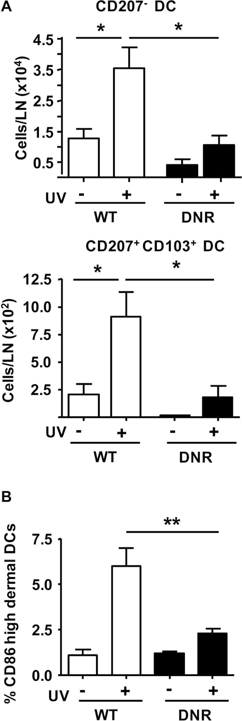
Fig. 5.
Blockade of TGFβ signaling in dermal CD11c+ cells suppresses UV-induced activation and migration. (A) Numbers of CD207− and CD207+ CD103+ DCs in SDLNs of WT or CD11c-DNR transgenic mice (DNR) 72h after UVB IR. (B) Expression of activation marker CD86 in WT and CD11c-DNR dDCs 24h after UVB IR. Mean fluorescence intensity of CD86 was established by flow cytometry and the percentage of CD86High subset of dDCs was plotted for the different treatment groups. n = 2–3 for controls and n = 3–4 for UVB IR groups. Error bars = ±SEM. *P < 0.05 relative to indicated group; **P < 0.01 relative to indicated group.

