Abstract
Brachydactyly type A2 (BDA2, MIM 112600) is characterized by the deviation and shortening of the middle phalange of the index finger and the second toe. Using genome-wide linkage analysis in a Chinese BDA2 family, we mapped the maximum candidate interval of BDA2 to a ∼1.5 Mb region between D20S194 and D20S115 within chromosome 20p12.3 and found that the pairwise logarithm of the odds score was highest for marker D20S156 (Zmax = 6.09 at θ = 0). Based on functional and positional perspectives, the bone morphogenetic protein 2 (BMP2) gene was identified as the causal gene for BDA2 in this region, even though no point mutation was detected in BMP2. Through further investigation, we identified a 4,671 bp (Chr20: 6,809,218–6,813,888) genomic duplication downstream of BMP2. This duplication was located within the linked region, co-segregated with the BDA2 phenotype in this family, and was not found in the unaffected family members and the unrelated control individuals. Compared with the previously reported duplications, the duplication in this family has a different breakpoint flanked by the microhomologous sequence GATCA and a slightly different length. Some other microhomologous nucleotides were also found in the duplicated region. In summary, our findings support the conclusions that BMP2 is the causing gene for BDA2, that the genomic location corresponding to the duplication region is prone to structural changes associated with malformation of the digits, and that this tendency is probably caused by the abundance of microhomologous sequences in the region.
Introduction
The term brachydactyly (BD) is derived from the ancient Greek (brachy-: short; dactylos: digit). This term indicates the shortening of digits due to abnormal development of the phalanges and/or the metacarpals and may also be accompanied by other hand malformations, such as syndactyly, polydactyly, reduction defects, or symphalangism [1]. One of the most commonly used classifications of brachydactyly based on anatomical grounds was provided by Bell [2] and further elaborated by Temtamy and McKusick [3]. BD can occur either as an isolated malformation or as part of a complex malformation syndrome. The isolated BDs are classified clinically into BDA-E and three other subgroups [3]. BD types have certain overlapping features, including hypoplasia/aplasia of phalanges and abnormal interdigital joint formation, suggesting that the formations of phalanges and joints are linked on a developmental and molecular basis.
In recent years, most of the isolated brachydactylies have been characterized at the molecular level, and these studies have provided new insights into the mechanisms of joint and digit development [4]. BDA1 (MIM 112500) is characterized by the shortening or absence of the middle phalanges and is associated with mutations in the Indian hedgehog gene (IHH) or chromosome 5p13.3-p13.2 [5], [6], [7], [8], [9]. BDB1-2 (MIM 113000; MIM 611377) is characterized by terminal deficiency of the fingers and toes and mutations that have been identified in an orphan receptor tyrosine kinase ROR2 or in NOGGIN (NOG) [10], [11], [12], [13]. BDC (MIM 113100) can present with a range of anomalies (from shortening to hyperphalangy of the middle phalanges of the index, middle, and little fingers) and is caused by mutations affecting growth/differentiation factor 5 (GDF5) [14]. Two mutations of HOXD13 are associated with distinctive limb phenotypes in which brachydactylies of specific metacarpals, metatarsals, and phalangeal bones are the most constant features and exhibit overlap with brachydactyly types D (MIM 113200) and E (MIM 113300) [15].
BDA2 (MIM 112600) was described first by Mohr and Wiredt in a large Norwegian kindred of Danish descent [16]. Patients afflicted with BDA2 display hypoplasia/aplasia of the middle phalanx of the second and, sometimes, fifth fingers. Common to all BDA2 families are the deviation and shortening of the middle phalange of the index finger and/or the second toe. BDA2 is genetically heterogeneous. Lehmann et al. found that BDA2 is caused by missense mutations in bone morphogenetic receptor 1 b (BMPR1B), a type I transmembrane serine-threonine kinase, and that three different missense mutations exist in three German families [17], [18]. The second BDA2-causing gene, GDF5, was identified in the descendants of the original BDA2 family by Seemann et al. and Kjaer et al. [19], [20], [21]. Recently, we discovered a Chinese BDA2 family, but no mutation was found either in the BMPR1B or GDF5 loci in the genetic analysis, suggesting additional genetic heterogeneity of this trait. Therefore, we performed a genome-wide scan using 403 microsatellite markers in our BDA2 family.
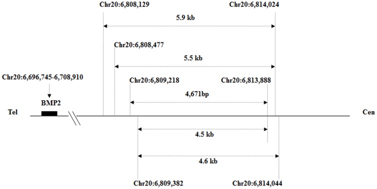
More recently, duplications downstream of BMP2 (NM 001200) were identified in two studies; one study comprised samples from two European families (∼5.9 kb and ∼5.5 kb), and the other study comprised samples from a Chinese family (∼4.6 kb) [22], [23]. In our current study, we found a 4,671 bp duplication downstream of BMP2 (Chr20: 6,809,218–6,813,888) associated with BDA2 in our Chinese family that partially overlaps with the previously reported duplications but has a different breakpoint flanked by the microhomologous sequence GATCA. Our finding supports the hypothesis that the genomic location corresponding to the duplication region is prone to structural changes associated with malformation of the digits and that this tendency is probably caused by the abundance of microhomologous sequences in the region.
Results
Clinical phenotypes
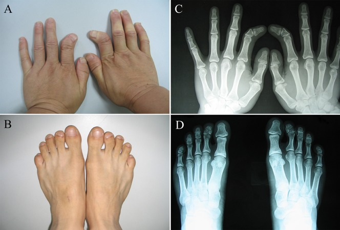
In this six-generation family, 19 affected and 10 unaffected individuals were clinically examined, and a subset of the affected individuals was selected for radiography. All of the affected individuals had the typical phenotypes of BDA2 characteristic of medially deviated and shortened index fingers and second toes with abnormal interdigital joint formation. Most of the affected adult individuals exhibited short stature. Triangular-shaped middle phalanges were observed in all of the affected individuals, as demonstrated in the photograph and X-rays of two affected adult (Figure 1). No sex-correlated characteristics were observed in the family.
Figure 1. Phenotype of BDA2.
A and B: Short and medially deviated index finger (above) and second toe (below). C and D: Radiograph of the triangular-shaped middle phalanx of the index finger (above) and the second toe (below).
The common characteristic of BDA2 caused by variants in different genes is that almost all of the patients showed shortened index fingers. The phenotypic differences consisted of some abnormal symptoms in other phalanges or metacarpals in some patients and a few severely affected individuals, indicating overlap with another type of brachydactyly, symphalangism, or syndactyly. In addition, few mutation carriers appeared to be clinically normal (Table S1). No variant-specific phenotypes were observed in the affected individuals.
Genetic analyses of BMPR1B and GDF5
The sequence analyses of the known BDA2 causing genes, BMPR1B and GDF5, in four affected family members (III:2, IV:4, IV:12, and V:12 in the family) revealed no mutations. In the two-point linkage analysis, the LOD scores were calculated for microsatellite markers in the BMPR1B and GDF5 loci, but none of the LOD scores exceeded 2.0 (at θ = 0) (data not shown).
Genome-wide scan and linkage analyses
A two-point LOD score for 21 markers on chromosome 20p12.3 for the pedigree are summarized in Table 1. Seventeen adjacent markers provided a positive LOD score (θ = 0), and the locus for BDA2 was mapped between markers D20S867 and D20S851 within a region of ∼5.2 Mb, based on the UCSC Human Genome (Mar 2006). The pairwise logarithm of the odds scores was highest for marker D20S156 (Zmax = 6.09 at θ = 0).
Table 1. Two-Point LOD Score Obtained from the Linkage Analysis between the BDA2 Locus and Chromosome 20p12.3 in the Pedigree.
| Marker | Physical | LOD Score AT θ = | Z max | θ | ||||||
| Locationa | 0.0 | 0.01 | 0.05 | 0.1 | 0.2 | 0.3 | 0.4 | |||
| D20S842 | 2634204 | −2.62 | 1.78 | 2.82 | 2.98 | 2.58 | 1.83 | 0.92 | 2.98 | 0.1 |
| D20S867 | 3624718 | −2.77 | 1.76 | 2.74 | 2.84 | 2.41 | 1.69 | 0.87 | 2.84 | 0.1 |
| D20S889 | 3894953 | 3.48 | 3.41 | 3.12 | 2.75 | 1.99 | 1.21 | 0.48 | 3.48 | 0 |
| D20S849 | 5142034 | 1.54 | 2.48 | 2.81 | 2.69 | 2.13 | 1.44 | 0.70 | 2.81 | 0.05 |
| D20S882 | 5583098 | 5.79 | 5.68 | 5.25 | 4.70 | 3.53 | 2.30 | 1.03 | 5.79 | 0 |
| D20S95 | 5664250 | 2.85 | 3.78 | 4.06 | 3.85 | 3.09 | 2.12 | 1.03 | 4.06 | 0.05 |
| D20S905 | 5811610 | 3.69 | 3.64 | 3.39 | 3.06 | 2.33 | 1.51 | 0.66 | 3.69 | 0 |
| D20S194 | 6090696 | 2.68 | 3.58 | 3.84 | 3.63 | 2.94 | 2.09 | 1.10 | 3.84 | 0.05 |
| D20S192 | 6645008 | 2.61 | 2.56 | 2.37 | 2.11 | 1.56 | 1.01 | 0.48 | 2.61 | 0 |
| D20S156 | 6648994 | 6.09 | 5.98 | 5.55 | 5.00 | 3.81 | 2.54 | 1.21 | 6.09 | 0 |
| D20S892 | 6698074 | 5.36 | 5.27 | 4.88 | 4.38 | 3.32 | 2.20 | 1.05 | 5.36 | 0 |
| D20S846 | 6712933 | 4.37 | 4.29 | 3.98 | 3.58 | 2.73 | 1.83 | 0.91 | 4.37 | 0 |
| D20S59 | 6739353 | 5.88 | 5.77 | 5.34 | 4.78 | 3.59 | 2.31 | 0.99 | 5.88 | 0 |
| D20S448 | 7110405 | 3.07 | 3.02 | 2.80 | 2.49 | 1.80 | 1.09 | 0.45 | 3.07 | 0 |
| D20S602 | 7274075 | 3.98 | 3.88 | 3.52 | 3.09 | 2.25 | 1.40 | 0.58 | 3.98 | 0 |
| D20S900 | 7309583 | 1.88 | 1.86 | 1.75 | 1.56 | 1.13 | 0.65 | 0.21 | 1.88 | 0 |
| D20S115 | 7607867 | 2.40 | 2.39 | 2.28 | 2.07 | 1.52 | 0.87 | 0.28 | 2.40 | 0 |
| D20S907 | 8005228 | 2.48 | 2.43 | 2.20 | 1.91 | 1.33 | 0.76 | 0.29 | 2.48 | 0 |
| D20S879 | 8512858 | 2.18 | 2.20 | 2.18 | 2.05 | 1.63 | 1.12 | 0.56 | 2.20 | 0.01 |
| D20S851 | 8809809 | −3.31 | 0.31 | 0.98 | 1.15 | 1.03 | 0.72 | 0.36 | 1.15 | 0.1 |
| D20S186 | 11471795 | −7.09 | −0.75 | 1.0 | 1.48 | 1.52 | 1.12 | 0.56 | 1.52 | 0.2 |
UCSC Browser, Mar 2006; http://genome.ucsc.edu/cgi-bin/hgGateway.
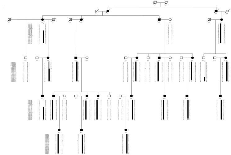
The subsequent haplotypes were constructed using the CYRILLIC 2.1 software to define the interval of the linked region (Figure 2). The markers (19 total) used are listed in Table 1, and the haplotypes were checked through visual inspection. The recombination events between the BDA2 phenotype and the markers spanning the region of interest defined the smallest cosegregating region that included critical meiotic recombinants in the pedigree. Careful examination of the haplotypes confirmed that the disease-associated alleles co-segregated with the phenotype of BDA2 in the pedigree. A recombination event in individual II:2 placed the disease locus distal at D20S194 because the affected individual did not inherit the disease-linked D20S194 alleles observed in the family. The other recombination event in individual IV:13 placed the disease locus close to D20S115 because this unaffected male shared the disease-linked alleles of D20S115 with his affected mother. Hence, the maximal interval of linkage with the BDA2 phenotype is bordered by D20S194 (telomeric) and D20S115 (centromeric) within a region of ∼1.5 Mb. Inspection of this interval revealed only one gene, BMP2.
Figure 2. Pedigree structure and haplotypes of the family.
The marker order was determined from the Marshfield map and the UCSC Human Genome database (Mar 2006). The open symbols indicate the unaffected individuals, the blackened symbols indicate the affected individuals, the squares indicate the males, and the circles indicate the females. The blackened bars indicate the chromosome region shared by the affected members of the pedigree.
Mutation and duplication analyses of BMP2
The sequence analysis of BMP2 (i.e., all exons, introns, promoter region, 5′UTR, 3′UTR, and evolutionary conserved regions) showed no mutation.
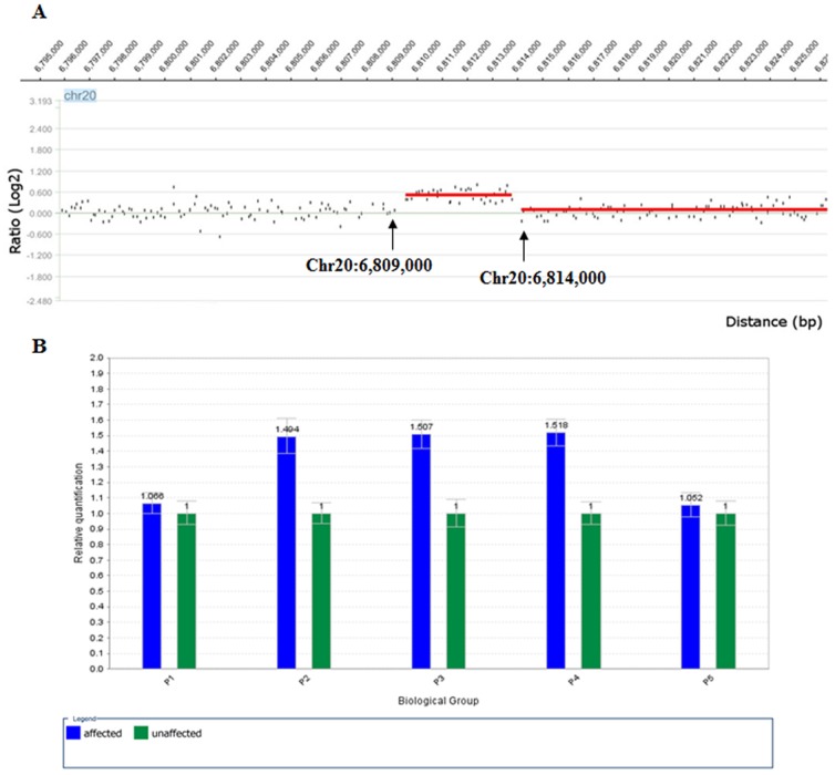
Using the Chromosome 20 array, a ∼5.0 kb duplication (Chr20: 6,809,000–6,814,000) was identified in an affected family member (IV:12) downstream of BMP2 (∼110 kb; Figures 3A and 4A).
Figure 3. Microduplication on 20p12.3.
A. Genomic profile of the microduplication as detected on the NimbleGen Human CGH 385K Chromosome 20 Tiling Array. The detected breakpoints are indicated by arrows. The duplication comprises ∼4.6 kb. The x axis shows the genomic positions on chromosome 20, and the y axis shows the log2 ratio. B. Microduplication confirmed by quantitative real-time PCR (Q-PCR) utilizing a ViiATM 7 Real-Time PCR system. The mean values of the relative quantification were exported from the ViiATM 7 software 1.0. The mean values and standard deviations (error bars) for each target amplicon relative to albumin, which was used as a two-copy reference gene, were calculated for eight affected (blue bars) and eight unaffected (green bars) individuals. The primers P1–P5 (P2–P4 are within the duplicate region, and P1 and P5 flank the region) were designed by Su et al. (2011). One duplicated allele plus one normal allele resulted in three copies of the amplicons of primers P2–P4 in the affected individuals and in a ratio of 1.5 relative to the two copies of the healthy control. The localization of the Q-PCR amplicons is illustrated in Figures 4A and 4B.
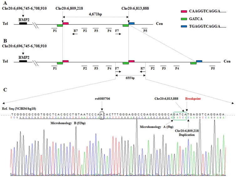
Figure 4. Results of long PCR, breakpoint identification, and positions of primers used.
A. Structure of duplicated region in the unaffected members. F7 and R7: primers used in the breakpoint identification. P1–P5 are the primer positions used in the Q-PCR. The red and green bars indicate the breakpoint region. The numbers above the BMP2 denote the genomic position. Tel: telomere; Cen: centromere. B. Structure of duplicated region in patients. The duplicated region is between Chr20: 6,809,218 and 6,813,888. PCR using primers F7 and R7 amplified the 655 bp sequences spanning the breakpoint. C. Sequencing results of the breakpoint PCR product. The nucleotides of microhomology A (5 bp) are marked by a green box, and the nucleotides of upstream microhomology B (52 bp), including the rs6085706 SNP, are underlined. In addition, the microhomologous nucleotides GTGACC (7 bp) [22] and TTGCAGTGAGC (11 bp) [23] reported previously were also observed downstream of microhomology A (data not shown).
Utilizing Q-PCR with three primer pairs (P2–P4) within the duplicated region and two primers (P1 and P5) within the flanking sequences (Figures 3B and 4B), this duplication was confirmed in eight affected family members (IV:3, IV:4, IV:7, IV:12, IV:15, V:9, V:10, and VI:1) and was not found in eight unaffected family members (III:7, IV:1, IV:6, IV:8, IV:13, V:3, V:7, and V:8).
Using primers F7 and R7, a 655 bp PCR fragment was only produced in the patients (Figures 4B and 5). Moreover, through Q-PCR and breakpoint-PCR, this duplication was not found in 150 healthy control individuals excluding a common copy number variant (CNV). Through the direct sequencing of this 655 bp fragment in eight affected family members (IV:3: File S1; IV:4: File S2; IV:7: File S3; IV:12: File S4; IV:15: File S5; V:9: File S6; V:10: File S7; and VI:1: File S8), we identified a 4,671 bp duplication (Chr20: 6,809,218–6,813,888) downstream of BMP2 and found that this duplication is flanked by the microhomologous sequences GATCA on the 5′ end (Chr20: 6,809,213–6,809,217) and 3′ end (Chr20: 6,813,884–6,813,888). Furthermore, in addition to the rs6085706 SNP, another microhomology (52 bp) could also be identified at the 5′ (Chr20: 6,809,160–6,809,211) and 3′ (Chr20: 6,813,831–6,813,882) flanking region of the 4,671 bp duplicated sequence. In addition, the microhomologous nucleotides GTGACC (7 bp) [22] and TTGCAGTGAGC (11 bp) [23] reported previously were also observed downstream of GATCA. The comparison with the previously reported duplications showed that the duplication identified in this study does have a different breakpoint, overlaps with the other three duplications, and differs in its 5′ and 3′ ends (Figure 6). Based on the above-described findings, we conclude that the fragment was duplicated and is associated with BDA2.
Figure 5. Agarose gel electrophoresis of PCR product with the breakpoint primers F7 and R7.
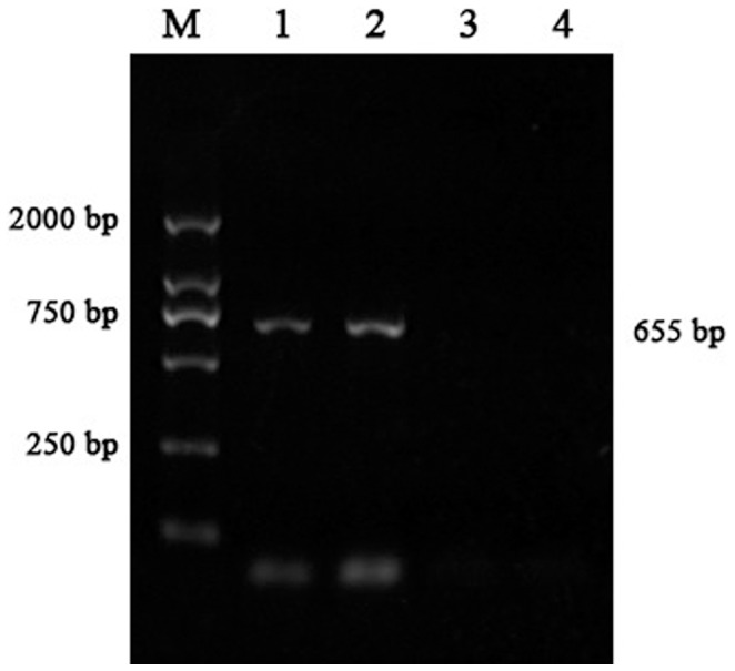
There is a 655-bp band in the patient lanes (1 and 2) but no product in the lanes of the unaffected family member (3 and 4). M: marker.
Figure 6. The chromosomal positions of the three previously reported duplications (Chr20: 6,808,129–6,814,024; Chr20: 6,808,477–6,814,024; and Chr20: 6,809,382–6,814,044) are indicated by the horizontal arrows.
Comparison with the reported duplications shows that our duplication (Chr20: 6,809,218–6,813,888) overlaps with them but has different ends. Furthermore, the causal sequences of BDA2 could be narrowed down to 4,507 bp (Chr20: 6,809,382–6,813,888).
Discussion
The family described in the current study has typical characteristics of BDA2 [1], [24], [25]. We excluded BMPR1B and GDF5, which were found to cause BDA2 in the previous studies [17], [18], [19], [20], [21], and mapped BDA2 to the locus within chromosome 20p12.3 because a strong linkage was observed between the BDA2 locus and the microsatellite markers in this chromosome region. The recombination events in individuals II:2 and IV:13 of the pedigree reduced the interval to a region of ∼1.5 Mb between D20S194 and D20S115.
Two factors enabled us to identify BMP2 as the BDA2-causing gene in chromosome 20p12.3. First, the results of genome-wide scan showed that BMP2 is the only known gene in the candidate interval, i.e., this gene is located at 6.697–6.709 Mb within the candidate interval (6.65–7.61 Mb on chromosome 20p12.3). It has been estimated that ∼25% of the human genome consists of long regions that contain no protein-coding sequences [26]. Some of these long regions contain regulatory sequences that act as large distances to control the expression of neighboring genes, and the sequence elements play critically important and conserved biological roles [27], [28], [29], [30]. Disruption of the long-range control of gene expression may cause disease [31]. BMP2 was embedded in a long region with no known genes ∼0.6 Mb upstream and ∼1.1 Mb downstream that was linked with the phenotype of BDA2 in the pedigree. It has been reported that the transcription of BMP2 is regulated by an enhancer region located 156.3 kb from the promoter of BMP2 and that its noncoding sequence variants may influence BMP2 transcription and thus may contribute to bone mass and osteoporosis [32], [33].
Second, BMP2 has a functional role in the BMP signaling pathway similar to that of the previously identified genes, i.e., BMPR1B and GDF5, that cause BDA2 disease [17], [18], [19], [20], [21], [34]. The proteins of the transforming growth factor β (TGF-β) superfamily are involved in the process of bone and joint development, cell proliferation, and differentiation. The superfamily encompasses extracellular ligands, including bone morphogenetic proteins (BMPs) and growth and differentiation factors (GDFs) [35]. The BMP type I receptors (BMPR1A and BMPR1B) and one type II receptor (BMPR2) are the receptors of BMPs and are needed to form a functional complex to initiate further signaling events [36], [37]. Specific mutations in GDF5 that result in a selective inactivation of the binding to BMPR1B are a cause of BDA2 [19], [20], similarly to dominant negative mutations in the GDF5 high-affinity receptor BMPR1B itself [17], [18]. BDA2 has also been shown to be caused by mutations in GDF5 that interfere with cleavage of the GDF5 prodomain from the mature peptide [21]. BMP2 activates the BMP pathway by binding to the receptor complex [38] and plays an essential role in cartilage differentiation and bone formation, similarly to GDF5 and BMPR1B [17], [19], [39], [40], [41]. It is known that the global loss of BMP2 in an animal results in an embryonic lethality and that BMP2 plays a center role in skeletal development, such as chondrogenesis, cartilage and bone development, the formation of bone mineral density, and limb patterning [39], [42], [43]. Therefore, our linkage results and the reported functional studies strongly support the conclusion that BMP2 is the causal gene of the BDA2 phenotype presented in the family analyzed in the present study.
Through further investigation, we identified a 4,671 bp (Chr20: 6,809,218–6,813,888) genomic duplication downstream of BMP2. This duplication is located within the linked region and was cosegregated with the BDA2 phenotype in this family but not the unaffected family members nor the unrelated control individuals. Recently, Dathe et al. reported two microduplications (∼5.5 and ∼5.9 kb) (Chr20: 6,808,477–6,814,024 and Chr20: 6,808,129–6,814,024) in a noncoding sequence ∼110 kb downstream of BMP2 in two European BDA2 families. Their study found that the duplication contains highly conserved sequences that constitute a cis-regulatory element regulating BMP2 expression in the limb and that this regulating element duplication can be considered a mutational mechanism for BDA2 [22]. More recently, Su et al. identified a duplication of ∼4.6 kb (Chr20: 6,809,382–6,814,044) in the same region in a Chinese family affected with BDA2 [23]. Our work shows a 4,671 bp duplicated sequence in the affected individuals and a 4,507 bp overlapping region (Chr20: 6,809,382–6,813,888) with the previous studies. A microhomology GATCA is located at the 5′ and 3′ flanking region of this duplicated sequence. The generation of the nonrecurring rearrangement agrees well with the replication-based model called fork stalling and template switching proposed by Lee et al. [44]. According to this hypothesis, the microhomology identified in the present study may confuse the DNA replication machinery, causing the replication fork to stall or pause, and lead to a switch of the lagging strand to another active replication fork in physical proximity before resuming replication of the original DNA template, thereby resulting in duplication formation. Furthermore, a longer microhomology (52 bp) was identified upstream of GATCA, and microhomologies GTGACC (7 bp) [22] and TTGCAGTGAGC (11 bp) [23], which are associated with the duplications identified in two other BDA2 families, were also observed downstream of GATCA in our study. Therefore, these findings support the conclusion that this region of the genome is prone to structural changes associated with malformation of the digits and that this tendency is probably caused by the abundance of microhomologous sequences in the region.
In a functional study, Dathe et al. observed that a 5.5 kb tandem duplication in BMP2 may increase the expression of BMP2 in the limb of transgenic mouse. They speculated that the overexpression of BMP2 may result in a deregulation of the fine-tuned BMP pathway by increasing the BMPR1A signal and in a relative decrease of BMPR1B signaling, which is compatible with the previously proposed molecular pathology of BDA2 [22]. Nevertheless, Su et al. revealed reduced activity of conserved 2.1 kb sequences in the duplication region in osteosarcoma U-2OS and HeLa cells through luciferase activity assays [23]. The discrepant expression caused by the duplication downstream of the BMP2 gene in vivo and in vitro may be due to the complex regulation of BMP2 and the other malformations observed in addition to BDA2 in some of the family members. It has been found that BMP2 contains two promoters and that the transcription-initiation site is not common between the human and mouse BMP2 genes, although the sequence of the BMP2 gene is well conserved in the promoter region between the two species [45]. In the 2T3 osteoblast cell line, a proximal promoter fragment can be autoregulated by BMP2 [46]. The studies of a ∼2.7 kb BMP2 promoter fragment have suggested that NF-κB and Gli2 can bind separately to it and thus drive BMP2 expression in different cells [47], [48]. Moreover, the 3′-UTR of BMP2 contains essential regulatory elements, and many serial regulatory elements are located within the large conserved non-coding regions of BMP2 [49], [50]. Therefore, the expression of BMP2 regulated by the duplication sequences may depend on different experimental conditions and/or interaction with other cis-regulatory elements.
BDA2 has genetic heterogeneity. The previous and current studies on BDA2 families show that three missense mutations in BMPR1B, two missense mutations in GDF5, and three duplication elements approximately 110 kb downstream of BMP2 can separately cause the phenotype of BDA2 [17], [18], [19], [20], [21], [22], [23]. Despite the complex molecular mechanisms, these findings suggest that these three genes are all involved in the BMP signaling pathway and that the cause of BDA2 is a shift in the signaling intensity from BMPR1B to BMPR1A [25]. BMP signaling has functions in the growth plate at various stages of differentiation for cartilage and bone formation because it can interact with the BDA1-causing gene IHH, some TGF-β proteins, and other extracellular and nuclear factors in endochondral ossification and thereby form a molecular regulatory network [42], [51], [52], [53], [54]. Under different conditions, BMP2 can mediate BMP signaling through the canonical SMAD pathway to induce SMAD target genes, through a mitogen-activated protein kinase (MAPK) pathway to result in the induction of alkaline phosphatase (ALP) activity, or through a protein kinase C (PKC) signaling pathway to increase Bax/Bcl-2 caspase activity [37], [38], [55]. Therefore, there is no simple interpretation for BMP2 function in the phenotypes of BDA2, and further efforts to clarify the regulation and the role of BMP2 in the BMP signaling pathway and molecular network are warranted.
In summary, we identified a 4,671 bp duplication with a novel breakpoint flanked by the microhomology GATCA downstream of BMP2 in a Chinese family affected with BDA2. Our findings support the conclusion that the genomic location that corresponds to the duplication region is prone to structural changes associated with malformation of the digits and that this tendency is probably caused by the abundance of microhomologous sequences in the region. Additionally, our study further exemplifies the peculiar nature of the balance of expression of the BMP2 gene during development, and the genomic location corresponding to the duplication region is critical for the function of this gene. Moreover, our results indicate that such duplications may be considered a regulatory mutational mechanism. Therefore, similar duplications may conceivably be implemented into bioinformatic search tools to detect variants as an additional method to associate a genetic mutation with a given phenotype.
Materials and Methods
Subjects
The study was approved by the genetic research ethics committees of Shanghai Jiaotong University. A Chinese family with BDA2 was recruited for the study (see Figure 2). This family was from Anhui province in eastern China and does not have any distant relatives. The informed consent was written and obtained from all participants.
Genetic analyses of BMPR1B and GDF5
Samples of peripheral blood DNA were collected from all available family members, and the DNA was isolated using standard procedures. We amplified the coding region, the splice junctions, and the promoter sequence of the BMPR1B and GDF5 genes using standard protocol polymerase chain reaction (PCR). The PCR products were analyzed using 1.5% agarose gel electrophoresis. The purified PCR products were bidirectionally sequenced using PCR primers as sequencing primers and the Applied Biosystems Prism BigDye terminator cycle sequencing reaction kit. The products were evaluated using an Applied Biosystems 3100 DNA sequencer. In addition, we genotyped the flanking polymorphic microsatellite markers of BMPR1B and GDF5 and performed a two-point linkage analysis using the LINKAGE 5.2 software program assuming a genetic model featuring autosomal dominance, a disease-allele frequency of 0.0001, an evenly shared allele frequency, a zero phenocopy rate, no sex difference, and full penetrance [56].
Genome-wide scan and linkage analyses
We performed a genome-wide scan of the family using a set of 403 polymorphic microsatellite markers (ABI Prism Linkage Mapping Set version 2.5) with an average marker density of 10 centimorgans according to the Marshfield genetic linkage map. Additional markers were used for the fine mapping. The products of the PCR assays with fluorescently labeled primers were analyzed by automated capillary genotyping on MegaBACE 1000 (Amersham Pharmacia Biotech). A two-point linkage analysis was performed using the LINKAGE 5.2 software program [18], [56]. The haplotypes were reconstructed using the CYRILLIC version 2.1 software.
Mutation analyses of BMP2
Following the procedures described above, we amplified and sequenced the BMP2 gene (i.e., all exons, introns, promoter region, 5′UTR, and 3′UTR). The evolutionary conserved regions that were sequenced were Chr20: 6,811,205–6,813,071. The following evolutionary conserved sequences were identified using the publicly available web-browser program ECR [57]. The PCR products were analyzed and sequenced by standard protocols as described above.
Duplication analyses of BMP2
A CGH array of human chromosome 20 was performed using a custom Roche array (NimbleGen Human CGH 385K Chromosome 20 Tiling Array). The labeling and hybridization were performed following the protocols provided by the manufacturers (NimbleGen Hybridization System 4). The array was analyzed using the NimbleGen MS 200 Microarray Scanner and NimbleScan software. A graphical overview was obtained using the SignalMap software (v1.9.0.05, NimbleGen Systems Inc). Quantitative real-time PCR (Q-PCR) with five primer pairs (P1–P5) was performed using a ViiATM 7 Real-Time PCR system and the ViiATM 7 software 1.0 (Applied Biosystems) to confirm the duplication region. PCR with the primers F7 and R7 was performed to amplify the breakpoint region, and the products were sequenced as described above. The abovementioned primers were designed by Su et al. [23]. The sequences were compared with the data from the UCSC Genome Browser (http://genome.ucsc.edu).
Supporting Information
The summary of causing genes and phenotypes of BDA2.
(DOC)
Sequencing of the family number IV:3.
(AB1)
Sequencing of the family number IV:4.
(AB1)
Sequencing of the family number IV:7.
(AB1)
Sequencing of the family number IV:12.
(AB1)
Sequencing of the family number IV:15.
(AB1)
Sequencing of the family number V:9.
(AB1)
Sequencing of the family number V:10.
(AB1)
Sequencing of the family number V:I1.
(AB1)
Acknowledgments
We are deeply grateful to all of the families who participated in this study and to the physicians who helped us with the identification of the families.
Funding Statement
This work was supported by the National Natural Science Foundation of China (No. 30700453; No. 81121001; No. 31371298; No. 81301151), the 973 Program (2010CB529600), the National Key Technology R&D Program (2012BAI01B09), the Shanghai Municipal Commission of Science and Technology Program 09DJ1400601, the Shanghai Leading Academic Discipline Project (B205). The funders had no role in study design, data collection and analysis, decision to publish, or preparation of the manuscript.
References
- 1. Temtamy SA, Aglan MS (2008) Brachydactyly. Orphanet J Rare Dis 3: 15. [DOI] [PMC free article] [PubMed] [Google Scholar]
- 2.Bell J (1951) On brachydactyly and symphalangism. In Treasury of Human Inheritance Volume 5. London: Cambridge University Press: pp.1–31.
- 3.Temtamy S, McKusick V (1978) The Genetics of Hand Malformations New York: Alan R Liss, INC. [PubMed]
- 4. Kornak U, Mundlos S (2003) Genetic disorders of the skeleton: a developmental approach. Am J Hum Genet 73: 447–474. [DOI] [PMC free article] [PubMed] [Google Scholar]
- 5. Gao B, Guo J, She C, Shu A, Yang M, et al. (2001) Mutations in IHH, encoding Indian hedgehog, cause brachydactyly type A-1. Nat Genet 28: 386–388. [DOI] [PubMed] [Google Scholar]
- 6. Gao B, Hu J, Stricker S, Cheung M, Ma G, et al. (2009) A mutation in Ihh that causes digit abnormalities alters its signalling capacity and range. Nature 458: 1196–1200. [DOI] [PubMed] [Google Scholar]
- 7. Guo S, Zhou J, Gao B, Hu J, Wang H, et al. (2010) Missense mutations in IHH impair Indian Hedgehog signaling in C3H10T1/2 cells: Implications for brachydactyly type A1, and new targets for Hedgehog signaling. Cell Mol Biol Lett 15: 153–176. [DOI] [PMC free article] [PubMed] [Google Scholar]
- 8. Ma G, Yu J, Xiao Y, Chan D, Gao B, et al. (2011) Indian hedgehog mutations causing brachydactyly type A1 impair Hedgehog signal transduction at multiple levels. Cell Res 21: 1343–1357. [DOI] [PMC free article] [PubMed] [Google Scholar]
- 9. Armour CM, McCready ME, Baig A, Hunter AG, Bulman DE (2002) A novel locus for brachydactyly type A1 on chromosome 5p13.3–p13.2. J Med Genet 39: 186–188. [DOI] [PMC free article] [PubMed] [Google Scholar]
- 10. Oldridge M, Fortuna AM, Maringa M, Propping P, Mansour S, et al. (2000) Dominant mutations in ROR2, encoding an orphan receptor tyrosine kinase, cause brachydactyly type B. Nat Genet. 24: 275–278. [DOI] [PubMed] [Google Scholar]
- 11. Schwabe GC, Tinschert S, Buschow C, Meinecke P, Wolff G, et al. (2000) Distinct mutations in the receptor tyrosine kinase gene ROR2 cause brachydactyly type B. Am J Hum Genet. 67: 822–831. [DOI] [PMC free article] [PubMed] [Google Scholar]
- 12. Akbarzadeh S, Wheldon LM, Sweet SM, Talma S, Mardakheh FK, et al. (2008) The deleted in brachydactyly B domain of ROR2 is required for receptor activation by recruitment of Src. PLoS One 3: e1873. [DOI] [PMC free article] [PubMed] [Google Scholar]
- 13. Lehmann K, Seemann P, Silan F, Goecke TO, Irgang S, et al. (2007) A new subtype of brachydactyly type B caused by point mutations in the bone morphogenetic protein antagonist NOGGIN. Am J Hum Genet 81: 388–396. [DOI] [PMC free article] [PubMed] [Google Scholar]
- 14. Polinkovsky A, Robin NH, Thomas JT, Irons M, Lynn A, et al. (1997) Mutations in CDMP1 cause autosomal dominant brachydactyly type C. Nat Genet. 17: 18–19. [DOI] [PubMed] [Google Scholar]
- 15. Johnson D, Kan SH, Oldridge M, Trembath RC, Roche P, et al. (2003) Missense mutations in the homeodomain of HOXD13 are associated with brachydactyly types D and E. Am J Hum Genet. 72: 984–997. [DOI] [PMC free article] [PubMed] [Google Scholar]
- 16.Mohr O, Wriedt C (1919) A New Type of Hereditary Brachyphalangy in Man Washington: Carnegie Inst: pp.5–64.
- 17. Lehmann K, Seemann P, Stricker S, Sammar M, Meyer B, et al. (2003) Mutations in bone morphogenetic protein receptor 1B cause brachydactyly type A2. Proc Natl Acad Sci U S A 100: 12277–12282. [DOI] [PMC free article] [PubMed] [Google Scholar]
- 18. Lehmann K, Seemann P, Boergermann J, Morin G, Reif S, et al. (2006) A novel R486Q mutation in BMPR1B resulting in either a brachydactyly type C/symphalangism-like phenotype or brachydactyly type A2. Eur J Hum Genet 14: 1248–1254. [DOI] [PubMed] [Google Scholar]
- 19. Seemann P, Schwappacher R, Kjaer KW, Krakow D, Lehmann K, et al. (2005) Activating and deactivating mutations in the receptor interaction site of GDF5 cause symphalangism or brachydactyly type A2. J Clin Invest 115: 2373–2381. [DOI] [PMC free article] [PubMed] [Google Scholar]
- 20. Kjaer KW, Eiberg H, Hansen L, van der Hagen CB, Rosendahl K, et al. (2006) A mutation in the receptor binding site of GDF5 causes Mohr-Wriedt brachydactyly type A2. J Med Genet 43: 225–231. [DOI] [PMC free article] [PubMed] [Google Scholar]
- 21. Ploger F, Seemann P, Schmidt-von Kegler M, Lehmann K, Seidel J, et al. (2008) Brachydactyly type A2 associated with a defect in proGDF5 processing. Hum Mol Genet 17: 1222–1233. [DOI] [PubMed] [Google Scholar]
- 22. Dathe K, Kjaer KW, Brehm A, Meinecke P, Nurnberg P, et al. (2009) Duplications involving a conserved regulatory element downstream of BMP2 are associated with brachydactyly type A2. Am J Hum Genet 84: 483–492. [DOI] [PMC free article] [PubMed] [Google Scholar]
- 23. Su P, Ding H, Huang D, Zhou Y, Huang W, et al. (2011) A 4.6 kb genomic duplication on 20p12.2–12.3 is associated with brachydactyly type A2 in a Chinese family. J Med Genet 48: 312–316. [DOI] [PubMed] [Google Scholar]
- 24. Fitch N (1979) Classification and identification of inherited brachydactylies. J Med Genet 16: 36–44. [DOI] [PMC free article] [PubMed] [Google Scholar]
- 25. Mundlos S (2009) The brachydactylies: a molecular disease family. Clin Genet 76: 123–136. [DOI] [PubMed] [Google Scholar]
- 26. Venter JC, Adams MD, Myers EW, Li PW, Mural RJ, et al. (2001) The sequence of the human genome. Science 291: 1304–1351. [DOI] [PubMed] [Google Scholar]
- 27. Nobrega MA, Ovcharenko I, Afzal V, Rubin EM (2003) Scanning human gene deserts for long-range enhancers. Science 302: 413. [DOI] [PubMed] [Google Scholar]
- 28. Kimura-Yoshida C, Kitajima K, Oda-Ishii I, Tian E, Suzuki M, et al. (2004) Characterization of the pufferfish Otx2 cis-regulators reveals evolutionarily conserved genetic mechanisms for vertebrate head specification. Development 131: 57–71. [DOI] [PubMed] [Google Scholar]
- 29. Uchikawa M, Takemoto T, Kamachi Y, Kondoh H (2004) Efficient identification of regulatory sequences in the chicken genome by a powerful combination of embryo electroporation and genome comparison. Mech Dev 121: 1145–1158. [DOI] [PubMed] [Google Scholar]
- 30. Ovcharenko I, Loots GG, Nobrega MA, Hardison RC, Miller W, et al. (2005) Evolution and functional classification of vertebrate gene deserts. Genome Res 15: 137–145. [DOI] [PMC free article] [PubMed] [Google Scholar]
- 31. Kleinjan DA, van Heyningen V (2005) Long-range control of gene expression: emerging mechanisms and disruption in disease. Am J Hum Genet 76: 8–32. [DOI] [PMC free article] [PubMed] [Google Scholar]
- 32. Chandler RL, Chandler KJ, McFarland KA, Mortlock DP (2007) Bmp2 transcription in osteoblast progenitors is regulated by a distant 3′ enhancer located 156.3 kilobases from the promoter. Mol Cell Biol 27: 2934–2951. [DOI] [PMC free article] [PubMed] [Google Scholar]
- 33. Styrkarsdottir U, Cazier JB, Kong A, Rolfsson O, Larsen H, et al. (2003) Linkage of osteoporosis to chromosome 20p12 and association to BMP2. PLoS Biol 1: E69. [DOI] [PMC free article] [PubMed] [Google Scholar]
- 34. Sammar M, Stricker S, Schwabe GC, Sieber C, Hartung A, et al. (2004) Modulation of GDF5/BRI-b signalling through interaction with the tyrosine kinase receptor Ror2. Genes Cells 9: 1227–1238. [DOI] [PubMed] [Google Scholar]
- 35. Allendorph GP, Vale WW, Choe S (2006) Structure of the ternary signaling complex of a TGF-beta superfamily member. Proc Natl Acad Sci U S A 103: 7643–7648. [DOI] [PMC free article] [PubMed] [Google Scholar]
- 36. Kawabata M, Chytil A, Moses HL (1995) Cloning of a novel type II serine/threonine kinase receptor through interaction with the type I transforming growth factor-beta receptor. J Biol Chem 270: 5625–5630. [DOI] [PubMed] [Google Scholar]
- 37. Derynck R, Zhang YE (2003) Smad-dependent and Smad-independent pathways in TGF-beta family signalling. Nature 425: 577–584. [DOI] [PubMed] [Google Scholar]
- 38. Nohe A, Hassel S, Ehrlich M, Neubauer F, Sebald W, et al. (2002) The mode of bone morphogenetic protein (BMP) receptor oligomerization determines different BMP-2 signaling pathways. J Biol Chem 277: 5330–5338. [DOI] [PubMed] [Google Scholar]
- 39. Tsuji K, Bandyopadhyay A, Harfe BD, Cox K, Kakar S, et al. (2006) BMP2 activity, although dispensable for bone formation, is required for the initiation of fracture healing. Nat Genet 38: 1424–1429. [DOI] [PubMed] [Google Scholar]
- 40. Storm EE, Kingsley DM (1999) GDF5 coordinates bone and joint formation during digit development. Dev Biol 209: 11–27. [DOI] [PubMed] [Google Scholar]
- 41. Baur ST, Mai JJ, Dymecki SM (2000) Combinatorial signaling through BMP receptor IB and GDF5: shaping of the distal mouse limb and the genetics of distal limb diversity. Development 127: 605–619. [DOI] [PubMed] [Google Scholar]
- 42. Long F, Ornitz DM (2013) Development of the endochondral skeleton. Cold Spring Harb Perspect Biol 5: a008334. [DOI] [PMC free article] [PubMed] [Google Scholar]
- 43. Bandyopadhyay A, Tsuji K, Cox K, Harfe BD, Rosen V, et al. (2006) Genetic analysis of the roles of BMP2, BMP4, and BMP7 in limb patterning and skeletogenesis. PLoS Genet 2: e216. [DOI] [PMC free article] [PubMed] [Google Scholar]
- 44. Lee JA, Carvalho CM, Lupski JR (2007) A DNA replication mechanism for generating nonrecurrent rearrangements associated with genomic disorders. Cell 131: 1235–1247. [DOI] [PubMed] [Google Scholar]
- 45. Sugiura T (1999) Cloning and functional characterization of the 5′-flanking region of the human bone morphogenetic protein-2 gene. Biochem J 338 (Pt 2): 433–440. [PMC free article] [PubMed] [Google Scholar]
- 46. Ghosh-Choudhury N, Choudhury GG, Harris MA, Wozney J, Mundy GR, et al. (2001) Autoregulation of mouse BMP-2 gene transcription is directed by the proximal promoter element. Biochem Biophys Res Commun 286: 101–108. [DOI] [PubMed] [Google Scholar]
- 47. Feng JQ, Xing L, Zhang JH, Zhao M, Horn D, et al. (2003) NF-kappaB specifically activates BMP-2 gene expression in growth plate chondrocytes in vivo and in a chondrocyte cell line in vitro. J Biol Chem 278: 29130–29135. [DOI] [PubMed] [Google Scholar]
- 48. Zhao M, Qiao M, Harris SE, Chen D, Oyajobi BO, et al. (2006) The zinc finger transcription factor Gli2 mediates bone morphogenetic protein 2 expression in osteoblasts in response to hedgehog signaling. Mol Cell Biol 26: 6197–6208. [DOI] [PMC free article] [PubMed] [Google Scholar]
- 49. Fritz DT, Liu D, Xu J, Jiang S, Rogers MB (2004) Conservation of Bmp2 post-transcriptional regulatory mechanisms. J Biol Chem 279: 48950–48958. [DOI] [PubMed] [Google Scholar]
- 50. Abrams KL, Xu J, Nativelle-Serpentini C, Dabirshahsahebi S, Rogers MB (2004) An evolutionary and molecular analysis of Bmp2 expression. J Biol Chem 279: 15916–15928. [DOI] [PubMed] [Google Scholar]
- 51. Kronenberg HM (2003) Developmental regulation of the growth plate. Nature 423: 332–336. [DOI] [PubMed] [Google Scholar]
- 52. Yoon BS, Lyons KM (2004) Multiple functions of BMPs in chondrogenesis. J Cell Biochem 93: 93–103. [DOI] [PubMed] [Google Scholar]
- 53. Mackie EJ, Ahmed YA, Tatarczuch L, Chen KS, Mirams M (2008) Endochondral ossification: how cartilage is converted into bone in the developing skeleton. Int J Biochem Cell Biol 40: 46–62. [DOI] [PubMed] [Google Scholar]
- 54. Minina E, Kreschel C, Naski MC, Ornitz DM, Vortkamp A (2002) Interaction of FGF, Ihh/Pthlh, and BMP signaling integrates chondrocyte proliferation and hypertrophic differentiation. Dev Cell 3: 439–449. [DOI] [PubMed] [Google Scholar]
- 55. Hay E, Lemonnier J, Fromigue O, Marie PJ (2001) Bone morphogenetic protein-2 promotes osteoblast apoptosis through a Smad-independent, protein kinase C-dependent signaling pathway. J Biol Chem 276: 29028–29036. [DOI] [PubMed] [Google Scholar]
- 56. Xing QH, Wang MT, Chen XD, Feng GY, Ji HY, et al. (2003) A gene locus responsible for dyschromatosis symmetrica hereditaria (DSH) maps to chromosome 6q24.2–q25.2. Am J Hum Genet 73: 377–382. [DOI] [PMC free article] [PubMed] [Google Scholar]
- 57. Ovcharenko I, Nobrega MA, Loots GG, Stubbs L (2004) ECR Browser: a tool for visualizing and accessing data from comparisons of multiple vertebrate genomes. Nucleic Acids Res 32: W280–286. [DOI] [PMC free article] [PubMed] [Google Scholar]
Associated Data
This section collects any data citations, data availability statements, or supplementary materials included in this article.
Supplementary Materials
The summary of causing genes and phenotypes of BDA2.
(DOC)
Sequencing of the family number IV:3.
(AB1)
Sequencing of the family number IV:4.
(AB1)
Sequencing of the family number IV:7.
(AB1)
Sequencing of the family number IV:12.
(AB1)
Sequencing of the family number IV:15.
(AB1)
Sequencing of the family number V:9.
(AB1)
Sequencing of the family number V:10.
(AB1)
Sequencing of the family number V:I1.
(AB1)


