Abstract
For some traits, the human genome is more closely related to either the bonobo or the chimpanzee genome than they are to each other. Therefore, it becomes crucial to understand whether and how morphostructural differences between humans, chimpanzees and bonobos reflect the well known phylogeny. Here we comparatively investigated intra and extra labyrinthine semicircular canals orientation using 260 computed tomography scans of extant humans (Homo sapiens), bonobos (Pan paniscus) and chimpanzees (Pan troglodytes). Humans and bonobos proved more similarities between themselves than with chimpanzees. This finding did not fit with the well established chimpanzee – bonobo monophyly. One hypothesis was convergent evolution in which bonobos and humans produce independently similar phenotypes possibly in response to similar selective pressures that may be associated with postural adaptations. Another possibility was convergence following a “random walk” (Brownian motion) evolutionary model. A more parsimonious explanation was that the bonobo-human labyrinthine shared morphology more closely retained the ancestral condition with chimpanzees being subsequently derived. Finally, these results might be a consequence of genetic diversity and incomplete lineage sorting. The remarkable symmetry of the Semicircular Canals was the second major finding of this article with possible applications in taphonomy. It has the potential to investigate altered fossils, inferring the probability of post-mortem deformation which can lead to difficulties in understanding taxonomic variation, phylogenetic relationships, and functional morphology.
Introduction
Phenotypic traits have been used for decades for the purpose of reconstructing the evolutionary history of humans (Homo sapiens) and their closest relatives [1], chimpanzee and bonobo species (Pan troglodytes and Pan paniscus, respectively). More recently, phenotypic data have been supplemented by growing evidence from the genome wide sequencing analysis [2]–[4].
The comparison between human and chimpanzee genomes revealed genetic differences accumulated since these species diverged from their common ancestor [5]. The hominin-Pan split date was lately recalibrated to at least 7–8 million years [6]. It is often argued that chimpanzee subspecies and bonobos carry no or marginal genetic differences, when compared to the corresponding differences seen in humans from different continents [7]–[9]. Besides, common chimpanzees show the greatest population stratification when compared to all other great ape lineages, while humans and western chimpanzees show a remarkable dearth of genetic diversity when compared to other great apes. It was also found that the rate of gene loss in the human branch is not different as compared to other internal branches in the great ape phylogeny [10]. Recently, more extensive comparisons revealed that bonobo and chimpanzee genomes were not necessarily more closely related to each other than to the human one [2].
The phenotype of bonobos has received less attention than that of chimpanzees, despite several studies investigating dental development [11] and morphology [12], [13], craniometry [14], [15], intralabyrinthine angles [16]–[19], cranial development and vascularization [20]–[22], endocranial ontogeny and morphology [23]. For some traits, bonobos appear to be less diverse than chimpanzees in both their phenotype [12], [14], [20], and DNA [9], [24]–[27]. Interestingly, the variation of some phenotypic traits has been shown to correlate more closely with genetic data than others [5].
For instance, the inner ear morphology has proven to be useful to assess diversity among extant and fossil primates [18], [28]. However, a few studies have focused on variability within the genus Pan and have compared such variations with the extant human figures [16], [18], [19], [29]. Studies from the early 70's [16]–[18] included large samples focused on angles taken from 2D radiographs. They concluded that the bonobo and the chimpanzee labyrinths are more similar to each other than to the human one.
Here, we investigated differences in the orientation of semicircular canals (SCC) starting from the null hypothesis (H0) that not only Homo and Pan differ in SCC orientation, but also that the two Pan species are more similar as compared to humans. Based on a sample of 260 medical X-ray computed tomography (CT) scans, we applied a mathematical model verified with microcomputed tomography (μCT) scans [30] and measurement error quantification. We subsequently investigated how these features discriminate the three species and we discussed our results in the context of their evolutionary relationships. Besides, we explored the existence of eventual sexual dimorphism related to human SCCs.
Material and Methods
Ethics statement
The human CT scans were provided from medical CT. The data reported here involved no experimentation on human subjects but only reprocessing of existing anonymized scan data. The use of these data for the present purpose was in respect of bioethical laws in France. Written consent was given by the patients for their information to be stored in the hospital database and used for research purpose. The “comité de protection des personnes – Bordeaux” (French IRB) approved the use of these data for the present purpose.
The Pan sample was composed of a set of inner ear reconstructed from dry skulls. We obtained permission from the Royal Museum of Central Africa of Tervuren (Belgium) and the Museum of Comparative Zoology at Harvard University (Cambridge, MA, USA) to access the collections. The collections were elaborated in the early twentieth century from mostly wild-shot animals donated to the museums. These collections were widely used in several studies including Lieberman et al. [31] and Durrleman et al. [23]. The samples were donated to the museums, and the parties involved in the hunting of the animals held the proper permits.
Samples
Our sample was composed of 137 anonymized human clinical records (H. sapiens: 70 females and 67 males), 61 P. paniscus and 62 P. troglodytes (8 P. troglodytes verus and 54 P. troglodytes schweinfurthi) (see Table 1 and Table S1). The three species were represented by subadult and adult individuals.
Table 1. Description of the sample.
| Class | abbreviation | Human | Bonobo | Chimpanzee | Apes | Total |
| infant | NJ1 | 1 | 3 | 7 | 10 | 11 |
| infant stage 2 | J1 | 0 | 11 | 5 | 16 | 16 |
| young juvenile | J2 | 35 | 14 | 11 | 25 | 60 |
| old juvenile | J3 | 50 | 10 | 10 | 20 | 70 |
| sub-adult | A1 | 16 | 8 | 15 | 23 | 39 |
| adult | A2 | 35 | 15 | 14 | 29 | 64 |
| Total | 137 | 61 | 62 | 123 | 260 |
Number of individuals according to Age classes [30] and species.
The human sample was composed of patients from the Pasteur Hospital (Toulouse, France) and the Faculty of Dentistry at the University of Toulouse (France), scanned between 2007 and 2010. These individuals had been referred for cranial trauma, inflammation of maxillary sinuses or neonatal distress but were found to be free of reportable abnormalities having any direct or indirect impact on inner ear morphology. The pixel size ranged from 0.3 to 0.49 mm and the slice thickness from 0.3 to 0.8 mm (for detailed information see Table S1). The human CT scans were provided from medical CT.
The Pan sample was composed of wild animals. The pixel size ranged from 0.27 to 0.49 mm and the slice thickness from 0.5 to 1 mm (for detailed information see Table S1).
The Maturational Status (MS) was assessed using dental stages [32] and reported in Table S1.
Data collection
Data were saved initially as Digital Imaging and Communications in Medicine (DICOM) format files, and then as Tagged Image File Format (TIFF) files.
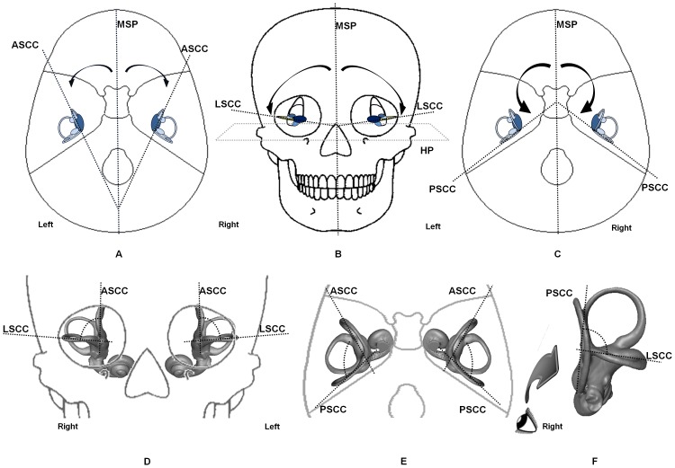
Thirty landmarks (Table 2) were placed on the CT images using Amira software to best represent each SCC, as well as the midsagittal (MSP) and horizontal (HP) planes of the skull, used as references (Table 3). Anterior, posterior and lateral SCCs were referred to as ASCC, PSCC and LSCC, respectively. Each SCC was represented by three landmarks located at the center of its lumen [33]. The vestibule was also represented by one single landmark (see Method S1 for detailed information). Each SCC plane coordinate was then calculated from its landmarks as well as from the vestibular one. Then we calculated the 3D angles between planes (Figure 1) using the dot product of the two plane normal vectors [30]. For the main results, significance level was set at p = 0.01.
Table 2. Definition of the landmarks used in the present study.
| N° | Name | Definition | Bookstein landmarks type |
| 1 | Frontal crest (Fc) | Summit of the Fc | II |
| 2 | Crista galli (Cr) | Summit of Cr | I |
| 3 | Internal occipital crest (iOc) | Medial most eminent point on the iOc | II |
| 4 | Vomer (Vm) | Point on the posterior border of the Vm | II |
| 5 | Nasopalatine foramen (NPf) | Central point of the NPf | I |
| 6 | Foramen Magnum (fMo) | Midpoint on the anterior border of the fMo | II |
| 7,8 | Infraorbital foramina (IOf) | Midpoint of the IOf | I |
| 9,10 | Supraorbital foramen (SOf) | Cranial part of the notch of the SOf | II |
| 11,12 | Vestibule (Vb) | Center of the lumen of the Vb | I |
| 13,16 | ASCC (middle) | Superior-most point at the center of the ASCC lumen | II |
| 14,17 | ASCC (anterior) | Anterior point at the center of the ASCC lumen before its ampulla | II |
| 15,18 | ASCC (posterior) | Posterior point at the center of the ASCC lumen before the common crus | II |
| 19,22 | LSCC (anterior) | Anterior point at the center of the LSCC lumen before its ampulla | II |
| 20,23 | LSCC (middle) | Lateral-most point at the center of the LSCC lumen | II |
| 21,24 | LSCC (posterior) | Posterior point at the center of the LSCC lumen before joining the vestibule | II |
| 25,28 | PSCC (inferior) | Inferior point at the center of the PSCC lumen before its ampulla | II |
| 26,29 | PSCC Right (middle) | Posterior-lateral-most point at the center of the PSCC lumen | II |
| 27,30 | PSCC Right (superior) | Superior point at the center of the PSCC lumen before the common crus | II |
Anterior, posterior and lateral SCCs (semicircular canals) were noted respectively ASCC, PSCC and LSCC. Landmarks of Type I were well defined locally; their homology from individual to another was strongly supported. Type II landmarks was corresponding to points which position was first defined locally using specific structures but it was also depending on less specific factors such as the maximum or minimum of a curve. When using type II landmarks the individual to individual homology was only supported geometrically, to calculate plane coordinates for example.
Table 3. Definition of the reference planes used in the study.
| planes | landmarks (Table 2) |
| Mid-Sagittal Plane | MSP: 1, 2, 3, 4, 5, 6 |
| Horizontal Plane | HP: 7, 8, 11, 12 |
| Semi Circular Canal planes | ASCC right: 11, 13, 14, 15 |
| ASCC left: 12, 16, 17, 18 | |
| LSCC right: 11, 19, 20, 21 | |
| LSCC left: 12, 22, 23, 24 | |
| PSCC right: 11, 25, 26, 27 | |
| PSCC left: 12, 28, 29, 30 |
Anterior, posterior and lateral SCC (semicircular canals) were noted respectively ASCC, PSCC and LSCC.
Figure 1. SCC angle representations.
(a) MSP/ASCC angles. (b) Orientation of LSCC with MSP and HP (c) MSP/PSCC angles (d) ASCC/LSCC angles (e) ASCC/PSCC angles (f) LSCC/PSCC angles.
Body size is important to consider when comparing the size of morphological structures between species. However it was not considered in our study as it has no real impact on angular measurements [19]. The radii of the SCC were not reported as they did not meet our validation criteria.
Statistics
Statistical tests were performed with R software. Before angular comparisons, the normality was tested using either Shapiro-Wilk's test (N<50) or D'Agostino's test (N>50). The normal probability plot and frequency histogram were also established to visually check the normality of the distribution. The equality of variances was tested with Levene's tests.
The angular measurements were compared using a one-way ANOVA and Tukey's HSD post-hoc test providing a correction due to multiple comparisons. The Kruskall-Wallis test for multiple comparisons was used when normality criteria were not completed. A between-group principal component analysis (bgPCA) [34] of angular measurements was also applied on angles presented in Table 4. The bgPCA was previously successfully used to discriminate labyrinthine shape differences between predefine groups of chimpanzees or fossil hominins [35], [36]. Measurements were tested also for sexual dimorphism, fluctuating asymmetry, anti-symmetry and directional asymmetry [37], [38] using a two-way anova and student's t-tests.
Table 4. Statistical comparisons of the SCC orientation between humans, chimpanzees and bonobos.
| Humans | ← p → | Chimpanzees | ← p → | Bonobos | ← p → | Humans | |
| Canal pair | N = 137 | H vs C | N = 62 | C vs B | N = 61 | B vs H | N = 137 |
| ASCC/LSCC* | 74.2±4.4° | <0.001 | 77.0±7.4° | NS | 74.8±7.1° | NS | 74.2±4.4° |
| ASCC/PSCC* | 111.2±6.4° | <0.001 | 105.7±7.6° | <0.001 | 109.5±6.1° | NS | 111.2±6.4° |
| LSCC/PSCC** | 88.2±6.2° | <0.001 | 80.1±10.8° | <0.001 | 86.9±10.5° | NS | 88.2±6.2° |
| LSCC right/left** | 19.7±8.9° | NS | 22.0±9.5° | NS | 21.3±15.2° | NS | 19.7±8.9° |
| ASCC right/PSCC left** | 10.6±5.4° | <0.001 | 19.2±9.5° | <0.001 | 12.8±7.8° | NS | 10.6±5.4° |
| ASCC left/PSCC right** | 11.4±6.4° | <0.001 | 18.0±10.6° | <0.001 | 14.3±9.2° | NS | 11.4±6.4° |
| MSP/ASCC* | 34.9±5.1° | <0.001 | 39.1±5.9° | NS | 38.1±4.6° | <0.001 | 34.9±5.1° |
| MSP/LSCC* | 80.9±5.5° | NS | 80.6±6.6° | NS | 82.9±8.6° | NS | 80.9±5.5° |
| MSP/PSCC* | 145.3±6.2° | <0.001 | 142.1±8.0° | <0.001 | 146.1±7.4° | NS | 145.3±6.2° |
| HP/LSCC* | 25.9±6.7° | <0.001 | 23.0±7.2° | <0.001 | 26.1±7.5° | NS | 25.9±6.7° |
Angles measurements and angles comparisons between species showing subtle differences between humans and bonobos and marked differences with chimpanzees (NS = not significant, H = humans, B = Bonobos, C = Chimpanzees). Anterior, posterior and lateral SCC were referred to as ASCC, PSCC and LSCC. (*) parametric tests were used since angular measurements showed normal distribution. (**) non parametric tests were used since angular measurements did not show normal distribution; however parametric tests results were consistent with those of non parametric tests. All angles in degrees.
Measurement error and validation
In order to validate the reproducibility of our results, we ran a test to assess the extent of intra-observer error. We used 60 randomly selected subjects. All measurements were taken twice by one observer (MEK), on separate days and without any knowledge on sex, age and species attributions [39]. Mean error was then analyzed using the angular mean error and a two-way ANOVA (sides*individuals) to compare Measurement Error relative to fluctuating asymmetry [40]. We did not find significant differences between the two sets of measurements over the 60 selected subjects (p>0.80) resulting in a mean error of 0.48°±0.30. ME was found to be significantly lower than fluctuating asymmetry (p<0.02).
Additionally, we ran other tests to validate the accuracy of angular calculation by using μCT as a comparison. To this end, Four P. troglodytes were scanned using both clinical Cone Beam Computed Tomography (K9500, Trophy, KODAK) and μCT (Xtreme CT by SCANCO - Switzerland). Non parametric Kruskal-Wallis tests were used to detect differences between conventional-CT and μCT groups. Since μCT images covered only the petrous bones, only inter-SCC angles were computed. No significant differences were detected between the two imaging procedures (p>0.63). Similar evidence resulted from an independent additional test run on three Papio anubis, and one Gorilla gorilla using the same procedure as the four P. troglodytes.
Results
Medical CT images are contentious to measure very small dimensions [41] due to their spatial resolution [42]. The intra-observer error was found negligible in the present study and no statistical differences were found between the CT and μCT imaging procedures providing an indication of the method reliability [30].
Intra-specific comparisons
The possible influence of sex was not assessed in Pan, as sex attribution was not available for all specimens. In humans, we found sexual dimorphism in ASCC/PSCC and LSCC/PSCC angles. Both of them were more open in females than in males (respectively +2.7°, p<0.001 and +2.9°, p<0.001). Previously, sexual dimorphism was already pointed out as reflecting allometry only [19].
No differences were found between MS confirming that SCC orientations undergo no further important changes after birth [19], [43].
Only chimpanzees proved evidence for directional asymmetry in the MSP/LSCC angle (right: 82.6±6.4°, left: 78.6±6.7°, p<0.002). In all other instances, we observed a remarkable symmetry (asymmetry ranging from 0.09% to 3.05%).
Inter-specific comparisons
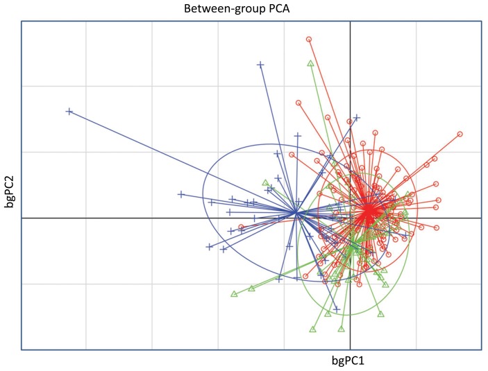
As shown in Table 4, significant differences were found between chimpanzees and the two other species. Humans and Bonobos shared more similarities. The bgPCA (Figure 2) highlight this result: the chimpanzee group was quite separated from both humans and bonobos, with a weak overlap seen on the graph, while a tighter overlap between humans and bonobos was observed.
Figure 2. Between group principal component analysis (bgPCA) of the angular measurements.
Humans (•) and bonobos (▴) were very closed to each other and distant from chimpanzees (+). The ellipses graphically represent the scatter plots encompassing approximately 67% of the subjects. The bgPCA showed a strait overlap of humans with bonobos and a weaker overlap of the latter two species with the chimpanzees.
Between humans and chimpanzees, MSP/ASCC and MSP/PSCC angles were significantly different (p<0.001), indicating in humans ASCC and PSCC respectively 4.2° and 3.2° closer to the MSP. Thus, the angle between ASCC and PSCC was more open in humans (+5.5°, p<0.001). LSCC was 2.9° more horizontal in chimpanzees (HP/LSCC was 2.9° more open in humans) (p<0.001), 2.8° closer to ASCC in humans (p<0.001), and 8.1° closer to PSCC in chimpanzees (p<0.001). The angle between ASCC and the contra lateral PSCC was 7.6° more open in humans than in chimpanzees (p<0.001). Between chimpanzees and Bonobos, LSCC was more horizontally oriented in chimpanzees (p<0.001). The ASCC/PSCC angle was 3.8° more open in bonobos (p<0.001). PSCC was more closely related to MSP in bonobos (4° closer, p<0.001). The angle between ASCC and the contra lateral PSCC was 5.1° flatter in bonobos (p<0.001). In chimpanzees, as compared to bonobos, LSCC was more horizontally oriented (p<0.001). Only the MSP/ASCC angle exhibited a significant difference (p<0.001) between humans and bonobos.
Discussion and Conclusions
Differences between humans and Pan species were previously noticed in semicircular canal size and shape [19], [35], [44], but not specifically between chimpanzees and bonobos, usually found to be similar (see Table S2). In this regard, we found two significantly distinct groups: humans and bonobos on one side and chimpanzees on the other side. This new finding could have three explanations. First, earlier studies excluded the vestibule from calculations of the SCC planes. Second, the present study used 3D angles instead of projected angles. Finally, previous intra-SCC angle measurements in Pan were based on very limited sample sizes. We used the vestibule as it contains the utricule and the saccule that are parts of the vestibular system. The fluids filling the vestibule and SCC play a major role in their physiology by the detection of head rotational movement which contributes to balance and maintaining visual fixation during head movements. Because the utricule and the saccule are parts of this system to detect motion and orientation, their use is functionally relevant in SCC study. The precision and reliability of the vestibule landmark is presented in additional data (Method S1).
A previous bgPCA analysis of overall labyrinth shape revealed significant discrimination among two subspecies of P. troglodytes [35] demonstrating subtle, yet significant, differences between P. troglogytes troglodytes and P. t. verus and a clear separation between humans and the two chimpanzees groups. However Gunz et al. [35] focused on the total morphological pattern of the labyrinth (through a Geometric Morphometric approach) whereas our study concentrated only on a limited number of angular measurements of the labyrinth. Despite this, our results demonstrate differences between the two species of Pan and are in line with Gunz et al. [35] study. We did not find a clear separation between the two subspecies of chimpanzees represented in our sample (P. t. schweinfurthi and P. t. verus), even though genetical and morphological data suggest that among subspecies of common chimpanzee, P.t. verus is the most distinct [24]. As regard the labyrinth, Gunz et al. [35] observed only subtle differences in canal radii. This may explain why our angular values failed to discriminate P. t. schweinfurthi and P. t. verus.
We found more similarities between humans and bonobos than between chimpanzees and bonobos. The differences recorded between the present study and that of Spoor and Zonneveld (1998) [44] could result from differences in resolution, as the smallest available slice thickness used by the latter (1.5 mm) was higher than our highest available slice thickness (1 mm) (Table S1). Also, we used a sample of Pan wild specimens of known geographical origin much larger than in Spoor and Zonneveld (1998) (61 vs 6 P. paniscus and 62 vs 7 P. troglodytes).
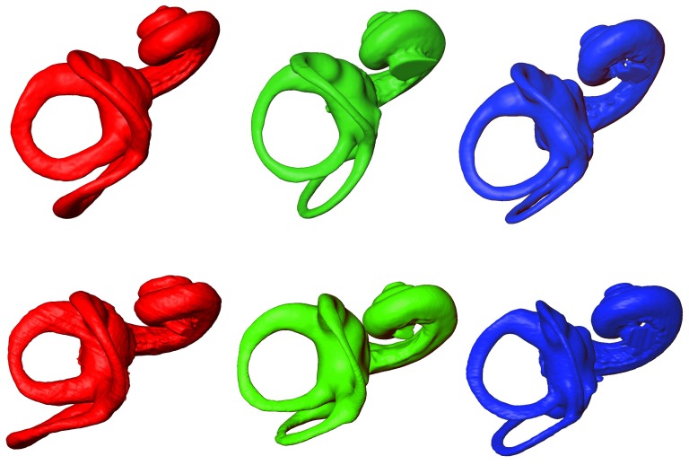
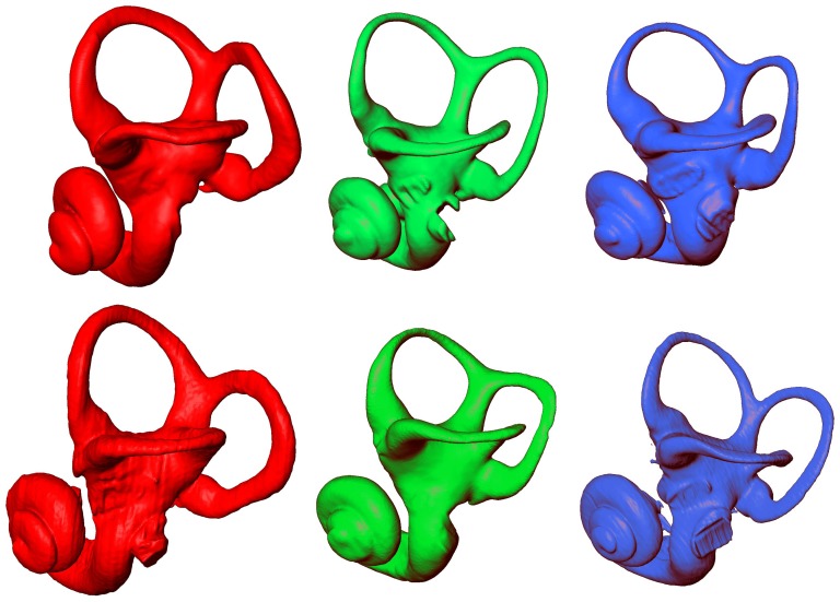
The extent of differences and similarities between P. paniscus, P. troglodytes, and H. sapiens were illustrated in Figure 3 and 4 by the μCT-based reconstruction of 2 humans, 2 chimpanzees and 2 bonobos labyrinths. A simple visual inspection (Figure 3 and 4) showed important intra-specific differences. We remarked morphological differences between Pan and humans. The thickness was greater in humans, especially for ASCC and PSCC whereas the 3D curvature did not clearly separate human and Pan species. However, these differences did not suffice to identify clear-cut inter-SCC angular inter-specific differences
Figure 3. MicroCT-based rendering of the left bony labyrinth superior aspect in: Homo sapiens (red, on the left), Pan paniscus (green, in the middle) and Pan troglodytes (blue, on the right).
Figure 4. MicroCT-based rendering of the left bony labyrinth lateral aspect in: Homo sapiens (red, on the left), Pan paniscus (green, in the middle) and Pan troglodytes (blue, on the right).
In the context of our observed significant intra-SCC angular differences between chimpanzees and bonobos, it is important to add that basicranial differences between the two species of Pan were demonstrated by Cousin et al. [29] using 83 and 179 specimens of P. troglodytes and P. paniscus respectively. In their comparative study, the skulls were oriented using the lateral semicircular canal (the so-called «vestibular orientation method»). They found inter-specific differences in basicranial geometry. For some measurements and at each stage of its growth, H. sapiens appeared closer in shape to one of the two Pan species than they were to each other. Bonobos and humans were closer in the angle between LSCC and a line rejoining Nasion-Vestibule (Na-Op/V). Chimpanzees and humans were closer in the angle between LSCC and Nasion-sella turcica. Even though we could not directly compare our results with those taken from Cousin et al. [29], more studies are needed to investigate in detail, with appropriate comparative samples, how and which labyrinthine features show different orientations when compared to basicranial structures in humans, chimpanzees and bonobos.
We observed that chimpanzees have more horizontally-oriented LSCC. However, to our knowledge, there is currently no satisfactory and consensual explanation based on comparative physiological studies between the Pan species and modern humans. There is evidence that the use of bipedality is different between the two species [45]. Using feet contact time and hind limb joint angles, Pontzer et al. [46] suggested that bipedal locomotion might have been relatively costly for bonobos as compared to chimpanzees. This cost may reflect a difference in gait mechanics whereas none of the two Pan species is more bipedal than the other [45]. There is still no detailed explanation based on the relationship between locomotor patterns and SCC orientation.
Directional asymmetry was also found in chimpanzees with a more open MSP/LSCC angle on the right side. While gait asymmetry is well documented in chimpanzees with predominance of the right to make ground contact first [47], data are missing on bonobos as well as for bipedality. Our unexpected results for more horizontally oriented LSCC in chimpanzees need to be confirmed by further studies.
The characterization of functional relationships between canal morphology and locomotion is limited by the lack of data, especially in vivo. Only the MSP/ASCC angle clearly discriminates humans from the two Pan species. This may be due to the fact that bonobos and chimps show anatomical features that favor versatility [48]. The more open MSP/ASCC angle in Pan may contribute to their greater locomotor repertoire, particularly to head rotation through a transverse axis. A second possible explanation may lie in gait modalities of the three species. Bipedal and quadrupedal walking among chimpanzees and bonobos is similar but drastically different from that in humans.
The labyrinth morphology is open to external developmental influences only prior to ossification of the otic capsule, in humans at the end of the second trimester of gestation [28]. No differences were found between MS confirming that SCC orientations undergo no further important changes after birth [19], [43].
Our main result about similarity between bonobos and humans, as compared to chimpanzees, does not fit with our null hypothesis. One explanation is convergent evolution in which bonobos and humans produce independently similar phenotypes due to selection. Such an homoplasic pattern has already been argued for genetic data [2], [49]. Another possibility is convergence following a “random walk” (Brownian motion) evolutionary model (e.g. [50], [51]). Moreover, as demonstrated by complete genome analysis of gorillas, chimpanzees and bonobos [2], [4], incomplete lineage sorting (ILS) [52] may influence phenotypic similarities that humans share with one of these three species but not the others.
Two species might share more similarities in a single trait whereas the phylogenetic tree reveals a different overall pattern of speciation. This might be a consequence of a complex intertwining of genetic diversity from ancestral population, selection and “Brownian walk”. Human and chimpanzee genomes reveals genetic differences accumulated since the divergence of these two species from their common ancestor [5], dating at least to 7–8 million years [6]. The genetically-based estimated time between P. troglodytes and P. paniscus split is 2.1–1.5 million years [53]. In order to identify the respective role of each process, it would be interesting to investigate the labyrinthine morphology in fossil specimens attributed to Ardipithecus (ARA-VP-6/500) [54], and Sahelanthropus (TM 266-01-060-1) [55].
A final and more parsimonious explanation is that the bonobo-human labyrinthine shared morphology, more closely represents the Pan-Homo ancestral condition with chimpanzees being subsequently derived. Interestingly, the MSP/LSCC angle does not show the same trend as the other angles, showing no significant differences between the three species. This may reflect a primitive shared condition.
The petrous bone is often well preserved in fossil specimens [44], [56]–[59]. However, deformation occurring from compaction and other diagenetic processes [60], [61] makes it sometimes difficult to infer phylogenetic relationships [62] as well as missing data on extant specimens. The remarkable symmetry of the SCC is another important result of this study and may have implications in taphonomy. Further observations on fossil hominids are needed to investigate the potential usefulness of asymmetry to evaluate post-mortem deformation.
Most studies which have attempted to find sexual dimorphism at the inner ear level provided inconsistent results. However our observed sexual dimorphism in ASCC/PSCC and LSCC/PSCC angles are in agreement with results obtained in modern humans either for the bony labyrinth as a whole [63] or on the cochlea only [64], [65].
In conclusion, we have used the largest known sample of individuals to measure the three dimensional orientation of semicircular canals in humans, bonobos and chimpanzees. We have demonstrated in this sample that bonobos SCC orientation is closer to humans than to chimpanzees. This finding may have crucial implications in hominid evolution that still need to be addressed. Additionally, the low intra-individual asymmetrical signal of the inner ear in our sample could open a most interesting track for the study of paleoanthropological records.
Supporting Information
Detailed information and acquisition modes from the 260 subjects of the present sample.
(DOCX)
Comparisons with previous studies related to Semicircular Canal orientation.
(DOCX)
Placement and reliability of the vestibular landmark.
(DOCX)
Acknowledgments
We thank W. Van Neer and E. Gilissen as the former and current curator of the Musée Royal de l'Afrique Centrale at Tervuren (Belgium) and J.L. Kahn of the Musée d'Anatomie Normale et Pathologique of the University of Strasbourg (France). We are also indebted to ME Rutzmoser of the Department of Mammalogy at the Museum of Comparative Zoology, Harvard University (Cambridge, MA, USA). We thank Jean-Noel Vergnes for his kind support in statistics. The helpful comments of the two referees and PLOS ONE academic editor improved remarkably the manuscript.
Funding Statement
The research was funded by the University of Toulouse (http://www.univ-tlse3.fr/) and the Molecular Anthropology and Image Synthesis laboratory (http://www.anthropobiologie.fr/). The funders had no role in study design, data collection and analysis, decision to publish, or preparation of the manuscript.
References
- 1. Wood B, Harrison T (2011) The evolutionary context of the first hominins. Nature 470: 347–352. [DOI] [PubMed] [Google Scholar]
- 2. Prüfer K, Munch K, Hellmann I, Akagi K, Miller JR, et al. (2012) The bonobo genome compared with the chimpanzee and human genomes. Nature 486(7404): 527–531. [DOI] [PMC free article] [PubMed] [Google Scholar]
- 3. Locke DP, Hillier LW, Warren WC, Worley KC, Nazareth LV (2011) Comparative and demographic analysis of orang-utan genomes. Nature 469(7331): 529–533. [DOI] [PMC free article] [PubMed] [Google Scholar]
- 4. Scally A, Dutheil JY, Hillier LW, Jordan GE, Goodhead I, et al. (2012) Insights into hominid evolution from the gorilla genome sequence. Nature 483(7388): 169–175. [DOI] [PMC free article] [PubMed] [Google Scholar]
- 5. Mikkelsen TS, Hillier LW, Eichler EE, Zody MC, Jaffe DB, et al. (2005) (The Chimpanzee Sequencing and Analysis Consortium) (2005) Initial sequence of the chimpanzee genome and comparison with the human genome. Nature 437: 69–87. [DOI] [PubMed] [Google Scholar]
- 6. Langergraber KE, Prüfer K, Rowney C, Boesch C, Crockford C, et al. (2012) Generation times in wild chimpanzees and gorillas suggest earlier divergence times in great ape and human evolution. Proc Natl Acad Sci 109(39): 15716–15721. [DOI] [PMC free article] [PubMed] [Google Scholar]
- 7. Enard W, Paabo S (2004) Comparative primate genomics. Annu Rev Genomics Hum Genet 5: 351–378. [DOI] [PubMed] [Google Scholar]
- 8. Prado-Martinez J, Sudmant PH, Kidd JM, Li H, Kelley JL, et al. (2013) Great ape genetic diversity and population history. Nature 499: 471–475. [DOI] [PMC free article] [PubMed] [Google Scholar]
- 9. Jensen-Seaman MI, Deinard AS, Kidd KK (2001) Modern African ape populations as genetic and demographic models of the last common ancestor of humans, chimpanzees, and gorillas. J Hered 92(6): 475–480. [DOI] [PubMed] [Google Scholar]
- 10. Kaessmann H, Wiebe V, Pääbo S (1999) Extensive nuclear DNA sequence diversity among chimpanzees. Science 286(5442): 1159–1162. [DOI] [PubMed] [Google Scholar]
- 11.Kinzey WG (1984) The dentition of the pygmy chimpanzee, Pan paniscus. In: The Pygmy Chimpanzee: Evolutionary Biology and Behavior (Ed. Susman RL), pp. 65–88. Plenum Press, New York.
- 12.Uchida A (1996) Craniodental variation among great apes. Peabody Mus Bull 4. Cambridge: Peabody Museum of Archaeology and Ethnology, Harvard University. 198p
- 13. Skinner MM, Gunz P, Wood BA, Boesch C, Hublin JJ (2009) Discrimination of extant Pan species and subspecies using the enamel–dentine junction morphology of lower molars. Am J Phys Anthropol 140: 234–243. [DOI] [PubMed] [Google Scholar]
- 14. Shea BT, Inouye SE (1993) Knuckle-walking ancestors. Science 259: 293–294. [PubMed] [Google Scholar]
- 15. Robinson C (2012) Geometric morphometric analysis of mandibular shape diversity in Pan . J Hum Evol 63: 191–204. [DOI] [PubMed] [Google Scholar]
- 16.Dardenne J (1970) Etude comparative des principaux paramètres sagittaux de la face et du crane, chez l'homme et les chimpanzés, par la méthode vestibulaire d'orientation. Thèse de doctorat. Université de Lille.
- 17.Deblock R (1973) Craniométrie comparée de Pan paniscus et Pan troglodytes. Thèse de doctorat. Université de Lille.
- 18. Fenart R, Dufresnoy P, Deblock R (1973) Use of boundary lines in the search for neurocranial sexual differences in man and chimpanzees. Arch Anat Pathol 21(4): 341–348. [PubMed] [Google Scholar]
- 19.Spoor F (1993) The comparative morphology and phylogeny of the human bony labyrinth. PhD thesis. Monograph Utrecht University.
- 20. Braga J (1995) Skeletal variation and measure of divergence among chimpanzees - contribution of the study of discrete traits. C R Acad Sci Paris II 320(10): 1025–1030. [Google Scholar]
- 21. Braga J (1998) Chimpanzee variation facilitates the interpretation of the incisive suture closure in South African Plio-Pleistocene hominids. Am J Phys Anthropol 105(2): 121–135. [DOI] [PubMed] [Google Scholar]
- 22. Braga J, Boesch C (1997) Further data about venous channels in South African Plio-Pleistocene hominids. J Hum Evol 33(4): 423–447. [DOI] [PubMed] [Google Scholar]
- 23. Durrleman S, Pennec X, Trouvé A (2011) Comparison of the endocranial ontogenies between chimpanzees and bonobos via temporal regression and spatiotemporal registration. J Hum Evol 62(1): 74–88. [DOI] [PubMed] [Google Scholar]
- 24. Morin PA, Moore JJ, Chakraborty R, Jin L, Goodall J, et al. (1994) Kin selection, social structure, gene flow, and the evolution of chimpanzees. Science 265: 1193–1201. [DOI] [PubMed] [Google Scholar]
- 25. Gonder MK, Locatelli S, Ghobrial L, Mitchell MW, Kujawski JT, et al. (2011) Evidence from Cameroon reveals differences in the genetic structure and histories of chimpanzee populations. Proc Natl Acad Sci 108(12): 4766–4771. [DOI] [PMC free article] [PubMed] [Google Scholar]
- 26. Deinard AS, Kidd K (2000) Identifying conservation units within captive chimpanzee populations. Am J Phys Anthropol 111(1): 25–44. [DOI] [PubMed] [Google Scholar]
- 27. Fischer A, Prüfer K, Good JM, Halbwax M, Wiebe V, et al. (2011) Bonobos Fall within the Genomic Variation of Chimpanzees. PLoS One 6(6): e21605. [DOI] [PMC free article] [PubMed] [Google Scholar]
- 28. Lebrun R, Ponce de León MS, Tafforeau P, Zollikofer CPE (2010) Deep evolutionary roots of strepsirrhine primate labyrinthine morphology. J Anat 216: 368–380. [DOI] [PMC free article] [PubMed] [Google Scholar]
- 29. Cousin RP, Fenart R, Deblock R (1981) Variation ontogéniques des angles basicraniens et faciaux: Etudes comparatives chez Homo et chez Pan . Bull Mem Soc Anthropol Paris 8(2): 189–212. [Google Scholar]
- 30. Bradshaw AP, Curthoys IS, Todd MJ, Magnussen JS, Taubman DS, et al. (2010) A mathematical model of human semicircular canal geometry: a basis for interpreting vestibular physiology. J Assoc Res Otolaryngol 11: 145–159. [DOI] [PMC free article] [PubMed] [Google Scholar]
- 31. Lieberman DE, Carlo J, Ponce de León M, Zollikofer CP (2007) A geometric morphometric analysis of heterochrony in the cranium of chimpanzees and bonobos. J Hum Evol 52(6): 647–662. [DOI] [PubMed] [Google Scholar]
- 32. Shea BT (1989) Heterochrony in Human Evolution: The Case for Neoteny Reconsidered. Yearb Phys Anthropol 32: 69–101. [Google Scholar]
- 33. Aoki S, Takei Y, Suzuki K, Masukawa A, Arai Y (2012) Planer orientation of the bilateral semicircular canals in dizzy patients. Auris Nasus Larynx 39(5): 451–454. [DOI] [PubMed] [Google Scholar]
- 34. Mitteroecker P, Bookstein F (2011) Linear Discrimination, Ordination, and the Visualization of Selection Gradients in Modern Morphometrics. Evol Biol 38(1): 100–114. [Google Scholar]
- 35. Gunz P, Ramsier M, Kuhrig M, Hublin JJ, Spoor F (2012) The mammalian bony labyrinth reconsidered, introducing a comprehensive geometric morphometric approach. J Anat 220: 529–543. [DOI] [PMC free article] [PubMed] [Google Scholar]
- 36. Braga J, Thackeray JF, Dumoncel J, Descouens D, Bruxelles L, et al. (2013) A new partial temporal bone of a juvenile hominin from the site of Kromdraai B (South Africa). J Hum Evol 65: 447–456. [DOI] [PubMed] [Google Scholar]
- 37.Palmer AR (1994) Fluctuating asymmetry analyses: A primer. In: Developmental Instability: Its Origins and Evolutionary Implications. (ed. MarKow TA), pp. 335–364. Kluwer, Dordrecht, The Netherlands.
- 38.Palmer AR, Strobeck C (2003) Fluctuating asymmetry analyses revisited. In: Developmental Instability (DI): Causes and Consequences, (ed. M Polak). pp. 279–319. Oxford University Press, Oxford.
- 39. Crespi B, Vanderkist B (1997) Fluctuating asymmetry in vestigial and functional traits of a haplodiploid insect. Heredity 79: 624–630. [Google Scholar]
- 40. Palmer AR, Strobeck C (1986) Fluctuating asymmetry: Measurement, analysis, patterns. Annu Rev Ecol Evol Syst 17: 391–421. [Google Scholar]
- 41. Lee JY, Shin KJ, Kim JN, Yoo JY, Song WC, et al. (2013) A morphometric study of the semicircular canals using micro-ct images in three-dimensional reconstruction. Anat Rec 296(5): 834–839. [DOI] [PubMed] [Google Scholar]
- 42. Spoor F, Zonneveld F (1995) Morphometry of the primate bony labyrinth: a new method based on high-resolution computed tomography. J Anat 186: 271–286. [PMC free article] [PubMed] [Google Scholar]
- 43. Richard C, Laroche N, Malaval L, Dumollard JM, Ch Martin et al. New insight into the bony labyrinth: a microcomputed tomography study. Auris Nasus Larynx 37(2): 155–161. [DOI] [PubMed] [Google Scholar]
- 44. Spoor F, Zonneveld F (1998) Comparative review of the human bony labyrinth. Am J Phys Anthropol 107(27): 211–251. [DOI] [PubMed] [Google Scholar]
- 45. Videan EN, McGrew WC (2001) Are bonobos (Pan paniscus) really more bipedal than chimpanzees (Pan troglodytes)? Am J Primatol 54: 233–239. [DOI] [PubMed] [Google Scholar]
- 46. Pontzer H, Raichlen DA, Rodman PS (2014) Bipedal and quadrupedal locomotion in chimpanzees. J Hum Evol 66: 64–82. [DOI] [PubMed] [Google Scholar]
- 47. Arcadi AC, Wallauer W (2011) Individual-level lateralization in the asymmetrical gaits of wild chimpanzees (Pan troglodytes): implications for hand preference and skeletal asymmetry? Behaviour 148: 1419–1441. [Google Scholar]
- 48. D'Août K, Vereecke E, Schoonaert K, De Clercq D, Van Elsacker L, et al. (2004) Locomotion in bonobos (Pan paniscus): differences and similarities between bipedal and quadrupedal terrestrial walking, and a comparison with other locomotor modes. J Anat 204(5): 353–61. [DOI] [PMC free article] [PubMed] [Google Scholar]
- 49. Patterson N, Richter DJ, Gnerre S, Lander ES, Reich D (2006) Genetic evidence for complex speciation of humans and chimpanzees. Nature 441(7097): 1103–1108. [DOI] [PubMed] [Google Scholar]
- 50. Hunt G (2007) The relative importance of directional change, random walks, and stasis in the evolution of fossil lineages. Proc Natl Acad Sci 104(47): 18404–18408. [DOI] [PMC free article] [PubMed] [Google Scholar]
- 51.Gómez-Robles A, Bermúdez de Castroc JM, Arsuagad JL, Carbonell E, P. Polly D (2013) No known hominin species matches the expected dental morphology of the last common ancestor of Neanderthals and modern humans. Proc Natl Acad Sci In press. doi:10.1073/pnas.1302653110. [DOI] [PMC free article] [PubMed]
- 52. Hobolth A, Dutheil JY, Hawks J, Schierup MH, Mailund T (2011) Incomplete lineage sorting patterns among human, chimpanzee, and orangutan suggest recent orangutan speciation and widespread selection. Genome Res 21(3): 349–356. [DOI] [PMC free article] [PubMed] [Google Scholar]
- 53. Stone AC, Battistuzzi FU, Kubatko LS, Perry GH Jr, Trudeau E, et al. (2010) More reliable estimates of divergence times in Pan using complete mtDNA sequences and accounting for population structure. Philos Trans R Soc Lond B Biol Sci 365(1556): 3277–3288. [DOI] [PMC free article] [PubMed] [Google Scholar]
- 54. Suwa G, Asfaw B, Kono RT, Kubo D, Lovejoy CO, et al. (2009) The Ardipithecus ramidus skull and its implications for hominid origins. Science 326(5949): 68e1–7 doi:10.1126/science.1175825 [PubMed] [Google Scholar]
- 55. Brunet M, Guy F, Pilbeam D, Mackaye HT, Likius A (2002) A new hominid from the Upper Miocene of Chad, Central Africa. Nature 418(6899): 801. [DOI] [PubMed] [Google Scholar]
- 56. Spoor F, Wood BA, Zonneveld F (1994) Implications of early hominid labyrinthine morphology for evolution of human bipedal locomotion. Nature 369: 645–648. [DOI] [PubMed] [Google Scholar]
- 57. Spoor F, Garland T, Krovitz G, Ryan TM, Silcox MT, et al. (2007) The primate semicircular canal system and locomotion. Proc Natl Acad Sci 104(26): 10808–10812. [DOI] [PMC free article] [PubMed] [Google Scholar]
- 58. Walker A, Ryan TM, silcox MT, Simons EL, Spoor F, et al. (2008) The semicircular canal system and locomotion: The case of extinct lemuroids and lorisoids. Evol Anthropol 17: 135–145. [Google Scholar]
- 59. Silcox MT, Bloch JI, Boyer DM, Godinot M, Ryan TM, et al. (2009) Semicircular canal system in early primates. J Hum Evol 56: 315–327. [DOI] [PubMed] [Google Scholar]
- 60.Shipman P (1981) Life history of a fossil: an introduction to taphonomy and paleoecology. Harvard University Press. Cambridge, Massachusetts and London
- 61.Lyman RL (1994) Vertebrate taphonomy. Cambridge Manuals in Archaeology. Cambridge University Press. Cambridge.
- 62. Boyd A, Motani R (2008) Three-Dimensional Re-Evaluation of the Deformation Removal Technique Based on “Jigsaw Puzzling”. palaeontologia electronica 11: 7A–7p Available: http://palaeo-electronica.org/2008_2/131/index.html. [Google Scholar]
- 63. Osipov B, Harvati K, Nathena D, Konstantinos Spanakis, Karantanas A, et al. (2013) Sexual dimorphism of the bony labyrinth: a new age-independent method. Am J Phys Anthropol 151(2): 290–301. [DOI] [PubMed] [Google Scholar]
- 64.Sato H, Sando I, Takahashi H (1991) Sexual dimorphism and development of the human cochlea. Computer 3-D measurement. Acta Otolaryngol 111: , 1037–1040. [DOI] [PubMed] [Google Scholar]
- 65. Marcus S, Whitlow CT, Koonce J, Zapadka ME, Chen MY, et al. (2013) Computed tomography supports histopathologic evidence of vestibulocochlear sexual dimorphism. Int J Pediatr Otorhinolaryngol 77(7): 1118–1122. [DOI] [PubMed] [Google Scholar]
Associated Data
This section collects any data citations, data availability statements, or supplementary materials included in this article.
Supplementary Materials
Detailed information and acquisition modes from the 260 subjects of the present sample.
(DOCX)
Comparisons with previous studies related to Semicircular Canal orientation.
(DOCX)
Placement and reliability of the vestibular landmark.
(DOCX)


