Abstract
Objectives
To evaluate the effect of passive ultrasonic agitation on the cleaning capacity of a hybrid instrumentation technique.
Materials and Methods
Twenty mandibular incisors with mesiodistal-flattened root shape had their crowns sectioned at 1 mm from the cementoenamel junction. Instrumentation was initiated by catheterization with K-type files (Denstply Maillefer) #10, #15, and #20 at 3 mm from the working length. Cervical preparation was performed with Largo bur #1 (Dentsply Maillefer) followed by apical instrumentation with K-type files #15, #20 and #25, and finishing with ProTaper F2 file (Denstply Maillefer). All files were used up to the working length under irrigation with 1 mL of 2.5% sodium hypochlorite (Biodynâmica) at each instrument change. At the end of instrumentation, the roots were randomly separated into 2 groups (n = 10). All specimens received final irrigation with 1 mL of 2.5% sodium hypochlorite. The solution remained in the root canals in Group 1 for one minute; and ultrasonic agitation was performed in Group 2 for one minute using a straight tip inserted at 1 mm from working length. The specimens were processed histologically and the sections were analyzed under optic microscope (×64) to quantify debris present in the root canal.
Results
The samples submitted to ultrasonic agitation (Group 2) presented significant decrease in the amount of debris in comparison with those of Group 1 (p < 0.05).
Conclusions
The hybrid instrumentation technique associated with passive ultrasonic agitation promoted greater debris removal in the apical third of the root canals.
Keywords: Ni-Ti, ProTaper, Sodium hypoclorite, Ultrasonic
Introduction
The aim of endodontic treatment is to eliminate the largest number of microorganisms present in the root canal system using different operative steps.1,2 Among these steps, biomechanical preparation is the most important because it must remove decomposed pulp remains and microorganisms from inside the canal lumen and root canal system.3,4 However, the cleaning and disinfection process is a challenge, not only due to the use of instruments, but also auxiliary chemical substances that eliminate irritant agents such as bacteria and their byproducts more effectively.1,2,3,4,5,6
Among the greatest difficulties is the complex and variable dental anatomy which may present accessory, secondary and reticular canals, dental canaliculi and multiple foraminal openings.2 In addition to the previously mentioned difficulties, mandibular incisors frequently present severely flattened roots in the mesiodistal direction.1,7 Due to this anatomical characteristic, areas may not be completely covered by instrumentation. Therefore, infected dentin walls in specific areas in the flattened region may remain untouched by files.1,2,7 Stainless steel manual instruments are used to clean such flattened region.1,2 However, despite advantages such as low cost and ease handling, these instruments have limited action when cleaning flat or curved root canals, and may promote iatrogenic occurrences during biomechanical preparation, such as the formation of zips, deviations and dentin compression.7,8
With the development of extremely flexible nickel-titanium (Ni-Ti) instruments, such difficulties have begun to be addressed as these instruments provide a more efficient preparation, with less risk of apical deviation and zip formation.2,3 Among the different brands of Ni-Ti instruments, the ProTaper system (Denstply Maillefer, Ballaigues, Switzerland) is one of the most popular systems in the market.5 The instruments of this system have different taper sizes and their function is to enlarge and shape the root canals.5,7 The system consists of three shaping files, SX, S1 and S2, and three finishing files, F1, F2 and F3.5,7 Due to their superelasticity, Ni-Ti files have a restricted area of action since the instruments cannot be pressed against root canal walls.8 Thereby, instrumentation needs to be complemented by a manual technique.1 Several combinations are possible in hybrid techniques, being the most used the initial catheterization using manual files, followed by cervical and apical preparation with Ni-Ti rotary system.1
Despite the constant development of endodontic instruments, their association with auxiliary irrigant solutions is still fundamental.6,9,10 Furthermore, ultrasound is an important auxiliary device in the cleaning process of the root canal system as it promotes a continuous flow of irrigant solutions through ultrasonic waves emitted inside the root canal.10 The procedure is called passive ultrasonic agitation and it optimizes cleaning and disinfection of the root canals by saturating them with the irrigating solution.3,10 Thus, the aim of this study was to evaluate, by means of histological analysis, the effect of passive ultrasonic agitation on the cleaning capacity of a hybrid instrumentation technique. The hypothesis tested was that different final irrigation protocols would present similar cleaning capacity.
Materials and methods
To conduct the study, twenty human mandibular incisors donated by the Human Tooth Bank of the State University of Amazonas (HTD-UEA), with prior approval of the Ethics Committee of the institution (protocol No 074/2012) were used. The teeth selected had mesiodistally-flattened roots and a single canal, which was previously confirmed by radiographic examination.
The crowns of the teeth were sectioned at 1 mm above the cementoenamel junction with diamond tips #2200 (KG Sorensen, Barueri, SP, Brazil). The roots were then embedded into silicon blocks (Silon2 APS, Dentsply, Petrópolis, RJ, Brazil) to standardize biomechanical preparation. The working length was determined by introducing a K-type file #10 (21 mm) (Dentsply Maillefer) into the canal until its active tip could be seen in the apical foramen and then withdrawn 0.5 mm. Catheterization and initial exploration were performed with K-type files #10, 15 and 20 at 3 mm short of working length. Cervical preparation was carried out with Largo bur #1 at 28 mm (Dentsply Maillefer) up to 7 mm inside the root canal. After cervical preparation, apical instrumentation was performed with K-type files # 15, 20 and 25 and finalized with ProTaper F2 rotary instrument, in the working length. The canal was irrigated with 1 mL of 2.5% sodium hypochlorite (Biodynâmica, Ibiporã, PR, Brazil) at each change of instrument, which was placed into the root canal using a thin Navitip needle (Ultradent, South Jordan, UT, USA) coupled to a plastic syringe.
At the end of instrumentation, the roots were randomly separated into two groups (n = 10), according to the final irrigation protocol. Final irrigation with 1 mL of 2.5% sodium hypochlorite using a thin Navitip needle coupled to a plastic syringe. In Group 1, the irrigant solution was not suctioned, and it remained into the root canal for 1 minute. In Group 2, the irrigant solution remained into the root canal and ultrasonic agitation was performed for 1 minute with the aid of a straight tip (TU13-Trinit, São Paulo, SP, Brazil), with a diameter compatible with the size of the last file used, which was mounted on dental ultrasound device at the power level No. 3 (Adiel VT 150, Ribeirão Preto, SP, Brazil). The ultrasonic tip was inserted at 1 mm from working length. At the end, the sodium hypochlorite solution was suctioned. Afterwards, the roots of the two groups were dried with F2 absorbent paper cones of the ProTaper system. It is valid to emphasize that all experimental procedures were performed by a single operator.
Histological processing was performed to analyze samples. First, the teeth were decalcified with ethylenediaminetetraacetic acid solution (EDTA, Merck, Darmstadt, Germany) for 24 hours. Next, the teeth were immersed in sodium sulfate solution for 12 hours to neutralize the EDTA, and then, washed in running water for 12 hours. Afterwards, the teeth were dehydrated by alcohol ascendant scale (70, 90, 95 and 100%, JT Baker, Phillipsburg, NJ, USA). The teeth were immersed into xylene for diaphanization, and embedded in paraffin. The paraffin blocks were sectioned in a microtome into semi-serial cuts 5 µm thick. The cuts were performed from the apical third up to 1 mm short of apical foramen. Two semi-serial cuts from the same tooth were placed on the same lamina, totaling 40 cuts on 20 laminae, which were then stained with hematoxylin and eosin (Merck).
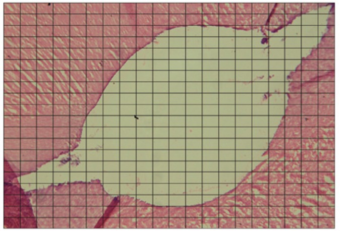
The histological images were blindly analyzed by a single examiner, under an optic microscope (Axiostar Plus, Carl Zeiss, Oberkachen, Germany) at ×64 magnification. An integration grid of 300 points (20 × 15) was superimposed on each image to calculate the total area of the apical third, as well as the percentage of the debris area, which were not removed during biomechanical preparation (Figure 1). The normal distribution of data was tested by the Kolmogorov-Smirnov test for homogeneity of variance. Student's t-test was used to compare the data using the GraphPad Prism 4.0 Software program (GraphPad Software, La Jolla, CA, USA) at a 5% significance level.
Figure 1.

Integration grid inserted onto histological image.
Results
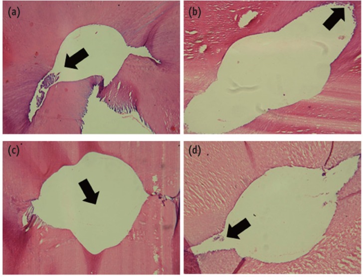
Histological images of Groups 1 and 2 can be seen in Figure 2. The mean values in percentage of the presence of debris in the root canals are shown in Table 1. The results demonstrated that the samples submitted to ultrasonic agitation (Group 2) after biomechanical preparation presented less amount of debris in the root canal than those of Group 1, with statistically significant difference (p < 0.05).
Figure 2.
Histological images from samples of Group 1 (a) and (b) and Group 2 (c) and (d). (a) Remaining pulp tissue in the region of the isthmus (arrow); (b) Debris in the root canal wall (arrow), where the instruments have not touched; (c) Root canal lumen (arrow), which is clean and free of debris; (d) Root canal lumen free of debris, with little remaining pulp tissue in the isthmus area. H. E. stain (×64).
Table 1.
Mean values (%) and standard deviation (SD) of debris remaining after biomechanical preparation

Different superscripts mean statistically significant difference (Student t-test, p < 0.05).
Discussion
The present study evaluated the cleaning capacity of a hybrid instrumentation technique submitted to different final irrigantion protocols. Based on the results obtained, the hypothesis tested was rejected since the protocols presented different behavior regarding debris removal. The biomechanical preparation using rotary Ni-Ti instruments promoted several conceptual changes, such as greater instrumentation efficiency and speed in the preparation of root canals.11 However, it must be consider that improvements obtained with this new technique cannot eliminate the need for irrigation during endodontic therapy.10 Irrigation and adequate choice of irrigant solutions are fundamental for organic material dissolution in areas where the endodontic instruments cannot reach.10,11 The hybrid technique is a combination of instrumentation techniques that uses Ni-Ti rotary systems prior to root canals exploration with manual files, enlarging the cervical and middle thirds, and consequently, leading to a lower concentration of forces on the rotary instruments, promoting better access to the apical foramen.5,12,13,14,15,16 The greatest concern regarding rotary instruments is their sudden fracture, which can occur without any permanent or visible deformation.12 This can be minimized by the technique proposed, as instrumentation is initiated with stainless steel files, which are more resistant than Ni-Ti ones.5,13 Therefore, these aspects justify the use of hybrid instrumentation as it minimizes working time, reduces cost and fatigue of the instruments.5
The use of ultrasound alone is not capable of completing efficiently all operating steps of endodontic treatment. However, it can be considered an important tool to help in the cleaning process, particularly the irrigation process, as it provides a continuous flow of the irrigant solution, improving debris and smear layer removal, and adequate cleaning of the root canal system.17,18,19,20 After shaping the root canal system, cleaning can be complemented by passive ultrasonic irrigation because it is more effective in removing remaining pulp tissue, dentin shavings and planktonic bacteria, as observed in the present study.20 Moreover, passive ultrasonic irrigation alone was not fully effective in cleaning the entire root canal.20 When distilled water was used as irrigant solution, it did not promote effective removal of the smear layer, but the opposite was found when the irrigant solution was 3% sodium hypochlorite. This observation corroborates the importance of using irrigant solutions with potential to dissolve organic material and, rather, provide antibacterial action against microorganisms, both planktonic bacteria, and mature biofilm.20,21 The action of sodium hypochlorite to dissolve organic material was increased when passive ultrasonic irrigation was performed, due to the agitation itself, and the temperature increase promoted by the ultrasonic waves.21 However, increase in temperature is not a concern, because, according to van der Sluis et al., there was an increase of only 0.6℃ on the external surface of the root during continuous agitation at initial temperature of the solution at 20℃.21 Thus, damage to the periodontal ligament and alveolar bone did not occur. Both in the isthmus and irregular areas of the root canal walls, this technique is more effective than conventional irrigation with a syringe, since the waves allow the agitation and penetration of the irrigant solution in a larger area of the root canals, as shown in the results of the present study. Thus, areas untouched by the manual and rotary instruments are affected by the irrigant solutions because agitation will reach the entire root canal system.20,21 Furthermore, the capacity of sodium hypochlorite to dissolve tissue, especially at high concentrations, increases the cleaning effectiveness in the root canal systems when combined with passive ultrasonic irrigation.20
However, several studies have conflicting results regarding smear layer removal probably due to the different concentrations of the irrigant solutions and intracanal activation time.9,10,21,22 There is no consensus regarding the application time of ultrasonic forces in the root canal.23 In the present study, the authors chose to use 60 seconds, and we found that this time was significant to reduce the amount of remaining debris. Since ultrasonic vibration leads to the agitation of the irrigant solution in all directions, it is important to assess whether there is any risk of apical leakage and consequent damage to the periodontal tissues and alveolar bone. However, Tasdemir et al. reported that the risk of apical leakage of the irrigant solution is reduced when passive ultrasound irrigation is used.23
Conclusions
Despite the limitations of the present in vitro study, it may be concluded that passive ultrasonic agitation promoted greater debris removal in the apical third of root canals evaluated, making it an effective adjunct in endodontic treatment.
Acknowledgement
The authors deny any conflicts of interest related to this study. Also, the authors would like to thank to FAPEAM for financial support.
Footnotes
No potential conflict of interest relevant to this article was reported.
References
- 1.Gonçalves LC, Junior EC, da Frota MF, Marques AA, Garcia Lda F. Morphometrical analysis of cleaning capacity of hybrid instrumentation in mesial flattened root canals. Aust Endod J. 2011;37:99–104. doi: 10.1111/j.1747-4477.2010.00255.x. [DOI] [PubMed] [Google Scholar]
- 2.Arruda MP, Carvalho Junior JR, Miranda CE, Paschoalato C, Silva SR. Cleaning of flattened root canals with different irrigating solutions and nickel-titanium rotatory instrumentation. Braz Dent J. 2009;20:284–289. doi: 10.1590/s0103-64402009000400004. [DOI] [PubMed] [Google Scholar]
- 3.Ferreira RB, Alfredo E, Porto de Arruda M, Silva-Sousa YT, Sousa-Neto MD. Histological analysis of the cleaning capacity of nickel-titanium rotatory instrumentation with ultrasonic irrigation in root canals. Aust Endod J. 2004;30:56–58. doi: 10.1111/j.1747-4477.2004.tb00182.x. [DOI] [PubMed] [Google Scholar]
- 4.Sasaki EW, Versiani MA, Perez DE, Sousa-Neto MD, Silva-Sousa YT, Silva RG. Ex vivo analysis of the debris remaining in flattened root canals of vital and nonvital teeth after biomechanical preparation with Ni-Ti rotatory instruments. Braz Dent J. 2006;17:233–236. doi: 10.1590/s0103-64402006000300011. [DOI] [PubMed] [Google Scholar]
- 5.Farid H, Khan FR, Rahman M. ProTaper rotary instrument fracture during root canal preparation: a comparison between rotary and hybrid techniques. Oral Health Dent Manag. 2013;12:50–55. [PubMed] [Google Scholar]
- 6.Nadalin MR, Perez DE, Vansan LP, Paschoala C, Sousa-Neto MD, Saquy PC. Effectiveness of different final irrigation protocols in removing debris in flattened root canals. Braz Dent J. 2009;20:211–214. doi: 10.1590/s0103-64402009000300007. [DOI] [PubMed] [Google Scholar]
- 7.Rasquin LC, de Carvalho FB, Lima RK. In vitro evaluation of root canal preparation using oscillatory and rotary systems in flattened root canals. J Appl Oral Sci. 2007;15:65–69. doi: 10.1590/S1678-77572007000100014. [DOI] [PMC free article] [PubMed] [Google Scholar]
- 8.Kottoor J, Velmurugan N, Gopikrishna V, Krithikadatta J. Effects of multiple root canal usage on the surface topography and fracture of two different Ni-Ti rotary file systems. Indian J Dent Res. 2013;24:42–47. doi: 10.4103/0970-9290.114942. [DOI] [PubMed] [Google Scholar]
- 9.Baratto-Filho F, de Carvalho JR, Jr, Fariniuk LF, Sousa-Neto MD, Pécora JD, da Cruz-Filho AM. Morphometric analysis of the effectivenes of diferent concentrations of sodium hypochlorite associated with rotary instrumentation for root canal cleaning. Braz Dent J. 2004;15:36–40. doi: 10.1590/s0103-64402004000100007. [DOI] [PubMed] [Google Scholar]
- 10.Ribeiro EM, Silva-sousa YT, Souza-Gabriel AE, Sousa-Neto MD, Lorencentti KT, Silva SR. Debris and smear removal in flattened root canals after use of different irrigant agitation protocols. Microsc Res Tech. 2012;75:781–790. doi: 10.1002/jemt.21125. [DOI] [PubMed] [Google Scholar]
- 11.Marchesan MA, Arruda MP, Silva-Sousa YT, Saquy PC, Pecora JD, Sousa-Neto MD. Morphometrical analysis of cleaning capacity using nickel-titanium rotary instrumentation associated with irrigating solutions in mesio-distal flattened root canals. J Appl Oral Sci. 2003;11:55–59. doi: 10.1590/s1678-77572003000100010. [DOI] [PubMed] [Google Scholar]
- 12.da Frota MF, Filho IB, Berbert FL, Sponchiado EC, Jr, Marques AA, Garcia Lda F. Cleaning capacity promoted by motor-driver or manual instrumentation using Protaper Universal system: histological analysis. J Conserv Dent. 2013;16:79–82. doi: 10.4103/0972-0707.105305. [DOI] [PMC free article] [PubMed] [Google Scholar]
- 13.Zmener O, Pameijer CH, Alvarez Serrano S, Hernandez SR. Cleaning efficacy using two engine-driven systems versus manual instrumentation in curved root canals: a scanning electron microscopic study. J Endod. 2011;37:1279–1282. doi: 10.1016/j.joen.2011.05.036. [DOI] [PubMed] [Google Scholar]
- 14.Moura-Netto C, Palo RM, Camargo CH, Pameijer CH, Bardauil MR. Micro-CT assessment of two different endodontic preparation systems. Braz Oral Res. 2013;27:26–30. doi: 10.1590/s1806-83242012005000029. [DOI] [PubMed] [Google Scholar]
- 15.Yoo YS, Cho YB. A comparison of the shaping ability of reciprocating NiTi instruments in simulated curved canals. Restor Dent Endod. 2012;37:220–227. doi: 10.5395/rde.2012.37.4.220. [DOI] [PMC free article] [PubMed] [Google Scholar]
- 16.Lim YJ, Park SJ, Kim HC, Min KS. Comparison of the centering ability of Wave·One and Reciproc nickel-titanium instruments in simulated curved canals. Restor Dent Endod. 2013;38:21–25. doi: 10.5395/rde.2013.38.1.21. [DOI] [PMC free article] [PubMed] [Google Scholar]
- 17.Grecca FS, Garcia RB, Bramante CM, Moraes IG, Bernardineli N. A quantative analyses of rotary ultrasonic and manual techniques to treat proximally flattened root canals. J Appl Oral Sci. 2007;15:89–93. doi: 10.1590/S1678-77572007000200003. [DOI] [PMC free article] [PubMed] [Google Scholar]
- 18.Esposito PT, Cunningham CJ. A comparison of canal preparation with nickel-titanium and stainless steel instruments. J Endod. 1995;21:173–176. doi: 10.1016/S0099-2399(06)80560-1. [DOI] [PubMed] [Google Scholar]
- 19.Arya A, Bali D, Grewal MS. Histological analysis of cleaning efficacy of hand and rotary instruments in the apical third of the root canal: a comparative study. J Conserv Dent. 2011;14:237–240. doi: 10.4103/0972-0707.85797. [DOI] [PMC free article] [PubMed] [Google Scholar]
- 20.Mozo S, Llena C, Forner L. Review of ultrasonic irrigation in endodontics: increasing action of irrigating solutions. Med Oral Patol Oral Cir Bucal. 2012;17:e512–e516. doi: 10.4317/medoral.17621. [DOI] [PMC free article] [PubMed] [Google Scholar]
- 21.van der Sluis LW, Versluis M, Wu MK, Wesselink PR. Passive ultrasonic irrigation of the root canal: a review of the literature. Int Endod J. 2007;40:415–426. doi: 10.1111/j.1365-2591.2007.01243.x. [DOI] [PubMed] [Google Scholar]
- 22.Gu LS, Kim JR, Ling J, Choi KK, Pashley DH, Tay FR. Review of contemporary irrigant agitation techniques and devices. J Endod. 2009;35:791–804. doi: 10.1016/j.joen.2009.03.010. [DOI] [PubMed] [Google Scholar]
- 23.Tasdemir T, Er K, Celik D, Yildirim T. Effect of passive ultrasonic irrigation on apical extrusion of irrigating solution. Eur J Dent. 2008;2:198–203. [PMC free article] [PubMed] [Google Scholar]


