Abstract
Achromatopsia is a progressive autosomal recessive retinal disease characterized by early loss of cone photoreceptors and later rod photoreceptor loss. In most cases, mutations have been identified in CNGA3, CNGB3, GNAT2, PDE6C or PDE6H genes. Owing to this genetic heterogeneity, mutation-independent therapeutic schemes aimed at preventing cone cell death are very attractive treatment strategies. In pde6cw59 mutant zebrafish, cone photoreceptors expressed high levels of receptor-interacting protein kinase 1 (RIP1) and receptor-interacting protein kinase 3 (RIP3) kinases, key regulators of necroptotic cell death. In contrast, rod photoreceptor cells were alternatively immunopositive for caspase-3 indicating activation of caspase-dependent apoptosis in these cells. Morpholino gene knockdown of rip3 in pde6cw59 embryos rescued the dying cone photoreceptors by inhibiting the formation of reactive oxygen species and by inhibiting second-order neuron remodelling in the inner retina. In rip3 morphant larvae, visual function was restored in the cones by upregulation of the rod phosphodiesterase genes (pde6a and pde6b), compensating for the lack of cone pde6c suggesting that cones are able to adapt to their local environment. Furthermore, we demonstrated through pharmacological inhibition of RIP1 and RIP3 activity that cone cell death was also delayed. Collectively, these results demonstrate that the underlying mechanism of cone cell death in the pde6cw59 mutant retina is through necroptosis, whereas rod photoreceptor bystander death occurs through a caspase-dependent mechanism. This suggests that targeting the RIP kinase signalling pathway could be an effective therapeutic intervention in retinal degeneration patients. As bystander cell death is an important feature of many retinal diseases, combinatorial approaches targeting different cell death pathways may evolve as an important general principle in treatment.
Keywords: RIP3, RIP1, Pde6c, retina, zebrafish, achromatopsia
Photoreceptor cell death is a common phenotypic end point associated with inherited retinal degenerations.1 Although in many cases the causative genetic defects are known, the underlying mechanisms by which photoreceptor degeneration and cell death occurs is not fully understood. Previous studies have identified caspase-dependent apoptosis as a critical factor in retinal diseases,1, 2 however, more recently caspase-independent mechanisms of apoptosis have been proposed.3 For example, DNA damage leading to excessive activation of DNA repair enzymes such as poly-ADP-ribose polymerase (Parp) causes an increase in oxidative stress, mitochondrial damage and subsequent apoptotic cell death in the rd1, P23H and S334ter rodent models of retinal degeneration.4, 5 Similarly, activation of the calcium-dependent cysteine protease calpain can result in an increase in reactive oxygen species (ROS), mitochondrial damage and lysosomal membrane disruption leading to apoptotic cell death in the retina.5 Recently, the necroptosis mechanism of cell death6, 7 has been implicated in other types of retinal defects such as retinal detachment,8 retinal ischemia9 and ocular coloboma.10 In these cases, either cell death was delayed by the inhibitor necrostatin-1, which targets receptor interacting protein kinase 1 (RIP1)11 or the presence of elevated receptor interacting protein kinase 3 (RIP3) was detected, suggesting that cell death in the retina maybe dependent on these key regulators of necroptosis.12, 13
Congenital achromatopsia (ACHM) is a progressive autosomal recessive cone disorder14 characterized by photophobia, nystagmus, poor visual acuity, eccentric fixation and severe deficits in color discrimination.15 Complete and incomplete forms of ACHM have been described relating to the severity of the disease. Mutations in five genes (CNGA3, CNGB3, GNAT2, PDE6C, PDE6H) that encode proteins of the phototransduction pathway have been implicated in the pathogenesis of ACHM and result in clinically indistinguishable forms of the disease.16, 17, 18 However, the cause of ACHM in 10–20% patients remains unresolved, implying further heterogeneity. A zebrafish model of human achromatopsia (pde6cw59) has been recently identified displaying rapid degeneration of cone photoreceptors caused by a mutation in the cone phosphodiesterase α-subunit gene (pde6c).19 In this blind mutant, cone outer segments appear to form normally by 3 days post fertilization (d.p.f.), after which there is a rapid loss of cone cells. In addition, genetically normal rod photoreceptors degenerate in the vicinity of the dying cones in the central retina, a so-called bystander effect.20 This bystander cell death effect is seen in human retinal disease21 and mouse models of retinal degeneration,22 and represents a critical challenge to overcome in designing gene replacement strategies for retinal degeneration.
To further evaluate the pde6cw59 mutant as a model for retinal degeneration, and in particular to get a better understanding of the mechanistic framework driving cell death in the different photoreceptor cell types, we investigated caspase-dependent and -independent pathways and their contribution to pathogenesis.
Results
Pde6cw59−/− retinal phenotype
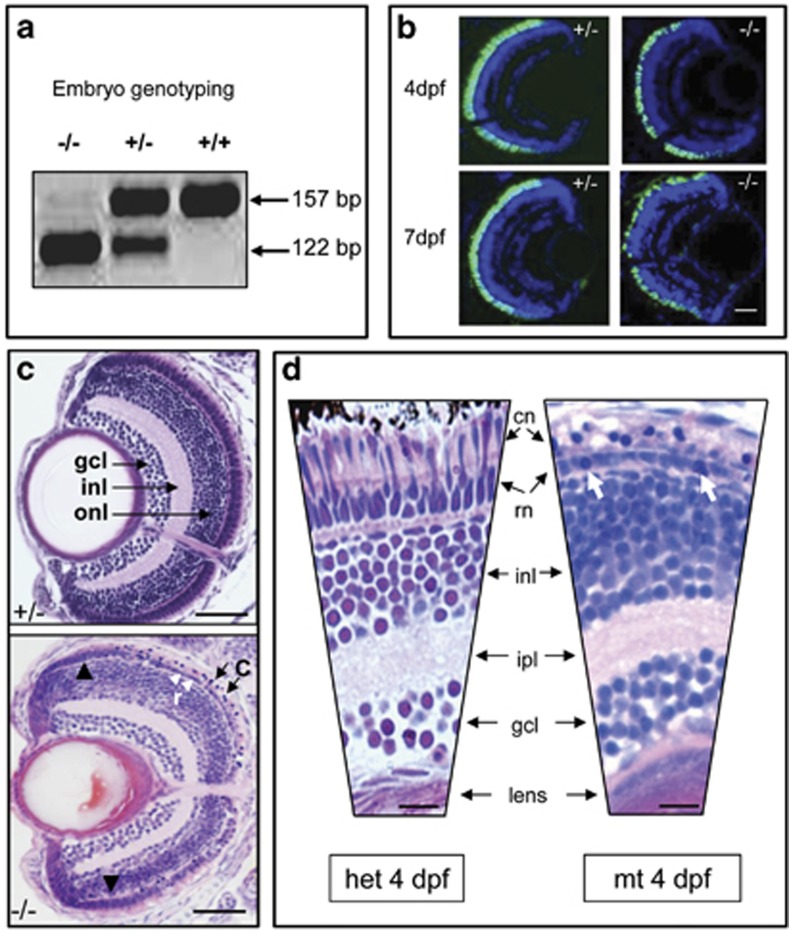
A stable transgenic (Tg) zebrafish line Tg(TαCP:EGFP) expressing enhanced green fluorescent protein (EGFP) in cones cells via a cone transducin promoter fragment23 was crossed to heterozygous carriers of the pde6cw59 mutation to obtain heterozygous Tg(TαCP:EGFP)-pde6cw59 carrier zebrafish. As it not possible to visually distinguish between homozygous and heterozygous Tg(TαCP:EGFP)-pde6cw59 embryos during early development, a simple PCR protocol for genotype analysis from fin DNA was used (Figure 1a). Up to 3 d.p.f., the development of the Tg(TαCP:EGFP)-pde6cw59 mutant retina is equivalent to wild-type/heterozygous eyes; however, from days 4 to 7 there is a decline in the number of Tg GFP-expressing cone photoreceptors (Figure 1b) and condensed cone nuclei are clearly visible by histology (Figure 1c). There is a single layer of rods remaining in the central area of degenerating cones, whereas the newly formed photoreceptors at the peripheral margins are preserved (Figure 1c). In the central retina, some of the rods nuclei are condensed and rounded-up, suggesting that there is an adverse effect on the genotypically normal rod photoreceptors (Figure 1d).
Figure 1.
Genotyping and retinal histology for mutant pde6cw59 zebrafish. (a) Genotyping for mutation status using BsaX1 restriction digest of pde6c PCR amplicon. Wild-type (+/+) band is 157 bp and the mutant (−/−) band is 122 bp. (b) Heterozygous (+/−) and mutant pde6c phenotype on transgenic background of GFP-expressing cone photoreceptors at 4 and 7 days post fertilization (d.p.f.) showing degeneration of cones. Scale bar=20 μm. (c) Comparison of sections through heterozygous (+/−) and mutant (–/–) retinas at 7 d.p.f. C, degenerating cones (black arrows); gcl, ganglion cell layer; inl, inner nuclear layer; onl, outer nuclear layer; r, single layer of rods (white arrowheads); black arrowheads, preserved peripheral retina. Scale bar=50 μm. Dorsal retina is at the top of each panel. (d) Higher magnification of heterozygous (het) versus mutant (mt) retina at 4 d.p.f. cn, cone nuclei; ipl, inner plexiform layer; rn, rod nuclei. Scale bar=10 μm. White arrows: condensed rod nuclei
Cell death in the retina
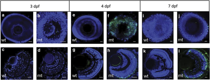
As there is rapid cone degeneration in the pde6cw59 mutant retina, we examined larvae from 3 to 7 d.p.f. using terminal deoxynucleotidyl transferase dUTP nick end labeling (TUNEL) staining as a marker for cell death on the non-Tg background. At 3 d.p.f., very few TUNEL-positive cells were detected either in whole-mount wild-type or mutant eyes (Figures 2a and b) or in sections through the retina (Figures 2c and d). At 4 d.p.f. during the height of cone degeneration, many TUNEL-positive cells were observed in whole-mount mutant eyes compared with wild-type (Figures 2e and f) and this was also seen in sections through the retina (Figures 2g and h). At 7 d.p.f., there were still TUNEL-positive cells present in the mutant whole-mount eyes compared with wild type (Figures 2i and j) and in corresponding retinal sections (Figures 2k and l).
Figure 2.
Time course of TUNEL staining in whole-mount and retinal sections. Representative images of whole-mount eyes and corresponding sections at the denoted ages of 3, 4 or 7 days post fertilization (d.p.f.). (a, b, e, f, i, k) Whole-mount eyes. (c, d, g, h, k, l) Retinal sections. mt, pde6cw59−/− mutant; wt, wild type. Scale bar is 20 μm. TUNEL-positive cells, green labelling; 4′,6-Diamidino-2-phenylindole counterstain for cell nuclei
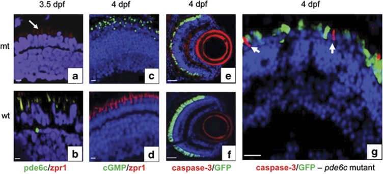
Although we observed a high level of TUNEL-positive cells corresponding to the time at which there is most cone degeneration at 4 d.p.f., this does not inform on the underlying mechanism of cell death. We initially confirmed the molecular and biochemical abnormalities in this model and then examined markers of caspase-dependent and caspase-independent cell death. In the pde6cw59 mutant retina at 3.5 d.p.f., there were still some double cones present detected by zebrafish double-cone photoreceptor antibody (zpr1) immunohistochemistry (Figure 3a), but they were not immunoreactive for pde6c (compare Figures 3a and b) confirming the underlying genetic defect. Based on other phosphodiesterase mutants, mutation of the pde6c gene would be predicted to lead an accumulation of cyclic guanosine monophosphate (cGMP).24 We observed a dramatic accumulation of cGMP in the pde6cw59 mutant retina at 4 d.p.f., demonstrating the biochemical consequence of pde6c gene mutation (compare Figures 3c and d) and also the absence of zpr1 cone photoreceptor staining in the mutant (Figure 3c). If cone cell death was mediated through a caspase-dependent mechanism, then we would expect to see activation of caspase-3. At 4 d.p.f., activated caspase-3 labelling was only present in the Tg(TαCP:EGFP)-pde6cw59 mutant retina (compare Figures 3e and f), and at higher resolution, caspase-3 labelling was specifically in rod cells and no co-localization with GFP-expressing cones (Figure 3g). This suggested that caspase-dependent cell death was not contributing to cone photoreceptor cell death in the mutant retina, but rather to the dying rod photoreceptors.
Figure 3.
Expression of cell death markers in the retinal sections. (a, c, e, g) Mutant pde6c (mt) retinal images. (b, d, f) Wild-type (wt) retinal images. (a and b) Double immunostaining with zpr1 cone-specific marker in red and pde6c in green. (c and d) Double immunostaining for cGMP labelling in green and zpr1 in red. (e and f) Activated caspase-3 localization in red on GFP-expressing cone transgenic background. (g) Higher magnification of retina in image (e) showing no overlap of caspase-3 red staining in GFP-expressing cones (green). All sections counterstained with DAPI. Scale bar is 5 μm, except in e and f, where it is 20 μm
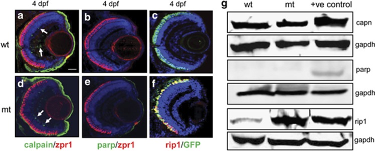
As a number of different caspase-independent mechanisms of cell death have been reported in rodent models of retinal degeneration, we tested specific markers for these processes in the pde6cw59 zebrafish. In the pde6cw59 mutant retina, calpain activity was not detected in cone photoreceptors, although a low level of expression was observed in a few cells of the inner retina of both wild-type and mutant eyes at 4 d.p.f. (Figures 4a and d). Interestingly, the developing cornea in both wild-type and mutant eyes showed high levels of expression of calpain. Similarly, Parp staining was not present in the retina of either wild-type or mutant eyes (Figures 4b and e), neither was LC3 (mammalian homolog of Atg8 autophagy protein) staining25 for autophagic vacuoles (data not shown). In contrast, high levels of rip1 staining co-localized with the dying GFP-expressing cone photoreceptor cells (Figure 4f). Confirmation of the immunohistochemical data was carried out by western blot analysis, which demonstrated that calpain levels were similar in both wild-type and mutant samples, whereas Parp levels were not detectable in either wild-type or mutant samples (Figure 4g). However, there was a sixfold increase in rip1 protein in the mutant eyes compared with wild-type levels (Figure 4g). These data suggested that the major cell death pathway activated in cone photoreceptors in the pde6cw59 mutant was necroptosis.
Figure 4.
Expression patterns of caspase-independent cell death markers. (a–c) Wild-type (wt) retinal images. (d–f) Mutant pde6c-/- (mt) retinal images. (a and d) Double labelling of calpain in green and zpr1 cone-specific marker in red. A few cells express calpain in the inner retina (white arrows). Scale bar=20 μm. (b ande) Double labelling of parp in green and zpr1 in red. (c and f) rip1 co-labelling in red on GFP-expressing cone transgenic background. (g) Western blot of calpain (capn), parp and rip1 at 7 d.p.f. Positive (+ve) controls: for parp and capn, mouse E14.5 eye tissue; for rip1, HEK293 cell line. Gapdh used to normalize gel loading
Rip3-dependent cone photoreceptor cell death
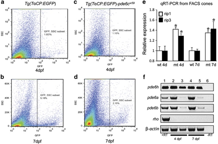
As RIP3 protein is the key activator of necroptosis, but available RIP3 antibodies do not recognize zebrafish rip3 protein, we took advantage of fluorescent-activated cell sorting (FACS) from wild-type embryos on the Tg background and Tg(TαCP:EGFP)-pde6cw59 mutant embryos to isolate populations of GFP-positive cone cells (Figures 5a–d). From 300 embryos at 4 d.p.f. and 150 embryos at 7 d.p.f., we obtained approximately 70 000–100 000 EGFP-positive cells that were zpr1 positive and isolated 250 ng of cone photoreceptor total RNA. Real-time reverse transcriptase-PCR (RT-PCR) analysis of rip1 and rip3 at 4 and 7 d.p.f. revealed that both genes were significantly upregulated in the mutant retina at both time points (Figure 5e, P<0.05, n=9).
Figure 5.
Flow cytometry sorting of fluorescent cone photoreceptors for gene expression analysis. (a and b) FACS plots for transgenic Tg(TαCP:EGFP) embryos that are wild type at the pde6c+/+ locus showing the side scatter (SSC) and EGFP profiles. (c and d) FACS plots for mutant Tg(TαCP:EGFP)-pde6cw59−/− cone photoreceptors. (e) Quantification of real-time RT-PCR expression data for rip1 and rip3 from EGFP-positive cells at 4 and 7 d.p.f. in photoreceptors from wild-type (wt) and mutant (mt) embryos. Data presented as mean±S.E.M. *P<0.05, n=9. (f) Relative RT-PCR analysis for rod photoreceptor genes (pde6a, pde6c, rho), cone pde6h gamma subunit and β-actin internal control from whole adult retinal RNA (lanes 1 and 6); FACS-sorted mutant cone RNA (lanes 2 and 4); FACS-sorted wild-type cone RNA (lanes 3 and 5). RT, reverse transcriptase
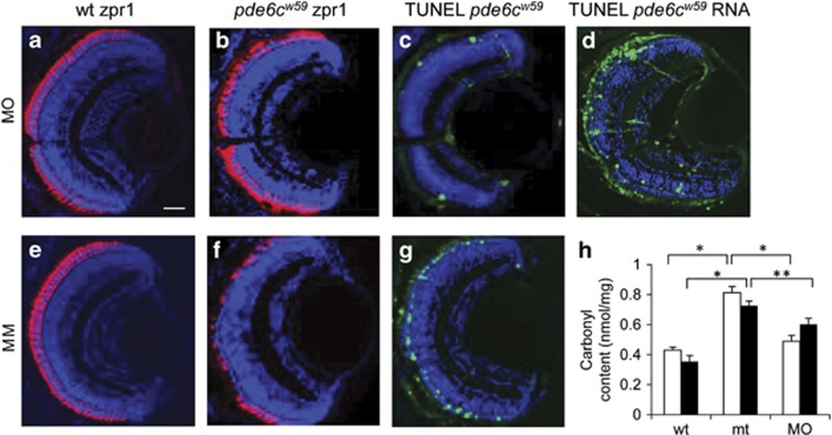
As rip3 expression was upregulated, we reasoned that morpholino knockdown of rip3 in the mutant embryos could prevent necroptosis in the dying cones cells. Injection of either rip3 antisense or mismatch morpholinos into wild-type embryos had no effect on wild-type zpr1 cone-specific staining (Figures 6a and e). When mutant pde6cw59 embryos were injected with an antisense morpholino to rip3, we found that cone cell staining was similar to wild-type embryos when analyzed by zpr1 immunohistochemistry (Figure 6b) compared with mismatch control injections, where a reduction in zpr1 cone cell labelling was clearly evident (Figure 6f). TUNEL assays in rip3 morphants demonstrated significantly fewer TUNEL-positive cells in the retina (Figure 6c) compared with the mismatch control injected embryos (Figure 6g; Supplementary Figure 1). Furthermore, when rip3 RNA was co-injected with the rip3 morpholino, it reversed the TUNEL staining levels (Figure 6d) to that seen in mismatch-treated pde6cw59 larvae (Figure 6g), showing specificity of the morpholino. As Rip3 can promote necroptosis through the production of ROS in the mitochondria,26 we examined carbonyl adduct formation derived from protein oxidation in pde6cw59 mutant and rip3 morphant pde6cw59 larvae. At both 4 and 7 d.p.f., the carbonyl content in pde6cw59 embryos was significantly increased (P<0.0001; n=8) compared with wild-type eyes (Figure 6h). However, in rip3 morphants, the carbonyl content production was significantly reduced compared with pde6cw59 mutant eyes (P<0.001; n=8). This demonstrates that activation of necroptosis leading to increased ROS production is rip3-dependent in the pde6cw59 mutant and that rip3 deficiency suppresses ROS production and consequently oxidative damage to the retina.
Figure 6.
Phenotype at 4 d.p.f. after morpholino knockdown of rip3 in wild-type (wt) and pde6cw59 mutant (mt) embryos. (a–d) rip3 morpholino (MO) injection. (e–g) rip3 mismatch morpholino (MM) injection. (a and e) Control injections of MO or MM in wt embryos. Zpr1 cone-specific labelling in red. Scale bar=20 μm. (b) rip3 MO knockdown in mt embryos showing increased zpr1 labelling in red. (c) TUNEL labelling in rip3 MO knockdown mt embryos. (d) TUNEL labelling in mt embryos co-injected with rip3 MO and rip3 RNA. (f) rip3 MM knockdown in mt embryos showing decreased zpr1 labelling in red. (g) TUNEL labelling in mt embryos injected with rip3 MM. (h) ELISA quantitation of carbonyl content in wt, mt and MO-injected embryos at 4 d.p.f. (white bars) or 7 d.p.f. (black bars). Mean±S.E.M., n=8, *P<0.0001; **P<0.001
As we found cone cell death to be mediated through the necroptosis pathway, we next asked what signalling mechanisms could lead to activation of necroptosis. cGMP-dependent protein kinase G (PKG) is one of the major cellular targets for cGMP in the retina,27 therefore, we examined the expression of pkg1 in mutant pde6cw59 retina and found this gene was upregulated from 3 d.p.f. (Supplementary Figure 2a). These results support a recent report showing that PKG was increased in a mouse model of retinal degeneration.28 In addition, as Rip3 is known to be induced by tumor necrosis factor α (TNFα),29 we examined the spatiotemporal expression pattern of TNFα in mutant pde6cw59 retina. TNFα expression began at 3 d.p.f. before cone cell death (Supplementary Figure 3) and was highly elevated at 4 d.p.f., co-incident with increased Rip1 upregulation (Supplementary Figure 3) and Rip3 upregulation (Supplementary Figure 2b).
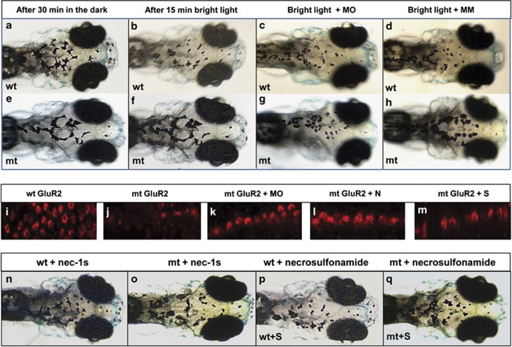
To determine whether the retina was functionally responsive to light after rip3 knockdown, we examined the visual background adaptation (VBA) response in morphant larvae. VBA is a neuroendocrine response used for camouflage where dark-adapted fish disperse their melanin granules (Figure 7a) and thus appear darker, whereas light-adapted fish aggregate their melanin into small granules (Figure 7b) and therefore appear more transparent.30 As this response is driven through the retino-thalamic tract, blind fish are unable to exhibit this response (Figures 7e and f). After 30 min of dark adaptation followed by 15 min of bright light exposure, 7 d.p.f. wild-type larvae injected with either rip3 morpholino or the mismatch control display small melanin granules as expected (Figures 7c and d). In contrast, pde6cw59 mutants injected with the mismatch control morpholino display larger diffuse melanin granules indicating visual impairment (Figure 7h). However, in rip3 morphant pde6cw59 larvae, the melanin granules have reverted to the small wild-type appearance (Figure 7g), demonstrating that the VBA response is normalized. These data confirm that cone cell death in the pde6cw59 mutants is rip3 dependent and that knockdown of rip3 remarkably restores the responsiveness of the retina to light.
Figure 7.
Visual background adaptation (VBA) in response to bright light stimulation at 7 d.p.f. Upper panels, wild-type (wt) embryos; lower panels, pde6cw59 mutant (mt) embryos. Representative images showing the distribution of melanin after dark adaptation (a and e); after light adaptation (b and f); after rip3 morpholino (MO) injection (c and g); after mismatch control morpholino (MM) injection (d and h). (a and b) Images taken from the same wild-type larva before and after light adaptation. (e and f) Taken in the same mutant larva before and after light adaptation. (i) Labelling of GluR2 receptors in wt retina (i); mt retina (j); in rip3 morphant retina (k); in retina treated with necrostatin-1s (l); in retina treated with necrosulfonamide (m). VBA response in wt (n) or mt larvae (o) treated with necrostatin-1s (N) showing small pigment granules. VBA response in wt (p) or mt larvae (q) treated with necrosulfonamide (S) showing small pigment granules
How this recovery of response to light occurs seems initially counterintuitive, as rip3 knockdown would not replace the absence of pde6c α-subunit protein required for phototransduction. Furthermore, pde6cw59 mutants also display significant remodelling of second-order neurons (horizontal cells)31 that could limit communication with the inner retina and thereby contribute to functional visual deficits. However, recently an elegant study in a mouse model lacking cone Pde6c showed that cones expressed the rod photoreceptor Pde6a/b subunit proteins and that these subunits were used instead for cone phototransduction.32 Therefore, the expression of rod phototransduction genes in FACS Tg(TαCP:EGFP) wild-type and mutant cones was examined. We found that wild-type cones expressed very low levels of pde6a and pde6b, whereas mutant cones had highly upregulated levels of these two genes at both 4 and 7 d.p.f. (Figure 5f). Rhodopsin, which is a rod-specific gene, is not expressed in the FACS RNA samples, indicating that no contaminating rods were present. Presence of the cone-specific gamma subunit (pde6h) verified the presence of cone photoreceptor cells in all samples. Furthermore, we examined the expression of two photoreceptor-specific transcription factors crx and nrl, which are required to drive expression of pde6a and pde6b33 and found they were expressed in mutant cone cells (Supplementary Figure 4). At 7 d.p.f. when nrl expression decreases in wild-type cones, it remains more highly expressed in mutant cones (Supplementary Figure 4). These results suggested that the VBA response observed in rip3 morphant pde6cw59 larvae most likely have occurred through upregulation of compensatory rod subunit proteins. To determine whether rip3 knockdown was involved in remodelling of the inner retina, we examined immunolabelling for glutamate receptor subunit 2 (GluR2), which detects glutamate receptors on horizontal cell processes that synapse with rods and cones. In wild-type retina, GluR2-labelled horizontal cells synapsing with cone pedicles in a rosette-like cluster in the outer plexiform layer (Figure 7i), whereas in pde6cw59 mutant retina only punctate staining of rod spherules was apparent (Figure 7j). In rip3 morphants, the GluR2 rosette staining pattern was present but there were fewer cones than in wild-type eyes (Figure 7k). In the presence of either necrostain-1s (Figure 7l) or necrosulfonamide (Figure 7m), rosette staining for GluR2 was present, suggesting that second-order remodelling in the inner plexiform layer was a direct consequence of rip3 signalling.
Pharmacological inhibition of cone and rod photoreceptor cell death
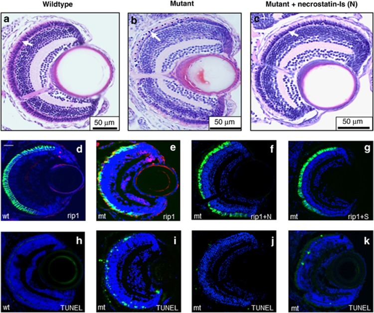
As RT-PCR revealed that both rip1 and rip3 were upregulated in pde6cw59 mutant cone photoreceptors, we took advantage of small-molecule drugs that target specifically RIP134 and RIP3 kinases35 as potential pharmacological approaches to inhibit cone cell death. Using dose-response analysis in wild-type zebrafish,10 the highest concentration of necrostatin-1s (RIP1 inhibitor) and necrosulfonamide (RIP3 inhibitor) that could be utilized without causing toxicity to embryos was 100 and 20 μM, respectively. When Tg(TαCP:EGFP)-pde6cw59 mutant embryos were treated with 100 μM necrostatin-1s from 10 h.p.f. to 7 d.p.f., the outer retina was preserved resembling wild-type retina, with only occasional condensed nuclei present (Figures 8a–c). Rip1 labelling of cone cells in necrostatin-1s-treated mutants was not detected (Figure 8f) as in wild-type retina (Figure 8d), compared with untreated embryos where intense rip1 labelling co-localized with EGFP-expressing cone cells (Figure 8e). Only occasional TUNEL-positive cells were present in treated embryos (Figure 8j) compared with untreated mutants (Figure 8i). Rip1 labelling was also absent in embryos treated with 20 μM necrosulfonamide used to inhibit rip3 (Figure 8g) with a concomitant reduction in TUNEL staining (Figure 8k) compared with untreated embryos. These data establish that pharmacological targeting of Rip1 and Rip3 kinases inhibits cone photoreceptor cell death in the pde6cw59 zebrafish model. To test if the pharmacologically treated embryos were also responsive to light, VBA analysis revealed that in the presence of either necrostatin-1s (Figure 7o) or necrosulfonamide (Figure 7q) melanin granule contraction was normalized compared with untreated larvae (Figure 7f).
Figure 8.
Effect of RIP signalling inhibitors on pde6cw59 mutant phenotype at 7 d.p.f. Retinal histology in (a) wild-type (wt), (b) untreated mutant (mt) and (c) mt retina from embryos treated with 100 mM necrostatin-1s (N). White arrows in a–c indicate the photoreceptor nuclear layer of the retina. Rip1 labelling (red) in wt retina (d), in mt retina co-localizing with EGFP-expressing cones that are dying (e), and in mt retina treated with necrostatin-1s (f). (g) rip1 labelling in the retina of pde6cw59 mutant embryos treated with 20 μM necrosulfonamide (S). TUNEL labelling in non-transgenic pde6cw59 mutant embryos (h–k) corresponding to the treatment panels (d–g) above. Scale bar in d=20 μm
Discussion
In this study, we report that cone-photoreceptor cell death in the retina of the blind pde6cw59 model of retinal degeneration occurs through a rip3-dependent pathway and that either genetic or pharmacological inhibition of necroptosis effectors rescues cone cell death and restores the responsiveness of the retina to light. In addition, we show that in genetically normal rod photoreceptors there is bystander cell death mediated through a caspase-dependent mechanism. Our data may provide an explanation of why using single inhibitors of cell death have had limited rescue effects and poor long-term outcomes when tested in other retinal degeneration models,36, 37 because the rod and cone photoreceptors are dying by different mechanisms so eventually all the photoreceptors are lost. The significance of our findings is that combination therapy directed at the different cell death mechanisms would be crucial in designing effective treatment strategies.
A surprise finding in the study was that the visual response in pde6cw59 larvae was restored by rip3 knockdown, despite the absence of a key phototransduction protein (pde6c). Recently, several studies have suggested that the linear rod and cone phototransduction pathways can in fact couple to one another in disease states. For instance, in a genetically derived all-cone retina mouse model lacking cone Pde6c (Nrl−/−;cpfl1), it was demonstrated that the cones instead expressed the rod Pde6 catalytic subunits conferring phototransduction capability to the cones.32 Similarly, in the rd10 mouse (which lacks the rod Pde6β gene), the cone Pde6α′ subunit was transgenically expressed in the mutant rd10 rods and this rescued the response to light, demonstrating that cone Pde6α′ can functionally substitute for Pde6αβ.38 These studies suggest that the rod and cone Pde6 catalytic subunits can functionally substitute for one another and our work further implies that the local environment can determine rod and cone coupling. Although the exact mechanism by which rip3 knockdown leads to upregulation of pde6a and pde6b remains to be determined, our data suggest that maintenance of high levels of the nrl transcription factor in cones is crucial to this process. We also found that the inner retina remodeling is inhibited when rip3 is knocked down, which suggests that these secondary changes are directly related to cone cell death.
Although the exact mechanism by which activation of the RIP signalling pathway leads to cell death remains to be fully characterized, upregulation of RIP1 and RIP3 in cones cells provides evidence that the necroptosis pathway is the major cause of cone photoreceptor loss in pde6c-deficient zebrafish retina. Identification of the substrates for RIP1 and RIP3 kinases may lead to identification of further key effectors of necroptosis. Recent studies have revealed that increased RIP3 stimulates the production of ROS39 because of increased oxidative phosphorylation and it is thought to result in a catastrophic overload to the metabolic state of the cell.29 Our studies showing overproduction of ROS in the mutant pde6cw59 retina supports this downstream mechanism, suggesting that inhibition of oxidative stress may be a valid approach for inhibiting cone photoreceptor cell death in this model. Targeting the reduction of superoxide-free radicals using antioxidants such as N-acetylcysteine has been found to protect cone photoreceptors from bystander oxidative stress in mouse models of rod photoreceptor degeneration.40
Further understanding of the detailed molecular mechanisms leading to necroptosis could reveal new targets that maybe developed as therapeutic interventions for many necroptosis-related diseases. Recently, four specific substrates for RIP3 have been identified (death-domain-associated protein 6 (Daxx), phosphoglycerate mutase family member 5 (PGAM5), DNA-dependent activator of IRF and sirtuin 2 (SIRT2)) as well as activation of a number of biochemical processes that are critical for necroptosis, which could each represent potential targets for inhibiting RIP3-dependent necroptosis in retinal degeneration. (i) RIP3-dependent phosphorylation of Daxx results in its nuclear export and subsequent retinal ganglion cell death upon a retinal ischemic injury insult.41 In RIP3-depleted cells, phosphorylation and nuclear export of Daxx is prevented thereby inhibiting retinal cell death. Thus, manipulating the nuclear trafficking of Daxx might be a possible option. (ii) The mitochondrial phosphatase PGAM5 was identified as a RIP3 substrate in HT-29 human cells,42 and these enzyme complexes with RIP1/RIP3/mixed lineage kinase-like in the necrosome. Inhibitors of PGAM5 might therefore prevent execution of necroptosis. (iii) DNA-dependent activator of IRF is thought to act as a DNA sensor that induces interferon-1 production and interacts with RIP3 during viral infection-induced necroptotic cell death,43 so would most likely be beneficial for retinal degeneration associated with an immune response. (iv) Most recently, SIRT2 has been shown to regulate the deacetylation of RIP1, which is required for stable RIP1–RIP3 complex formation through RIP homotypic interaction motifs, and this deacetylation is RIP3-dependent process.44 Using a small-molecule inhibitor of SIRT2 (known as AKG2), inhibition of dopaminergic neuronal cell death in a Drosophila model of Parkinson's disease has been demonstrated,45 and could therefore be relevant for retinal degeneration.
Other biochemical activities that have been implicated in necroptotic cell death, including activation of phospholipase activity,46 phosphorylation of signal transducer and activator of transcription 347 and mitochondrial membrane pore opening, regulated through cyclophilin D.48 Very recently, involvement of cyclophilin D in necroptosis has been shown to be RIP3-dependent and that it can be inhibited by cyclosporin A.49 This suggests that the neuroprotective effect provided by cyclosporin A that has been used previously to treat rats with diabetic retinopathy50 and that can prevent the loss of therapeutic cell transplants from dying in the retina51 could be in part the result of inhibiting RIP3-dependent necroptosis rather than due to its immunosuppressive activity alone.
A clearer picture is now emerging regarding photoreceptor cell death in retinal degenerative disorders. Rod cell death in animal models of retinitis pigmentosa was originally thought to be mainly caused by apoptotic mechanisms, however, there are now examples of activation of alternative cell death pathways contributing to the pathogenesis.3 Neuroprotection strategies targeting apoptotic rod cell death in animal models have proved effective,52 although such strategies have yet to be translated into effective clinical treatments.53 This may be in part due to the observation that if the caspase-dependent cell death pathway is blocked then necroptosis is activated as a second line of defense.11, 54 Less is known regarding cone cell death, however, loss of rod-derived growth factors, autophagy and oxidative stress are key contributors.4, 55 As bystander defects are a major challenge for designing treatment strategies for retinal degeneration,20 the different cell death mechanisms in the retina need to be identified, so that appropriate combinatorial strategies can be proposed. This is now possible based on a more sophisticated classification of cell death and an improvement in ways to detect the underlying cell signalling mechanisms.
The use of combination therapy in clinical ophthalmology is firmly established. For instance, treatment of recalcitrant glaucoma with combinations of drugs targeting different pathways provides synergistic effectiveness over individual treatment modalities.56 We recently used a combinatorial approach of aminoglycoside read-through to treat the underlying genetic defect in the rat S334ter model coupled with glial-derived neurotrophic factor neuroprotection, which fully rescued the retinal degeneration, whereas individually we found that these treatments had limited efficacy.57 In this study, we have shown that RIP3-dependent necroptosis is the underlying molecular cause of cone cell death in the pde6cw59 model, whereas rod cells are dying via caspase-dependent apoptosis. Thus, development of effective treatment strategies for retinal degeneration affecting both rods and cones requires components that target both the primary genetic deficit and the secondary bystander cell loss.
Materials and Methods
Zebrafish maintenance and genotyping
Research was authorized by the University of British Columbia Animal Care Committee in accordance with the principles and guidelines of the Canadian Council of Animal Care and the Association for Research in Vision and Ophthalmology statement for the use of animals in vision research. Wild-type AB, pde6cw59 19 and Tg[TαCP:GFP]23 zebrafish strains were maintained as inbred stocks and staged according to morphological criteria. To visualize dying cone cells, we crossed the pde6cw59 mutation onto the Tg[TαCP:GFP] background where the cone α-transducin promoter drives GFP expression in all cone photoreceptors. Embryos were raised at 28.5 °C on a 14-h light/10-h dark cycle in 100 mm2 Petri dishes containing standard embryonic medium 2 or embryonic medium 3 medium. High concentrations of paramecia (3–10 × normal) were required to raise pde6cw59 mutant embryos to adulthood as the larvae do not normally survive past 10 d.p.f.. To aid image analysis where required, 0.2 mM phenylthiourea (Sigma-Aldrich, Oakville, ON, Canada) was added to the embryos at 10 h post fertilization (h.p.f.) to inhibit pigment formation. Genomic DNA was isolated from caudal fin tissue using a hot sodium hydroxide methodology.58 After PCR amplification (pde6c primers: Forward: 5′-ttggcctctggaatactggctctc-3′ reverse: 5′-gtttgaccagaacccggaag-3′), the 157 bp products were digested with BsaX1 enzyme to determine genotype.
Histology, immunohistochemistry and TUNEL assay
Zebrafish embryos were fixed by immersion in 4% paraformaldehyde overnight at 4 °C. Fixed embryos were washed in phosphate-buffered saline (PBS) and then dehydrated through a graded ethanol series (50, 70, 90 and 100%), before being transferred to xylene and embedded in paraffin wax. Hematoxylin and eosin staining was carried out on 3–5 μm thickness wax sections. For immunohistochemistry, embryos were infiltrated with 30% sucrose overnight at 4 °C before embedding in Polyfreeze medium (Polysciences, Warrington, PA, USA). Immunolabelling was carried out using 6- to 10-μm thick frozen sections. The following primary antibodies were used: Pde6c polyclonal (1 : 50, Abcam, Eugene, OR, USA); Zpr-1 monoclonal (1 : 200, Zebrafish International Resource Center, Eugene, OR, USA). Zpr1 is a zebrafish cone-specific monoclonal antibody that was raised against zebrafish retinal extracts); cGMP polyclonal (1 : 200, Abcam); cleaved Parp polyclonal (1 : 200, Cell Signaling Technology, Danvers, MA, USA); calpain polyclonal (1 : 100, Novus Biologicals, Oakville, ON, Canada); cleaved caspase-3 monoclonal (1 : 200, Cell Signaling Technology); RIP1 polyclonal (1 : 100, Abcam); GluR2 monoclonal (1 : 75, Millipore, Billerica, MA, USA); Tnf-α. polyclonal (1 : 30, Anaspec, Fremont, CA, USA). Secondary antibodies used were: Alexa Fluor 488 goat anti-rabbit immunoglobulin G or Alexa Fluor 546 goat anti-mouse immunoglobulin G (Life Technologies, Burlington, ON, Canada). For cGMP staining, the larvae were incubated in PBS with the phosphodiesterase inhibitor 3-isobutyl-1-methylxanthine (0.8 mM) for 10 min at 4 °C before fixation. Sections were incubated overnight at 4 °C with primary antibody diluted in blocking buffer. After extensive washes in PBS-Tween 20, localization of antibody labelling was detected by after 1 h incubation with secondary antibodies diluted in PBS containing 2% normal goat serum. Nuclei were counter stained with 4′,6-diamidino-2-phenylindole and then images acquired using scanning laser microscopy. Transmitted light microscopy was used to capture images using a Zeiss Axioplan 2 microscope with D63ZNC DVC camera (Carl Zeiss, Toronto, ON, Canada). TUNEL staining in whole-mount eyes or retinal tissue sections was carried out as previously described.10 Images were acquired using a Zeiss 510 laser scanning confocal microscope (Carl Zeiss).
Immunoblotting
Briefly, 25 wild-type or mutant embryo eyes at 4 d.p.f. were snap frozen in liquid nitrogen and homogenized by sonication in lysis buffer (10 mM Tris pH 7.5, 10 mM NaCl, 1% SDS, 1 × Protease Inhibitor Cocktail (Roche, Mississauga, ON, Canada)). Insoluble material was removed by a 10-min centrifugation (25 000 × g). Protein concentration was determined by the DC protein assay (Bio-Rad, Hercules, CA, USA). Proteins (40 μg) were separated on a 10% SDS-polyacrylamide gel, transferred to Immobilon-FL membrane (Millipore), blocked in 5% non-fat milk powder in PBS/0.1% Tween-20 (PBST) for 2 h at RT and incubated overnight at 4 °C with primary antibody in the same buffer and diluted according to manufacturer's instructions. Following three washes in PBST, the membrane was incubated in the dark for 1 h with a Li-COR secondary antibody (IRDye 680LT goat anti-rabbit; Mandel Scientific, Guelph, ON, Canada), washed three times in PBST in the dark and protein bands were visualized using a Li-COR Odyssey detector (Mandel Scientific). NIH Image J software was used to quantify band intensities relative to glyceraldehyde-3-phosphate dehydrogenase (GAPDH) or β-actin loading controls (1 : 2000 GAPDH mouse monoclonal).
FACS for cones and qRT-PCR
To purify cone photoreceptors, we took advantage of FACS according to from previously published methods.59 Briefly, 150–300 Tg pde6c+/+ and Tg pde6cw59−/− embryos were dechorionated, euthanized in tricaine and the eyes removed. Eyes were incubated in 1 × trypsin-EDTA solution for 7 min and then the reaction stopped by addition of 5% heat-inactivated FBS (Sigma-Aldrich). After centrifugation at 200 × g for 7 min, the pellet was incubated with FACSmax cell dissociation solution (AMS Biotechnology, Lake Forest, CA, USA) for 10 min at 37 °C and then a 1-ml syringe plunger was used to carefully dissociate the cells. Cells were filtered through a 40-μm mesh cell strainer and then fluorescent sorted with a flow cytometer (BD Biosciences Influx Sorter; BD Biosciences, Mississauga, ON, Canada). RNA was isolated from the sorted cells using an RNeasy Micro kit (Qiagen, Mississauga, ON, Canada) and quantitated by NanoDrop spectrometry (Thermo Scientific, Wilmington, DE, USA). cDNA was amplified using QuantiTect Reverse Transcription Kit (Qiagen) and relative gene expression of rip1 (NM_001043350), rip3 (ENSDARG00000090965) and gapdh (NM_001115114) was quantified using the TaqMan primer and labelled probe protocol and the ViiA 7 Real-Time PCR system (Applied Biosystems, Burlington, ON, Canada). Gene primers are listed in Supplementary Table 1. All reactions were performed using the TaqMan Universal Master Mix (2X), FAM-labelled TaqMan gene expression assays for rip1 and rip3, VIC-labelled TaqMan endogenous control gapdh and 5 ng of cDNA. Thermocycling parameters were as follows: 2 min at 50 °C, 10 min at 95 °C, 40 cycles of 15 s at 95 °C, plus 60 s at 60 °C. Real-time PCR data were analyzed by the comparative CT method. Each reaction was undertaken on three occasions, and for each of these, individual samples were subdivided into three aliquots for measurement.
Morpholino knockdown and RNA rescue
Morpholino sequence directed against the transcriptional start site of zebrafish rip3 was designed by GeneTools: 5′-AGCGCATCTCTCCGCCGTCCATCGC-3′ (rip3 MO) and mismatch (rip3 MM) 5′-AGCCCATGTCTCCCCCCTCCATCCC-3′ and were modified at the 3′end with carboxyfluorescein. Morpholinos were injected into the yolk of one or two cell stage wild-type or pde6c mutant embryos as follows: 2.5 ng each of rip3 MO or rip3 MM oligonucleotides (±p53 morpholino to take into account off-target effects) were injected in 1 × Danieau buffer (58 mM NaCl, 0.7 mM KCl, 0.4 mM MgSO4, 0.6 mM Ca (NO3)2, 5 mM HEPES, pH 7.6) with 0.1% phenol red using a Nanoject II variable injector (Drummond Scientific, Broomall, PA, USA). The morphant embryos were grown at 28.5 °C and observed for morphological changes under a stereoscopic microscope and fixed in 4% paraformaldehyde at 4 or 7 d.p.f. For RNA rescue experiments, 15 ng rip3 RNA was co-injected with 2.5 ng rip3 morpholino. Zebrafish rip3 5′-capped mRNA was synthesized as previously described,10 and primers for cloning the full-length cDNA is in Supplementary Table 1.
Detection of ROS
For each time point (4 or 7 d.p.f.), 150 eyes were obtained from wild-type, pde6cw59 mutant or rip3-knockdown pde6cw59 embryos. Protein extracts from eyes were isolated by adding 100 μl of lysis buffer (50 mM Tris-HCl buffer pH 7.4, 0.1 mM EDTA, 0.1 mM ethylene glycol tetraacetic acid, 1 mM phenylmethyl sulfonyl fluoride, 2 μM bestatin, 1 μM pepstatin, 2 μM leupeptin) and then sonicating samples for five cycles (30 s on–30 s off), followed by 5 × freeze-thawing. Lysates were centrifuged at 10 000 × g for 10 min at 4 °C and protein concentration in lysate supernatants was determined using the DC Protein assay (Bio-Rad). Quantitation of carbonyl adducts of proteins in 10 μg eye extracts was determined using the OxiSelect Protein Carbonyl ELISA kit (Cell Biolabs, Inc., San Diego, CA, USA), according to manufacturer's instructions.
VBA assay
After morpholino injection, embryos were incubated at 28.5 C for 7 d.p.f. Embryos were then transferred to a Petri dish and placed inside a box for dark-adaptation lasting 30 min. After placing the dish on the base of the microscope stand, a bright light was shined onto them from above. Images were taken as soon as the light was switched on and again 15 min later to allow for full contraction of their melanophores as previously described.60
Drug efficacy and treatment
After dechorionation of wild-type embryos at 10 h.p.f., a range of doses of necrostatin-1s (Biovision, Milpitas, CA, USA) or necrosulfonamide (Toronto Research Chemicals, Inc., Toronto, ON, Canada) were added directly to the aquarium water and then embryos were raised to 6 d.p.f. Each day the aquaria water containing drugs was refreshed. For each drug dose, 20 embryos were used in three independent experiments. The number of embryos surviving at 6 days was counted and quantitative data were expressed as mean±S.E.M. Pde6cw59 mutant embryos were treated with either 100 μM necrostatin-1i or 20 μM necrosulfonamide from 10 h.p.f. with assessment of the phenotypic effect at 7 d.p.f. by histology and immunocytochemistry.
Acknowledgments
This work was supported by the Canadian Institutes of Health Research Team Grant (222728). We thank Dr. Susan Brockerhoff at University of Washington, USA, for kindly providing the pde6cw59 and Tg[TαCP:GFP] zebrafish lines and Dr. Anat Yanai for advice on FACS analysis.
Glossary
- cGMP
cyclic guanosine monophosphate
- CNGA3
cyclic nucleotide gated channel α3
- CNGB3
cyclic nucleotide gated channel β3
- Daxx
death-domain-associated protein 6
- EGFP
enhanced green fluorescent protein
- GAPDH
glyceraldehyde-3-phosphate dehydrogenase
- GluR2
glutamate receptor subunit 2
- GNAT2
guanine nucleotide binding protein, alpha transducing 2
- H&E
hematoxylin and eosin
- Nrl
neural retina-specific leucine zipper
- PBS
phosphate-buffered saline
- Pde6c
cone-specific cGMP phosphopdiesterase α-subunit
- PDE6H
cone-specific cGMP phosphopdiesterase γ-subunit
- PGAM5
phosphoglycerate mutase family member 5
- Rip1
receptor-interacting protein kinase 1
- Rip3
receptor-interacting protein kinase 3
- RT-PCR
reverse transcriptase-PCR
- STAT3
signal transducer and activator of transcription 3
- TαCP
α subunit of cone transducin
- Tg
transgenic
- TNFα
tumor necrosis factor α
- TUNEL
terminal deoxynucleotidyl transferase dUTP nick end labeling
- zpr1
zebrafish double-cone photoreceptor antibody
- Parp
poly-ADP-ribose polymerase
- d.p.f.
days post fertilization
- h.p.f
hours post fertilization
- FACS
fluorescent-activated cell sorting
- PKG
protein kinase G
- VBA
visual background adaptation
- SIRT2
sirtuin 2
The authors declare no conflict of interest.
Footnotes
Supplementary Information accompanies this paper on Cell Death and Differentiation website (http://www.nature.com/cdd)
Edited by N Bazan
Supplementary Material
References
- Chang GQ, Hao Y, Wong F. Apoptosis: final common pathway of photoreceptor cell death in rd, rds and rhodopsin mutant mice. Neuron. 1993;11:595–605. doi: 10.1016/0896-6273(93)90072-y. [DOI] [PubMed] [Google Scholar]
- Marigo V. Programmed cell death in retinal degeneration: targeting apoptosis in photoreceptors as potential therapy for retinal degeneration. Cell Cycle. 2007;6:652–655. doi: 10.4161/cc.6.6.4029. [DOI] [PubMed] [Google Scholar]
- Sanch-Pelluz J, Arango-Gonzalez B, Kustermann S, Romero FJ, van Veen T, Zrenner E, et al. Photoreceptor cell death mechanisms in inherited retinal degeneration. Mol Neurobiol. 2008;38:253–269. doi: 10.1007/s12035-008-8045-9. [DOI] [PubMed] [Google Scholar]
- Pacquet-Durand F, Silva J, Talukdar T, Johnson LE, Azadi S, van Veen T, et al. Excessive activation of poly(ADP-ribose) polymerase contributes to inherited photoreceptor degeneration in the retinal degeneration mouse 1. J Neurosci. 2007;27:10311–10319. doi: 10.1523/JNEUROSCI.1514-07.2007. [DOI] [PMC free article] [PubMed] [Google Scholar]
- Kaur J, Mencl S, Sahaboglu A, Farinelli P, van Veen T, Zrenner E, et al. Calpain and PARP activation during photoreceptor cell death in P23H and S334ter rhodopsin mutant rats. PLoS One. 2011;6:e22181. doi: 10.1371/journal.pone.0022181. [DOI] [PMC free article] [PubMed] [Google Scholar]
- Holler N, Zaru R, Micheau O, Thome M, Attinger A, Valitutti S, et al. Fas triggers an alternative, caspase-8-independent cell death pathway using RIP as effector molecule. Nat Immunol. 2000;1:489–495. doi: 10.1038/82732. [DOI] [PubMed] [Google Scholar]
- Hitomi J, Christofferson DE, Ng A, Yao J, Degterev A, Xavier RJ, et al. Identification of a molecular signaling network that regulates a cellular necrotic cell death pathway. Cell. 2008;135:1311–1323. doi: 10.1016/j.cell.2008.10.044. [DOI] [PMC free article] [PubMed] [Google Scholar]
- Trichonas G, Murakami Y, Thanos A, Morizane Y, Kayama M, Debouck CM, et al. Receptor interacting protein kinases mediate retinal detachment-induced photoreceptor necrosis and compensate for inhibition of apoptosis. Proc Natl Acad Sci USA. 2010;107:21695–21700. doi: 10.1073/pnas.1009179107. [DOI] [PMC free article] [PubMed] [Google Scholar]
- Huang JF, Shang L, Zhang MQ, Wang H, Chen D, Tong JB, et al. Differential neuronal expression of receptor interacting protein 3 in rat retina: involvement in ischemic stress response. BMC Neurosci. 2013;14:16. doi: 10.1186/1471-2202-14-16. [DOI] [PMC free article] [PubMed] [Google Scholar]
- Viringipurampeer IA, Ferreira T, DeMaria S, Yoon JJ, Shan X, Moosajee M, et al. Pax2 regulates a fadd-dependent molecular switch that drives tissue fusion during eye development. Hum Mol Genet. 2012;21:2357–2369. doi: 10.1093/hmg/dds056. [DOI] [PMC free article] [PubMed] [Google Scholar]
- Degterev A, Hitomi J, Germscheid M, Ch'en IL, Korkina O, Teng X, et al. Identification of RIP1 kinase as a specific cellular target of necrostatins. Nat Chem Biol. 2008;4:313–321. doi: 10.1038/nchembio.83. [DOI] [PMC free article] [PubMed] [Google Scholar]
- He S, Wang L, Miao L, Wang T, Du F, Zhao L, et al. Receptor interacting protein kinase-3 determines cellular necrotic response to TNF-alpha. Cell. 2009;137:1100–1111. doi: 10.1016/j.cell.2009.05.021. [DOI] [PubMed] [Google Scholar]
- Cho YS, Challa S, Moquin D, Genga R, Ray TD, Guildford M, et al. Phosphorylation-driven assembly of the RIP1-RIP3 complex regulates programmed necrosis and virus-induced inflammation. Cell. 2009;137:1112–1123. doi: 10.1016/j.cell.2009.05.037. [DOI] [PMC free article] [PubMed] [Google Scholar]
- Thiadens AA, Somervuo V, van den Born LI, van Schooneveld MJ, Kuijpers RW, van Moll-Ramirez N, et al. Progressive loss of cones in achromatopsia: an imaging study using spectral-domain optical coherence tomography. Invest Ophthamol Vis Sci. 2010;51:5952–5957. doi: 10.1167/iovs.10-5680. [DOI] [PubMed] [Google Scholar]
- Pokorny J, Smith VC, Pinckers AJ, Cozijnsen M. Classification of complete and incomplete autosomal recessive achromatopsia. Graefes Arch Clin Exp Ophthalmol. 1982;219:121–130. doi: 10.1007/BF02152296. [DOI] [PubMed] [Google Scholar]
- Thiadens AA, Slingerland NW, Roosing S, van Schooneveld MJ, van Lith-Verhoeven JJ, van Moll-Ramirez N, et al. Genetic etiology and clinical consequences of complete and incomplete achromatopsia. Ophthalmology. 2009;116:1984–1989. doi: 10.1016/j.ophtha.2009.03.053. [DOI] [PubMed] [Google Scholar]
- Thiadens AA, den Hollander AI, Roosing S, Nabuurs SB, Zekveld-Vroon RC, Collin RW, et al. Homozygosity mapping reveals PDE6C mutations in patients with early-onset cone photoreceptor disorders. Am J Hum Genet. 2009;85:240–247. doi: 10.1016/j.ajhg.2009.06.016. [DOI] [PMC free article] [PubMed] [Google Scholar]
- Kohl S, Coppieters F, Meire F, Schaich S, Roosing S, Brennenstuhl C, et al. A nonsense mutation in PDE6H causes autosomal recessive incomplete achromatopsia. Am J Hum Genet. 2012;91:527–532. doi: 10.1016/j.ajhg.2012.07.006. [DOI] [PMC free article] [PubMed] [Google Scholar]
- Stearns G, Evangelista M, Fadool JM, Brockerhoff SE. A mutation in the cone-specific pde6 gene causes rapid cone photoreceptor degeneration in zebrafish. J Neurosci. 2007;27:13866–13874. doi: 10.1523/JNEUROSCI.3136-07.2007. [DOI] [PMC free article] [PubMed] [Google Scholar]
- Ripps H. Cell death in retinitis pigmentosa: gap junctions and the ‘bystander' effect. Exp Eye Res. 2002;74:327–336. doi: 10.1006/exer.2002.1155. [DOI] [PubMed] [Google Scholar]
- Evans K, Duvall-Young J, Fitzke FW, Arden GB, Bhattacharya SS, Bird AC. Chromosome 19q cone-rod retinal dystrophy, Ocular phenotype. Arch Ophthalmol. 1995;113:195–201. doi: 10.1001/archopht.1995.01100020079033. [DOI] [PubMed] [Google Scholar]
- Delyfer MN, Léveillard T, Mohand-Saïd S, Hicks D, Picaud S, Sahel JA. Inherited retinal degenerations: therapeutic prospects. Biol Cell. 2004;96:261–269. doi: 10.1016/j.biolcel.2004.01.006. [DOI] [PubMed] [Google Scholar]
- Kennedy BN, Alvarez Y, Brockerhoff SE, Stearns GW, Sapetto-Rebow B, Taylor MR, et al. Identification of a zebrafish cone photoreceptor-specific promoter and genetic rescue of achromatopsia in the nof mutant. Invest Ophthalmol Vis Sci. 2007;48:522–529. doi: 10.1167/iovs.06-0975. [DOI] [PubMed] [Google Scholar]
- Farber DB, Lolley RN. Cyclic guanosine monophosphate: elevation in degenerating photoreceptor cells of the C3H mouse retina. Science. 1974;186:449–451. doi: 10.1126/science.186.4162.449. [DOI] [PubMed] [Google Scholar]
- Yokoyama T, Miyazawa K, Naito M, Toyotake J, Tauchi T, Itoh M, et al. Vitamin K2 induces autophagy and apoptosis simultaneously in leukemia cells. Autophagy. 2008;4:629–640. doi: 10.4161/auto.5941. [DOI] [PubMed] [Google Scholar]
- Vanlangenakker N, Vanden Berghe T, Bogaert P, Laukens B, Zobel K, Deshayes K, et al. cIAP1 and TAK1 protect cells from TNF-induced necrosis by preventing RIP1/RIP3-dependent reactive oxygen species. Cell Death Diff. 2011;18:656–665. doi: 10.1038/cdd.2010.138. [DOI] [PMC free article] [PubMed] [Google Scholar]
- Paquet-Durand F, Hauck SM, van Veen T, Ueffing M, Ekström P. PKG activity causes photoreceptor cell death in two retinitis pigmentosa models. J Neurochem. 2009;108:796–810. doi: 10.1111/j.1471-4159.2008.05822.x. [DOI] [PubMed] [Google Scholar]
- Xu J, Morris L, Thapa A, Ma H, Michalakis S, Biel M, et al. cGMP accumulation causes photoreceptor degeneration in CNG channel deficiency: evidence of cGMP cytotoxicity independently of enhanced CNG channel function. J Neurosci. 2013;33:14939–14948. doi: 10.1523/JNEUROSCI.0909-13.2013. [DOI] [PMC free article] [PubMed] [Google Scholar]
- Zhang DW, Shao J, Lin J, Zhang N, Lu BJ, Lin SC, et al. RIP3, an energy metabolism regulator that switches TNF-induced cell death from apoptosis to necrosis. Science. 2009;325:332–336. doi: 10.1126/science.1172308. [DOI] [PubMed] [Google Scholar]
- Balm PH, Groneveld D. The melanin-concentrating hormone system in fish. Ann NY Acad Sci. 1998;839:205–209. doi: 10.1111/j.1749-6632.1998.tb10760.x. [DOI] [PubMed] [Google Scholar]
- Saade CJ, Alvarez-Delfin K, Fadool JM. Rod photoreceptors protect from cone degeneration-induced retinal remodelling and restore visual responses in zebrafish. J Neurosci. 2013;35:1804–1814. doi: 10.1523/JNEUROSCI.2910-12.2013. [DOI] [PMC free article] [PubMed] [Google Scholar]
- Kolandaivelu S, Chang B, Ramamurthy V. Rod phosphodiesterase-6 (PDE6) catalytic subunits restore cone function in a mouse model lacking cone PDE6 catalytic subunit. J Biol Chem. 2011;286:33252–33259. doi: 10.1074/jbc.M111.259101. [DOI] [PMC free article] [PubMed] [Google Scholar]
- Nelson SM, Frey RA, Wardwell SL, Stenkamp DL. The developmental sequence of gene expression within the rod photoreceptor lineage in embryonic zebrafish. Dev Dyn. 2008;237:2903–2917. doi: 10.1002/dvdy.21721. [DOI] [PMC free article] [PubMed] [Google Scholar]
- Takahashi N, Duprez L, Grootjans S, Cauwels A, Nerinckx W, DuHadaway JB, et al. Necrostatin-1 analogues: critical issues on the specificity, activity and in vivo use in experimental disease models. Cell Death Dis. 2012;3:e437. doi: 10.1038/cddis.2012.176. [DOI] [PMC free article] [PubMed] [Google Scholar]
- Sun L, Wang H, Wang Z, He S, Chen S, Liao D, et al. Mixed lineage kinase domain-like protein mediates necrosis signalling downstream of RIP3 kinase. Cell. 2012;148:213–217. doi: 10.1016/j.cell.2011.11.031. [DOI] [PubMed] [Google Scholar]
- Drack AV, Dumitrescu AV, Bhattarai S, Gratie D, Stone EM, Mullins R, et al. TUDCA slows retinal degeneration in two different mouse models of retinitis pigmentosa and prevents obesity in Bardet-Biedl syndrome type 1 mice. Invest Ophthalmol Vis Sci. 2012;53:100–106. doi: 10.1167/iovs.11-8544. [DOI] [PMC free article] [PubMed] [Google Scholar]
- Komeima K, Rogers BS, Campochiaro PA. Antioxidants slow photoreceptor cell death in mouse models of retinitis pigmentosa. J Cell Physiol. 2007;213:809–815. doi: 10.1002/jcp.21152. [DOI] [PubMed] [Google Scholar]
- Deng W-T, Sakurai K, Kolandaivelu S, Kolesnikov AV, Dinculescu A, Li J, et al. Cone phosphodiesterase-6α' restores rod function and confers distinct physiological properties in the rod phosphodiesterase-6β-deficient rd10 mouse. J Neurosci. 2013;33:11745–11753. doi: 10.1523/JNEUROSCI.1536-13.2013. [DOI] [PMC free article] [PubMed] [Google Scholar]
- Goossens V, Grooten J, De Vos K, Fiers W. Direct evidence for tumor necrosis factor-induced mitochondrial reactive oxygen intermediates and their involvement in cytotoxicity. Proc Natl Acad Sci USA. 1995;92:8115–8119. doi: 10.1073/pnas.92.18.8115. [DOI] [PMC free article] [PubMed] [Google Scholar]
- Lee SY, Usui S, Zafar AB, Oveson BC, Jo YJ, Lu L, et al. N-acetylcysteine promotes long-term survival of cones in a model of retinitis pigmentosa. J Cell Physiol. 2011;226:1843–1849. doi: 10.1002/jcp.22508. [DOI] [PubMed] [Google Scholar]
- Lee YS, Dayma Y, Park MY, Kim KI, Yoo SE, Kim E. Daxx is a key downstream component of receptor interacting protein kinase 3 mediating retinal ischemic cell death. FEBS Lett. 2013;587:266–271. doi: 10.1016/j.febslet.2012.12.004. [DOI] [PubMed] [Google Scholar]
- Wang Z, Jiang H, Chen S, Du F, Wang X. The mitochondrial phosphatase PGAM5 functions at the convergence points of multiple necrotic cell death pathways. Cell. 2012;148:228–243. doi: 10.1016/j.cell.2011.11.030. [DOI] [PubMed] [Google Scholar]
- Upton JW, Kaiser WJ, Mocarski ES. DAI/ZBP1/DLM-1 complexes with RIP3 to mediate virus-induced programmed necrosis that is targeted by murine cytomegalovirus vIRA. Cell Host Microbe. 2012;11:290–297. doi: 10.1016/j.chom.2012.01.016. [DOI] [PMC free article] [PubMed] [Google Scholar]
- Narayan N, Lee IH, Borenstein R, Sun J, Wong R, Tong G, et al. The NAD-dependent deacetylase SIRT2 is required for programmed necrosis. Nature. 2012;492:199–204. doi: 10.1038/nature11700. [DOI] [PubMed] [Google Scholar]
- Outeiro TF, Kontopoulos E, Altmann SM, Kufareva I, Strathearn KE, Amore AM, et al. Sirtuin 2 inhibitors rescue α-synuclein-mediated toxicity in models of Parkinson's disease. Science. 2007;317:516–519. doi: 10.1126/science.1143780. [DOI] [PubMed] [Google Scholar]
- De Valck D, Vercammen D, Fiers W, Beyaert R. Differential activation of phospholipases during necrosis or apoptosis: a comparative study using tumor necrosis factor and anti-Fas antibodies. J Cell Biochem. 1998;71:392–399. [PubMed] [Google Scholar]
- Shulga N, Pastorino JG. GRIM-19 mediated translocation of STAT3 to mitochondria is necessary for TNF induced necroptosis. J Cell Sci. 2012;125:2995–3003. doi: 10.1242/jcs.103093. [DOI] [PMC free article] [PubMed] [Google Scholar] [Retracted]
- Nakagawa T, Shimizu S, Watanabe T, Yamaguchi O, Otsu K, Yamagata H, et al. Cyclophilin D-dependent mitochondrial permeability transition regulates some necrotic but not apoptotic cell death. Nature. 2005;434:652–658. doi: 10.1038/nature03317. [DOI] [PubMed] [Google Scholar]
- Tian W, Xu D, Han W, He H, Cai H, Chen H, et al. Cyclophilin D modulates cell death transition from early apoptosis to programmed necrosis induced by honokiol. Int J Oncol. 2013;42:1654–1663. doi: 10.3892/ijo.2013.1863. [DOI] [PubMed] [Google Scholar]
- Krügel K, Wurm A, Pannicke T, Hollborn M, Karl A, Wiedemann P, et al. Involvement of oxidative stress and mitochondrial dysfunction in the osmotic swelling of retinal glial cells from diabetic rats. Exp Eye Res. 2011;92:87–93. doi: 10.1016/j.exer.2010.11.007. [DOI] [PubMed] [Google Scholar]
- West EL, Pearson RA, Barker SE, Luhmann UF, Maclaren RE, Barber AC, et al. Long-term survival of photoreceptors transplanted into the adult murine neural retina requires immune modulation. Stem Cells. 2010;28:1997–2007. doi: 10.1002/stem.520. [DOI] [PMC free article] [PubMed] [Google Scholar]
- Bode C, Wolfrum U. Caspase-3 inhibitor reduces apoptotic photoreceptor cell death during inherited retinal degeneration in tubby mice. Mol Vis. 2003;9:144–150. [PubMed] [Google Scholar]
- Talcott KE, Ratnam K, Sundquist SM, Lucero AS, Lujan BJ, Tao W, et al. Longitudinal study of cone photoreceptors during retinal degeneration and in response to ciliary neurotrophic factor treatment. Invest Ophthalmol Vis Sci. 2011;52:2219–2226. doi: 10.1167/iovs.10-6479. [DOI] [PMC free article] [PubMed] [Google Scholar]
- Vercammen D, Brouckaert G, Denecker G, Van de Craen M, Declercq W, Fiers W, et al. Dual signaling of the Fas receptor: initiation of both apoptotic and necrotic cell death pathways. J Exp Med. 1998;188:919–930. doi: 10.1084/jem.188.5.919. [DOI] [PMC free article] [PubMed] [Google Scholar]
- Léveillard T, Mohand-Saïd S, Lorentz O, Hicks D, Fintz AC, Clérin E, et al. Identification and characterization of rod-derived cone viability factor. Nat Genet. 2004;36:755–759. doi: 10.1038/ng1386. [DOI] [PubMed] [Google Scholar]
- Gheith ME, Mayer JR, Siam GA. Monteiro de Barros DS, Thomas TL, Katz LJ. Managing refractory glaucoma with a fixed combination of bimatoprost (0.03%) and timolol (0.5%) Clin Ophthalmol. 2008;2:15–20. doi: 10.2147/opth.s1175. [DOI] [PMC free article] [PubMed] [Google Scholar]
- Gregory-Evans K, Po K, Francis Chang F, Gregory-Evans CY. Pharmacological enhancement of ex vivo gene therapy neuroprotection in a rodent model of retinal degeneration. Ophthal Genet. 2012;47:32–38. doi: 10.1159/000325730. [DOI] [PubMed] [Google Scholar]
- Meeker ND, Hutchinson SA, Ho L, Trede NS. Method for isolation of PCR-ready genomic DNA from zebrafish tissues. Biotechniques. 2007;43:610–614. doi: 10.2144/000112619. [DOI] [PubMed] [Google Scholar]
- Manoli M, Driever W.Fluorescence-activated cell sorting (FACS) of fluorescently tagged cells from zebrafish larvae for RNA isolation Cold Spring Harb Protoc 2012. e-pub ahead of print 1 August 2012;doi: 10.1101/pdb.prot069633 [DOI] [PubMed]
- Fleisch VC, Neuhauss SC. Visual behavior in zebrafish. Zebrafish. 2006;3:191–201. doi: 10.1089/zeb.2006.3.191. [DOI] [PubMed] [Google Scholar]
Associated Data
This section collects any data citations, data availability statements, or supplementary materials included in this article.


