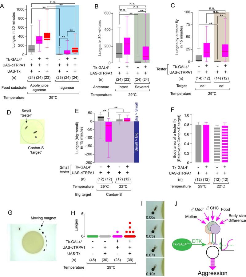
Figure 7. Activation of Tk-GAL41 neurons overrides the absence of aggression-promoting cues.
(A-C) Number of lunges during thermogenetic activation of Tk-GAL41 neurons, (A) on an apple juice agarose (left) or pure agarose (right) substrate; (B) with intact (left) or surgically removed (right) antennae; and (C) toward oe+ (left) or oe− (right) “target” males.
(D) Example image of a normal-sized “target” Canton-S male and a small “tester” male fly (70% the size of the target fly).
(E) Lunge number difference between smaller tester and larger target flies. Values in the shaded area indicate that the smaller testers performed more lunges than the larger targets.
(F) Relative body sizes (mean ± S.D.) of “tester”vs. target flies.
(G) Image of the “moving magnet” setup.
(H) Number of lunges (scatter plot) toward the magnet during thermogenetic activation of Tk-GAL41 neurons. Total recording time per session was 4 minutes (see Experimental Procedures).
(I) Example of a lunge performed by a male Tk-GAL41, UAS-dTRPA1; UAS-Tk fly toward a magnet.
(J) Schematic illustrating possible influence of Tk-GAL4FruM neurons in the male fly brain in relation to the processing of sensory cues regulating aggression.
For (A-C) and (E), ** p < 0.01, * p < 0.05 (Kruskal-Wallis and post-hoc Mann-Whitney U-tests). For (F), n.s.: p > 0.05 (one-way ANOVA and post hoc Student's t-test.
Also see Supplemental Movie S3.

