Dear Editor,
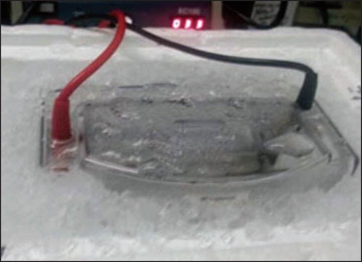
The article entitled ‘Western Blot: Technique, Theory, and Trouble Shooting’ was helpful and provided a detailed protocol for all stages of Western blotting.[1] However, there are concerns regarding Figure 1, which depicts the submersion of an electrophorator including power leads, in an ice water bath. This arrangement is a serious health and safety hazard due to the posed risk of electric shock and is therefore not recommended.
Figure 1.

A submersed electrophorator. Figure 7 from ‘Western Blot: Technique, Theory, and Trouble Shooting’.[1]
Although Western blotting involves passing an electric current through a liquid environment, it is not a dangerous technique if performed properly; however, it is easy to overlook the risks of each stage. Protein extraction often uses β-mercaptoethanol and sodium dodecyl sulfate (SDS), both of which are corrosive, acutely toxic and the former mutagenic. To limit risk they should be handled with care, in a fume hood with the use of personal protective equipment. Polyacrylamide gel electrophoresis (PAGE) is only unsafe if the equipment is not used correctly. Some tanks allow the unit to be plugged into the power pack without covering the buffer. The buffer should be covered at all times when attached to the power supply to prevent contact with the electrified liquid. The protein transfer stage should carry equal risks to PAGE, although many labs adopt the method depicted in Figure 1 of the aforementioned article,[1] which introduces further possibility of electric shock.
There are many safer methods for transferring proteins to a membrane.[2] Electroelution (wet, semi-dry, and dry) and diffusion transfer techniques all have their merits and applications, but it is important to note that method alone will not guarantee a successful blot. Effective protein transfer is also heavily reliant on the gel acrylamide percentage, the molecular weight of electrophoresed proteins, and the blotting membrane used.[3] A number of membranes have been developed in the past, but only nitrocellulose and polyvinylidene fluoride (PVDF) remain popular. PVDF offers a higher binding capacity than nitrocellulose,[4] but likewise has higher background binding.[1] PVDF is also physically stronger than nitrocellulose,[4] so may be preferable for stripping and reprobing.
The diffusion method is not widely utilized as it provides qualitative rather than quantitative transfer,[2] but is especially useful in producing up to 12 blots per SDS-PAGE gel for screening with multiple antibodies.[5] The technique involves sandwiching a single gel between two membranes and clamping it between glass plates to facilitate diffusion.[2] The process is repeated up to six times to produce up to 12 blots, as desired.[3]
Dry transfer is becoming increasingly popular due to the Life Technologies’ iBlot® systems, which boast a blotting time of only 7 min. The company claims their iBlot® systems produce superior protein transfer quality in comparison to wet and semi-dry methods.[6] Little data currently exists to support these claims.
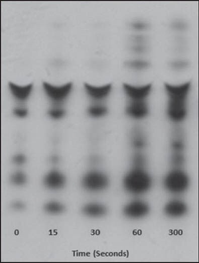
Semi-dry transfer involves soaking up to six layers of gel, membrane, and filter paper in transfer buffer and sandwiching them between two horizontal plate electrodes.[7] It is slower than dry blotting, but is generally more rapid and efficient than wet transfer,[8] and is especially suited to low molecular weight proteins.[3] Other advantages over wet transfer include more cost efficient electrodes, less complicated power packs, and the ability to blot several gels simultaneously.[4] Figure 2 shows the final product of western blotting using semi-dry protein transfer.
Figure 2.

A western blot of collagen-induced platelet activation over time using semi-dry protein transfer
Wet transfer involves immersing a gel-membrane sandwich in an upright tank of transfer buffer, usually with vertical platinum electrodes.[2] The ‘conventional’ wet transfer technique employs the use of a cooling system to reduce heat produced by the electrodes, although over the years modifications have been developed. One modification, the heat-mediated blotting method, is preferable over the conventional protocol due to enhanced transfer of both high and low molecular weight proteins.[9] The heated buffer is thought to increase permeability of the polyacrylamide gel, thus facilitating faster protein transfer.[3] Still if use of the ‘conventional’ wet transfer method is desirable, there are a number of companies that offer specialized wet transfer equipment. These include built-in cooling systems that eliminate the need for ice immersion. If this equipment is unavailable, the tank can be wrapped in ice packs and placed into a cold room, otherwise if an ice bath is absolutely necessary, ensure the level of ice is below that of the electrical components.
Successful protein blots can be produced via a range of methods, all of which have advantages and disadvantages, yet regardless of the technique, safety should always be a priority.
References
- 1.Mahmood T, Yang PC. Western Blot: Technique, theory, and trouble shooting. N Am J Med Sci. 2012;4:429–34. doi: 10.4103/1947-2714.100998. [DOI] [PMC free article] [PubMed] [Google Scholar]
- 2.Kurien BT, Scofield RH. Western blotting. Methods. 2006;38:283–93. doi: 10.1016/j.ymeth.2005.11.007. [DOI] [PubMed] [Google Scholar]
- 3.MacPhee DJ. Methodological considerations for improving Western blot analysis. J Pharmacol Toxicol Methods. 2010;61:171–7. doi: 10.1016/j.vascn.2009.12.001. [DOI] [PubMed] [Google Scholar]
- 4.Kurien BT, Scofield RH. Protein blotting: A review. J Immunol Methods. 2003;274:1–15. doi: 10.1016/s0022-1759(02)00523-9. [DOI] [PubMed] [Google Scholar]
- 5.Kurien BT, Scofield RH. Multiple immunoblots after non-electrophoretic bidirectional transfer of a single SDS–PAGE gel with multiple antigens. J Immunol Methods. 1997;205:91–4. doi: 10.1016/s0022-1759(97)00052-5. [DOI] [PubMed] [Google Scholar]
- 6.The iBlot® Dry Blotting System vs. Conventional Semi-Dry and Wet Transfer Systems. Paisley, UK. Life Technologies. 2013. [Accessed December 17, 2013]. at http://www.lifetechnologies.com/uk/en/home/life-science/proteinexpression-and-analysis/western-blotting/western-blot-transfer/iblot-dry-blottingsystem/iblot-dry-blottingcomparison-to-semi-dry-and-wet.html .
- 7.Kurien B, Scofield R. Introduction to Protein Blotting. In: Kurien B, Scofield R, editors. Protein blotting and detection. Vol. 3. New York: Humana Press; 2009. pp. 9–21. [Google Scholar]
- 8.Wisdom GB. Protein blotting. Methods Mol Biol. 1994;32:207–13. doi: 10.1385/0-89603-268-X:207. [DOI] [PubMed] [Google Scholar]
- 9.Kurien BT, Scofield RH. Heat-mediated, ultra-rapid electrophoretic transfer of high and low molecular weight proteins to nitrocellulose membranes. J Immunol Methods. 2002;266:127–33. doi: 10.1016/s0022-1759(02)00103-5. [DOI] [PubMed] [Google Scholar]


