
Figure 4. High content screening for assaying cytotoxic events triggered by nanoparticle interaction.
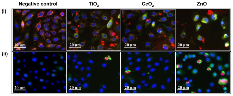
(i) BEAS-2B cells subjected to nanoparticles, stained with nucleic acid staining Hoechst 33342 and mitochondrial dye JC1. Healthy cells shows blue nuclei and red mitochondria (Negative control, TiO2 and CeO2) while the mitochondrial depolarization causes the cytoplasm to fluoresce green (green cells in ZnO). (ii) BEAS-2B cells treated with nanopaticles and stained with a dye cocktail of Hoechst 33342, fluo-4 (for intracellular Ca2+) and propidium iodide (for accessing membrane damage). Healthy cells shows blue nuclei, while damaged cells shows green cytoplasm and red nuclei (cells in ZnO group). Depending upon the color profile, the percentage of cells affected can be assayed and nanoparticles can be ranked for their cytotoxic potential.