Abstract
A 58-year-old Caucasian male presented to the urology clinic reporting an approximate one-year history of a persistent irritating, slowly progressive, glans penis redness. Biopsy revealed penile squamous cell carcinoma in situ. He underwent a partial glansectomy with circumcision and skin grafting. At three months follow-up there is no evidence of local disease recurrence. In western countries, primary malignant penile cancer is uncommon, with an incidence of less than 1 per 100,000 males. Squamous cell cancer accounts for more than 95% of cases of penile cancer. Squamous cell carcinoma in situ on the penile mucosa or transitional surfaces is also known as Erythroplasia of Queyrat. In the region, one third of penile squamous cell carcinoma in situ cases progress to invasive squamous cell carcinoma.
Key words: penile carcinoma in situ, penile cancer.
Case Report
A 58-year-old Caucasian male presented to the urology clinic reporting an approximate one-year history of a persistent irritating, slowly progressive, glans penis redness. The redness was refractory to topical steroid and anti-fungal treatments prescribed by his general practitioner. He had no lower urinary tract symptoms and was in excellent general health.
On clinical examination his abdomen was soft and non-tender. There was no palpable inguinal lymphadenopathy. Scrotal examination revealed normal testes. A distinct smooth, raised area of velvety appearing erythema, measuring approximately 3×3 cm, was visible over the glans penis involving the external urethral meatus (Figure 1).
Figure 1.
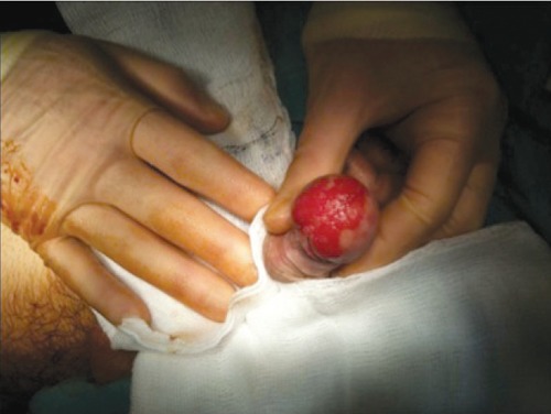
Antero-inferior view of the area of glans penis carcinoma in situ. The external urethral meatus is seen centrally to be involved in the disease process.
Differential diagnosis included balanitis (example: Zoon's balanitis), penile intraepithelial neoplasia/carcinoma in situ/Erythroplasia of Queyrat (EQ), squamous cell cancer of the penis, malignant melanoma of the penis and rarely basal cell cancer of the penis. Basal cell carcinoma of the penis is usually located along the penile shaft.
To ascertain the exact nature of the lesion, two punch biopsies were taken under local anaesthetic. Histological examination showed squamous cell carcinoma in situ. There was no evidence of an invasive malignancy.
He underwent local excision of the diseased skin. This was performed as a partial glansectomy with circumcision. The diseased skin area was resected in quadrants, with the margins and orientation of each quadrant being carefully labelled (Figure 2). A skin graft was harvested from the anterolateral aspect of his right thigh. The skin graft was modelled to fit the glans penis defect and perforated to allow for anastomosis of the spatulated distal penile urethra (Figures 3–5). A urethral catheter was left in situ for 5 days.
Figure 2.
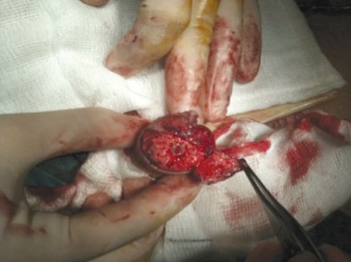
The right ventral and dorsal quadrants have been resected. The external urethral meatus is exposed.
Figure 3.
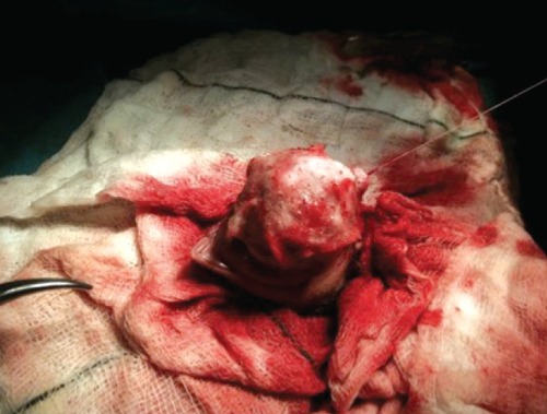
The skin graft is shown overlying the glans defect.
Figure 5.
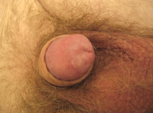
Lateral view of the glans penis three months post-operatively.
Figure 4.
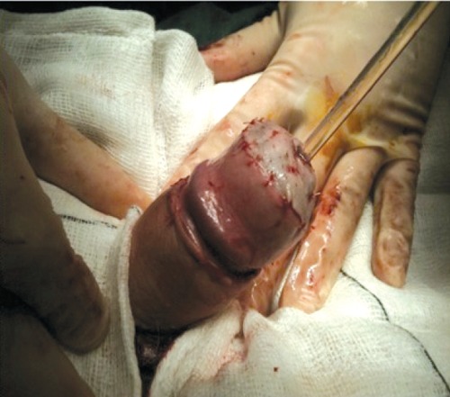
The remodelled skin graft is shown sutured in position with a urethral catheter bridging the remodelled external urethral meatus.
He made an excellent post-operative recovery. Histological examination of the foreskin showed chronic balanitis. Examination of the quadrants showed squamous cell carcinoma in situ. All peripheral and deep surgical margins were clear of disease. There was no evidence of Human Papilloma Virus (HPV).
At three months follow-up there was no evidence of stenosis of the remodelled external urethral meatus. There was no evidence of disease recurrence (Figure 6).
He shall be reviewed at six-monthly intervals for the first two years and yearly thereafter. If there is no recurrence at five years post diagnosis, he may be discharged from the urology service. The patient has been well educated on the need to attend for prompt evaluation if he detects any recurrence of a penile skin abnormality or lymphadenopathy.
Discussion
In western countries, primary malignant penile cancer is uncommon, with an incidence of less than 1 per 100,000 males.1 Penile cancer has a much higher incidence in non- western countries, being the most commonly diagnosed cancer in Uganda. Risk factors for the development of penile cancer include social and cultural habits, and hygienic and religious practices.2
Squamous cell cancer (SCC) accounts for more than 95% of cases of penile cancer. There are many histological sub-types of penile SCC;3 the usual type (accounting for almost 60% of cases), the verruciform type (including warty/condylomatous, papillary/not otherwise specified and verrucous types), the pseudohyperplastic type, the basaloid type, the sarcomatoid or spindle cell carcinoma, the adenosquamous type and the mixed type. Prognosis and metastatic potential relate to histological sub-type.
It is unclear what degree of penile SCC is preceded by a premalignant lesion.4 Squamous cell carcinoma in situ on the penile mucosa or transitional surfaces is also known as EQ. In the region, one third of penile squamous cell carcinoma in situ cases progress to invasive SCC.
Penile EQ mainly occurs on the glans penis, the prepuce, or the urethral meatus of elderly males.5 Typically it has a well-demarcated, velvety, shiny, bright red, plaque-like appearance. The exact aetiology is unknown.
Penile EQ may be treated with local resection, laser therapy, photodynamic and topical therapy with 5-fluorouracil or 5% imiquimod cream.6–8 The pathological assessment of surgical margins is critical in the attempt to reduce local recurrence rates.9 Penis-preserving strategies are recommended for treatment of small lesions. Total glansectomy and prepuce has the lowest recurrence rate at 2% for the treatment of small lesions.10 Diligent follow-up is necessary to enable early detection of recurrence, to assess treatment outcomes and to promote awareness amongst patients of the importance of continuous self-examination.
References
- 1.Barnholtz-Sloan JS, Maldonado JL, Powsang J, Giuliano AR. Incidence trends in primary malignant penile cancer. Urol Oncol. 2007;25:361–7. doi: 10.1016/j.urolonc.2006.08.029. [DOI] [PubMed] [Google Scholar]
- 2.Misra S, Chaturvedi A, Misra NC. Penile carcinoma: a challenge for the developing world. Lancet Oncol. 2004;5:240–7. doi: 10.1016/S1470-2045(04)01427-5. [DOI] [PubMed] [Google Scholar]
- 3.Micali G, Nasca MR, Innocenzi D, Schwartz RA. Penile cancer. J Am Acad Dermatol. 2006;54:369–91. doi: 10.1016/j.jaad.2005.05.007. [DOI] [PubMed] [Google Scholar]
- 4.Velazquez EF, Barreto JE, Rodriguez I, et al. Limitations in the interpretation of biopsies in patients with penile squamous cell carcinoma. Int J Surg Pathol. 2004;12:139–46. doi: 10.1177/106689690401200207. [DOI] [PubMed] [Google Scholar]
- 5.Wieland U, Jurk S, Weissenborn S, et al. Erythroplasia of queyrat: coinfection with cutaneous carcinogenic human papillomavirus type 8 and genital papillomaviruses in a carcinoma in situ. J Invest Dermatol. 2000;115:396–401. doi: 10.1046/j.1523-1747.2000.00069.x. [DOI] [PubMed] [Google Scholar]
- 6.Gerber GS. Carcinoma in situ of the penis. J Urol. 1994;151:829–33. doi: 10.1016/s0022-5347(17)35099-1. [DOI] [PubMed] [Google Scholar]
- 7.Micali G, Nasca MR, De Pasquale R. Erythroplasia of Queyrat treated with imiquimod 5% cream. J Am Acad Dermatol. 2006;55:901–3. doi: 10.1016/j.jaad.2006.07.021. [DOI] [PubMed] [Google Scholar]
- 8.Conejo-Mir JS, Munoz MA, Linares M, et al. Carbon dioxide laser treatment of erythroplasia of Queyrat: a revisited treatment to this condition. J Eur Acad Dermatol Venereol. 2005;19:643–4. doi: 10.1111/j.1468-3083.2005.01217.x. [DOI] [PubMed] [Google Scholar]
- 9.Minhas S, Kayes O, Hegarty P, et al. What surgical resection margins are required to achieve oncological control in men with primary penile cancer? BJU Int. 2005;96:1040–3. doi: 10.1111/j.1464-410X.2005.05769.x. [DOI] [PubMed] [Google Scholar]
- 10.Hadway P, Corbishley CM, Watkin NA. Total glans resurfacing for premalignant lesions of the penis: initial outcome data. BJU Int. 2006;98:532–6. doi: 10.1111/j.1464-410X.2006.06368.x. [DOI] [PubMed] [Google Scholar]


