

Abstract
A rare case of a posttraumatic extensive ganglion cyst of the anterolateral thigh with connection to the knee joint is presented. A 54-year-old man presented a palpable mass in the anterolateral region of his right thigh with a 15 months existing sense of fullness and tightness. He had an accident with his bicycle 21 months ago. Magnetic resonance imaging (MRI) was performed showing a cyst inside the quadriceps femoris muscle between vastus lateralis and intermedius with connection to recessus suprapatellaris and knee joint. In addition MRI detected a traumatic lesion in the quadriceps femoris tendon in the near of the knee joint. The ganglion cyst was 18 cm long and was excised completely. Intraoperatively, the knee joint connection was confirmed and excised as well. The ganglion cyst was filled with a gelatinous and viscous fluid.
Key words: post-traumatic, ganglion cyst, quadriceps femoris tendon, knee joint.
Introduction
Ganglion cysts are filled with gelatinous and viscous fluid in the neighbourhood of joints or tendon sheaths. They are frequently seen at joints and tendons of the wrist but are rare in the region of knee joint. The most common cysts in the knee region are popliteal also called Baker’s cysts.1,2 Ganglion cysts of the quadriceps femoris tendon are very rare.3,4 Ganglion cysts in general are described as idiopathic lesions. There are theories, which describe an increased demand of the joint as a cause for these cysts. However, traumatic events are also discussed to be a cause for occurrence of these lesions. A rare case of an extensive ganglion cyst with a length of 18 cm of the anterolateral thigh with connection to the knee joint is presented and discussed. The cyst appeared some months after an accident.
Case Report
A 54-year-old man presented palpable swelling in the anterolateral region of his right thigh causing a sense of fullness and tightness for 15 months. He had an accident with his bicycle 21 months ago without signs of injury on the initial medical examination. Magnetic resonance imaging (MRI) of the thigh and knee showed a non contrast-enhancing lesion, which appeared hyperintense in the T2-sequences inside the right quadriceps femoris muscle between vastus lateralis and intermedius with connection to the recessus suprapatellaris (Figure 1). In the quadriceps femoris tendon adjacent to the knee joint, a small traumatic injury of the tendon could be detected (Figure 2).
Figure 1.
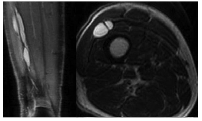
Preoperative magnetic resonance imaging; T2-weighted coronal plane (left picture) of the right thigh and knee show an extensive T2-hyperintense lesion inside the right quadriceps femoris muscle. Axial magnetic resonance imaging of the right thigh (right picture) shows the ganglion cyst with a septum membrane inside the cyst.
Figure 2.
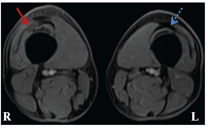
Preoperative magnetic resonance imaging of the right (R) and left (L) thigh (axial plane) detects traumatic lesion in the right quadriceps femoris tendon (red arrow) adjacent to the knee joint. Please note the difference to the normal tendon on the left side (blue dotted arrow), which appears as a hypointense curved structure similar to an eyebrow.
The lesion was 18 cm long and was excised completely by dissecting it from the muscle in microsurgical technique (Figure 3). The connection between the lesion and the knee joint could be demonstrated intraoperatively and was excised as well. The lesion was filled with a gelatinous and viscous fluid, which seemed to be under moderate pressure (Figure 3). There was no evidence of motor nerve fibers inside the lesion. Neurostimulation was used during surgery to avoid involuntary motor nerve. After operation the symptoms declined rapidly and the patient was discharged without neurological deficits. Histopathological results confirmed the diagnosis of a ganglion cyst. The follow-up time was 22 months without recurrence of the cyst.
Figure 3.
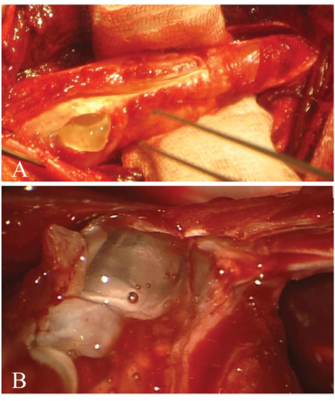
Intraoperative picture showing the ganglion cyst inside the right thigh with a length of 18 cm. The cyst was inside the quadriceps femoris muscle between vastus lateralis and intermedius and was excised completely under general anaesthesia. The ganglion cyst was filled with a gelatinous and viscous fluid. The fluid seemed to be under moderate pressure.
Discussion
Ganglion cysts are uncommon in the knee region. Cysts in this region are usually meniscal or popliteal cysts also called Baker’s cyst1,2,5,6 Baker's cysts are located between the tendon of the medial head of the gastrocnemius muscle and the semimembranosus muscle behind the knee joint. These cysts arise almost from any form of knee joint swelling such as arthritis or meniscal tears. Ganglion cysts are usually not associated with arthritis or meniscal tears and can occur in variable locations. Cysts with anterolateral location within quadriceps muscle are very rare.3,4 Particularly, a cyst of 18 cm in length, as described in our case, has not been reported yet.
Benign cysts like ganglion cysts can be very well detected on MRI as T2 weighted hyperintense defined lesions without contrast enhancement. A septum membrane inside the cyst visualized with MRI should be considered as a confirmation of the diagnosis. Ganglion cysts may have a definable connection to the joint, which can be visualized well on MRI.1 However, the detection of joint connection is not possible in all cases. The connection to a joint seems however to be the most important mechanism for the development of ganglion cysts. The degree of intraarticular pressure is an important factor as well, which causes the enlargement of the ganglion cyst. Furthermore a valve mechanism supports the development of these cysts. During surgery it is important to identify the connection to the joint and to remove it as well as the ganglion cyst itself. In addition, the relationship to the knee joint is important in order to evaluate the presence of associated intraarticular diseases.7,8
The decision for surgery should be considered in symptomatic cases or if the patient because of cosmetic reasons wishes it.
The cause of ganglion cysts is unknown. In our case, the palpable lesions occurred months after a bicycle accident, in which the knee was injured. An injury of the tendon of the quadriceps muscle in the near of the knee joint caused by the trauma could be demonstrated on MRI (Figure 2). Therefore, traumatic genesis can be an important factor for cyst formation, even in cases with bagatelle trauma. The development of ganglion cysts as a result of trauma should find more attention and should be taken into consideration in the differential diagnosis. In many cases, patients with ganglion cysts report that the joints related to them are under an increased amount of demand. That seems to be more typical in the wrist region. This increased demand may cause micro-traumas in the tendons of muscles, which can trigger the cyst formation. The connection to the joint, which may be degenerative in addition, may lead to entry of synovial fluid from the joint into the tendons and within the muscles or other structures. The intraarticular pressure and a valve mechanism may play a role in the development and extension of these cysts.
Conclusions
Trauma or micro-trauma may be an important cause of ganglion cyst formation. Surgery should be performed in symptomatic cases. Before surgery, MRI can confirm the cystic structure of these lesions. The relationship to the joint is important for surgery and for evaluation of the joint for the presence of associated intra-articular diseases.
References
- 1.Burk DL, Jr, Dalinka MK, Kanal E, et al. Meniscal and ganglion cysts of the knee: MR evaluation. AJR Am J Roentgenol. 1988;150:331–6. doi: 10.2214/ajr.150.2.331. [DOI] [PubMed] [Google Scholar]
- 2.Fukuda A, Kato K, Sudo A, Uchida A. Ganglion cyst arising from the posterolateral capsule of the knee. J Orthop Sci. 2010;15:261–4. doi: 10.1007/s00776-009-1434-8. [DOI] [PubMed] [Google Scholar]
- 3.Vayvada H, Tayfur V, Menderes A, et al. Giant ganglion cyst of the quadriceps femoris tendon. Knee Surg Sports Traumatol Arthrosc. 2003;11:260–2. doi: 10.1007/s00167-003-0364-9. [DOI] [PubMed] [Google Scholar]
- 4.Lakdawala A, Ireland J. Giant ganglion formation in the quadriceps femoris tendon following total knee replacement. Int J Clin Pract. 2004;58:990–1. doi: 10.1111/j.1742-1241.2004.00189.x. [DOI] [PubMed] [Google Scholar]
- 5.Vilchez F, Erquicia J, Pelfort X, Monllau JC. [Symptomatic ganglions in the knee joint. Report of three cases and literature review] Acta Ortop Mex. 2009;23:223–7. [PubMed] [Google Scholar]
- 6.Yu WD, Shapiro MS. Cysts and other masses about the knee: identifying and treating common and rare lesions. Phys Sportsmed. 1999;27:59–68. doi: 10.3810/psm.1999.07.920. [DOI] [PubMed] [Google Scholar]
- 7.Lu KH. Unusual solitary ganglion cysts of the anterior segment of the lateral meniscus. Arthroscopy. 2003;19:E16. doi: 10.1053/jars.2003.50067. [DOI] [PubMed] [Google Scholar]
- 8.Shetty GM, Nha KW, Patil SP, et al. Ganglion cysts of the posterior cruciate ligament. Knee. 2008;15:325–9. doi: 10.1016/j.knee.2008.02.010. [DOI] [PubMed] [Google Scholar]