Abstract
Solitary fibrous tumor is an uncommon neoplasm affecting adults and typically located in the pleura and can also occur in a large number of other extra thoracic sites. We present the case of a solitary fibrous tumor (SFT) of the retroperitoneum and describe their histopathological and immunohistochemical features. The identification of SFT in the retroperitoneum is of importance because its clinico-pathological behaviour is still unclear. The pathologist plays a fundamental role in establishing both the positive and differential diagnosis.
Key words: solitary fibrous tumor, retroperitoneum.
Introduction
Solitary fibrous tumor (SFT) is the current preferred term for an uncommon, but histomorphologically distinctive, spindle cell neoplasm of mesenchymal origin. While originally described as a pleural based lesion, it has subsequently been documented at a wide variety of extrapleural sites including the abdominal cavity, retroperitoneum, mediastinum, orbit, upper respiratory tract and soft tissues. It is now accepted that extrathoracic tumors are at least as common as thoracic lesions.1–4 The retroperitoneal location is unfrequented. In this location, we report the observation of a SFT which raised diagnosis problems to the pathologist.
Case Report
A 75-year-old man, who underwent surgical excision of prostatic adenoma 5 years ago, was seen to investigate an abdominal mass. On physical examination, a non painful hard mass was palpable in the left hypochondria. A computed tomography (CT) revealed an intraabdominal and pelvic tumor with spontaneous low density. After iodinated contrast injection, the mass demonstrated significant heterogeneous enhancement (Figure 1). This mass is located in projection to the pancreas tail and the left adrenal gland. The patient underwent complete surgical excision.
Figure 1.
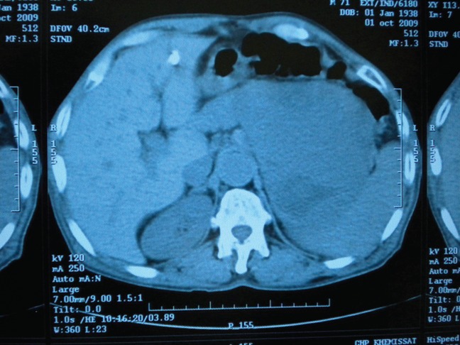
Contrast-enhanced pelvic computed tomography scan demonstrates a voluminous mass in projection to the pancreas tail and the left adrenal gland.
At our laboratory, we received a well circumscribed, lobulated and firm mass with a homogeneous tan-white, whorled cut surface with necrosis areas (Figure 2).
Figure 2.
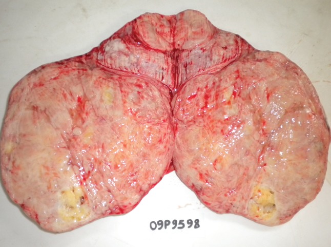
A well circumscribed gray mass with a lobulated appearance and necrosis areas.
Microscopically, it was a spindle cell proliferation, which was arranged haphazardly or in a short storiform pattern, and varying degrees of stromal collagenization. The cellularity varied from area to area, depending on the degree of stromal collagenization (Figure 3). In the less cellular areas, the stroma was dense sclerotic to markedly hyalinized. The vessels were frequently elongated, branching, and dilated, with a hemangiopericytoma-like growth pattern (Figure 3). The spindle tumor cells had fusiform or ovoid vesicular nuclei with finely dispersed chromatin, inconspicuous nucleoli, and scant, poorly defined, and slightly eosinophilic cytoplasm (Figure 4). Mitotic figures were rare less than 1 mitose per 10 in high power fields. The tumoral cells showed immunoreactivity for smooth muscle actin, CD34 (Figure 5) and Bcl2 (Figure 6). They were negative for S100-protein, CD99 and desmin. There were no complications in the immediate postoperative period.
Figure 3.
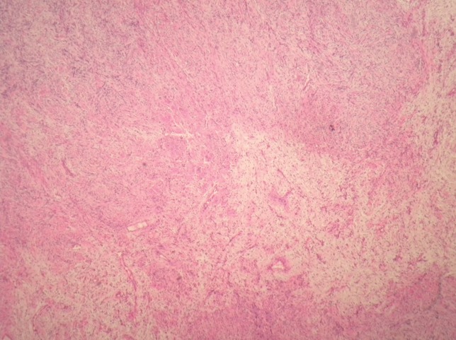
Architectural patternless pattern and branching haemangiopericytoma-like vascular pattern of SFT (HE×40).
Figure 4.
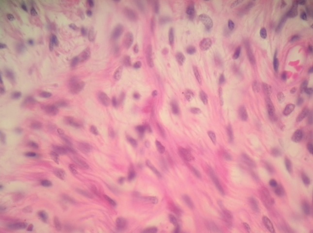
Bland spindle or polygonal cells with slightly eosinophilic cytoplasm (HE×400).
Figure 5.
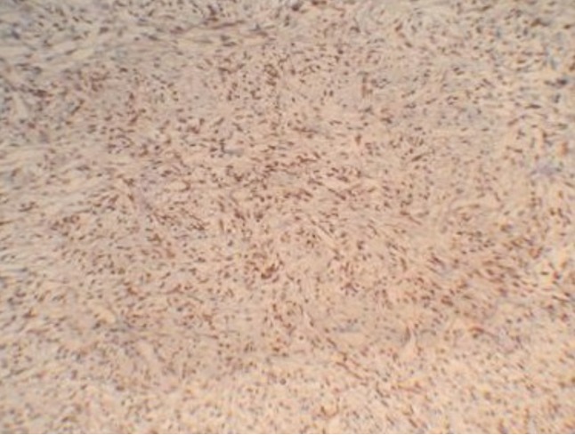
Diffuse immunoreactivity of tumor cells for CD34 (IHC×200).
Figure 6.
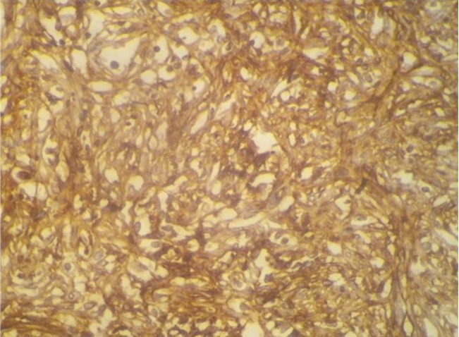
Diffuse immunoreactivity of tumor cells for Bcl2 (IHC×400).
Discussion
Solitary fibrous tumor was first documented by Klemperer and Rabin in 1931.1,2,5,6 SFT, synonymously referred to in the past as fibrous mesothelioma, localized fibrous mesothelioma, localized fibrous tumor, localized mesothelioma, pleural fibroma, solitary fibrous mesothelioma and submesothelial fibroma was for a long time considered to be exclusively pleural-based.1,2,6,7 Now, it has been reported in a wide variety of extrapleural sites including orbit, upper respiratory tract, salivary glands, thyroid, peritoneum, livery, retroperitoneum and pelvis, adrenal gland, kidney, spermafic cord, urinary bladder, prostate, uterine cervix, spinal cord, periosteum, and soft tissue.5 Retroperitoneum is a rare location. Less than 30 cases have been reported in the literature.2
SFT almost invariably occurs in adults of either sex, primarily between 40 and 70 years of age.1,2,5 In our case report, it's a 75-year-old male patient. Most lesions are asymptomatic and are only picked up as an incidental finding on clinical examination. Some patients may present osteoarthropathy and on rare occasions, symptomatic hypoglycemia as a result of the production of an insulin-like factor.1,2,7 On CT scanning, SFT shows a pattern of heterogeneous contrast enhacement as a mass that can envelop the kidney.7,8 Magnetic resonance imaging (MRI) of solitary fibrous tumor of the retroperitoneum demonstrates a signal isointense to muscle on T1-weighted sequences, a heterogeneous hypointense signal on T2 sequences and significant uptake after gadolinium injection. These imaging characteristics of solitary fibrous tumor of the retroperitoneum are very similar to those described for this tumor in a pleural location.8
These tumors are usually well circumscribed, unencapsulated lesions with a lobulated appearance. Lesional size is variable, ranging from 2–15 cm in diameter, although tumors as large as 30 cm have been documented and occasionally show cystic or necrotic changes on their cut surface as in the reported observation.1,2,5,7
Histomorphologically, SFT typically exhibits a patternless architecture characterized by the coexistence of hypo- and hypercellular areas separated by fibrous stroma having haemangiopericytoma-like branching blood vessels. The hypercellular areas are composed of bland spindle cells arranged in short intersecting fascicles, creating herringbone or storiform arrays. The hypocellular areas may be highly collagenized or less frequently present myxoid changes.4–7 Nuclei are generally bland, spindled in shape with only mild pleomorphism and minimal hyperchromasia. Mitoses are infrequent and usually number less than four per 10 high power fields. Cell borders are poorly defined and cytoplasm is scant and indistinct. This morphological aspect was observed in our case. Increased mitotic activity, cellularity, nuclear pleomorphism and the presence of lesional necrosis are ominous features and suggest an increased risk of local recurrence and metastasis.1,2,6 None of these elements was observed in our case.
Immuhistochemical studies are helpful in confirming the diagnosis of SFT. Anti CD34 antibody is a sensible marker (95% of SFT) but unspecific.2,5,6 Anti-CD99 antibody is positive in 70% of the cases. Anti- EMA, antibcl2, and anti-smooth muscle actin antibodies are positive only in 20–35% of the cases.2,6
The morphologic spectrum of SFT may mimic a number of benign and malignant spindle cell neoplasms including peripheral nerve sheath tumor, leiomyosarcoma, dedifferentiated liposarcoma, gastrointestinal stromal sarcoma (GIST), inflammatory myofibroblastic tumor and desmoplastic mesothelioma.1,2,4–6 Although the distinction is problematical with histology based examinations alone, sarcomas show cytologic features of malignancy, which are uncommon in SFT. Yet, some characteristics of SFT, such as short oval-spindled nuclei with bland chromatin, are occasionally found in synovial sarcoma, nerve sheath tumors, smooth muscle tumors, and desmoid tumors.4,9 Admixed various cellularity of polygonal cells is also observed in synovial sarcoma, peripheral nerve sheath tumors and smooth muscle tumors. The aforementioned neoplasms are rarely accompanied by the irregular distribution of dense collagen as found in SFTs. Additionally, immunoreactivity for CD34 confirms the diagnosis of SFT.4,9 Hemangiopericytoma and SFT share several histopathological features, and may occasionally be difficult to distinguish. In brief, hemangiopericytoma has more oval to rounded nuclei and less collagenized stromas.
Desmoid tumors can also mimic a SFT, but have more slender and elongated spindle cells on a myxoid background.
GISTs are also found as rare tumors in the retroperitoneum. However, they don't show a collagenous stroma, and most of them are specifically positive for CD117 staining.4,9
Close attention to the pattern as described, and the judicious use of an immunohistochemical panel (including pan keratins, EMA, S100-protein, CD34, smooth muscle actin, desmin, CD117, CD99 and calretinin can help in arriving at the correct diagnosis.
Most extrathoracic solitary fibrous tumors pursue a benign course, but may have the potential to recur or metastasize after complete resection even in the absence of atypical histological features. SFT is best regarded as a tumor with uncertain malignant potential and clinical follow-up is advised in all cases, focusing on the early detection of local recurrence and metastases.1,2,5,6
References
- 1.Graadt van Roggen JF, Hogendoorn PCW. Solitary fibrous tumour: the emerging clinicopathologic spectrum of an entity and its differential diagnosis. Curr Diagn Pathol. 2004;10:229–35. [Google Scholar]
- 2.Saint-Blancard P, Jancovici R. Tumeur fibreuse solitaire du rétropéritoine. Rev Med Interne. 2009;30:181–5. doi: 10.1016/j.revmed.2008.04.009. [DOI] [PubMed] [Google Scholar]
- 3.Cranshaw IM, Gikas PD, Fisher C, et al. Clinical outcomes of extra-thoracic solitary fibrous tumours. Eur J Surg Oncol. 2009;35:994–8. doi: 10.1016/j.ejso.2009.02.015. [DOI] [PubMed] [Google Scholar]
- 4.Takizawa I, Saito T, Kitamura Y, et al. Primary solitary fibrous tumor (SFT) in the retroperitoneum. Urol Oncol. 2008;26:254–9. doi: 10.1016/j.urolonc.2007.03.024. [DOI] [PubMed] [Google Scholar]
- 5.Hasegawa T, Matsuno Y, Shimoda T, et al. Extrathorac solitary fibrous tumor: their histological variability and potentially aggressive behavior. Human Pathol. 1999;30:1464–73. doi: 10.1016/s0046-8177(99)90169-7. [DOI] [PubMed] [Google Scholar]
- 6.Hannachi Sassi S, Charfi L, Mrad K, et al. Une tumeur rétropéritonéale inhabituelle. Ann Pathologie. 2007;27:385–6. doi: 10.1016/s0242-6498(07)78281-0. [DOI] [PubMed] [Google Scholar]
- 7.Travis William D, Brambilia E, Muller-Hermelink HK, Harris Curtis C. Pathology and genetics of tumors of the lung, pleura, thymus and heart. Lyon: IARC Press; 2004. World health organization Classification of tumors. [Google Scholar]
- 8.Casas JD, Balliu E, Sanchez MC, Mariscal A. Benign solitary fibrous tumor of the retroperitoneum: radiological features. CMIG Extra: Cases. 2004;28:50–3. [Google Scholar]
- 9.Enzinger FM, Weiss SW. 3rd ed. New York: Mosby; 1995. Soft tissue tumors; pp. 810–812. D, 9YO. [Google Scholar]


