Abstract
Clostridium perfringens epsilon toxin (ETX), one of the most potent toxins known, is a potential biological weapon; therefore, the development of an effective vaccine is important for preventing intoxication or disease by ETX. In this study, genetically detoxified epsilon toxin mutants were developed as candidate vaccines. We used site-directed mutagenesis to mutate the essential amino acid residues (His106, Ser111 and Phe199). Six site-directed mutants of ETX (mETXH106P, mETXS111H, mETXS111Y, mETXF199H, mETXF199E, mETXS111YF199E) were generated and then expressed in Escherichia coli. Both mETXF199E and mETXH106P with low or non-cytotoxicity that retained their immunogenicity were selected to immunize mice 3 times, and the mouse survival data were recorded after challenging with recombinant wild-type ETX. mETXF199E induces the same protection as mETXH106P, which was reported previously as a promising toxin mutant for vaccine, and both of them could protect immunized mice against a 100× LD50 dose of active wild-type recombinant ETX. This work showed that mETXF199E is another promising candidate vaccine against enterotoxemia and other diseases caused by ETX.
Keywords: Clostridium perfringens, cytotoxicity, enterotoxemia, site-directed mutagenesis, toxin mutant, vaccine, ε-toxin
Introduction
Epsilon toxin (ETX), which is produced by Clostridium perfringens types B and D, is considered a potential biological weapon.1 Its lethal activity is just below the botulinum and tetanus toxin, and the intraperitoneal injection dose of ETX that kills 50% of mice is 65–110 ng/kg.2,3 ETX can lead to fatal illnesses in livestock animals, especially induce enterotoxemia in sheep.1 Although very few ETX-mediated diseases have been reported in humans, evidence does suggest that the toxin may be toxic to humans because the human kidney cell lines G-402 and ACHN are sensitive to ETX.4-6 At present, vaccines against enterotoxemia caused by ETX are used in veterinary medicine.7 These vaccines are based on formaldehyde-treated bacterial culture filtrates or whole-cell cultures. However, the immunogenicity of ETX in these preparations varies, which may lead to safety problems.8,9 There is not yet a vaccine against ETX for humans. As such, it is significantly important to develop a viable and safe vaccine against ETX for livestock and human.
Chemical detoxification is a traditional method of toxin-based vaccines. Genetically detoxified toxins such as toxin mutants, which are not biologically active but retain immunogenicity, is a new and promising approach.7,10,11 This method has been widely used in the investigation of recombinant vaccines.7,11-13 In this study, one or more essential amino acid residues of a target toxin are selected and substituted to decrease the toxicity.
ETX contains three domains, domain I of ETX might have the function of binding to receptor and domain II has been predicted to be the channel-forming domain.1 Some essential amino acid residues in these two domains play important roles in the lethal activity of ETX. For example, it is previously reported that a group of amino acid residues (Tyr36, Tyr30, Tyr29, Tyr196 and Phe199) in domain I might have a receptor binding function.14 Also, recent research indicates that the amino acid motif including Tyr29, Tyr30, Tyr36, and Tyr196 is important for the ability of ETX to interact with cells.15 In addition, the molecule of ETX contains a unique Trp (Trp190) and two His residues (His106 and His149). A previous study shows that His106 is important for the biological activity, whereas His149 and Trp190 probably are involved in maintaining the structure of ETX, but they are not essential for the activity.10 A segment (His106 to Ala136 of the mature ETX) in domain II contains alternate hydrophobic-hydrophilic residues, which are characteristic of membrance-spanning β-hairpins, and forms two amphipathic β strands on ETX structure. Site-directed mutagenesis confirmed that this segment is involved in ETX channel activity in lipid bilayers.1,16 Paired cysteine substitutions were introduced to form a disulfide bond at I51/A114 and V56/F118 to yield the I51C/A114C and V56C/F118C mutant proteins, which lacked detectable cytotoxic activity could be candidate vaccines.17 Based on these amino acid residues, we ultimately chose His106, Ser111 and Phe199 as mutation sites.
ETX is secreted in an inactive form called prototoxin that has poor activity and is activated by proteases, for example, trypsin can cleave 13 N-terminal and 22 C-terminal residues to activate the prototoxin.2,18,19 The recombinant ETX (rETX), without 13 N-terminal and 23 C-terminal residues, has been successfully expressed in E. coli20 and has been illustrated to have similar cytotoxicity to native ETX activated by proteases.16 MDCK cells are commonly used in cytotoxicity assay of ETX among the four sensitive cell lines to ETX, including MDCK, mpkCCDcl4 cells, and G-402, ACHN cells.10,12,20,21
In this paper, our work focused on developing mutant-based ETX vaccines that could protect against ETX intoxication. We selected three amino acid residues (His106, Ser111 and Phe199) to construct mutant ETX (mETX) and selected the suitable one which has low or non-cytotoxicity and retained immunogenicity to be evaluated as candidate vaccine.
Results
Construction of the mETX
Some amino acid residues were substituted in ETX to obtain non-toxic mutants. The sequences of mETXs have been compared with the etx gene (GenBank Accession No.M80837) using software DNAMAN 7.0 (Lynnon Corporation). The His106 residue was changed to a proline, while the Ser111 residue was changed to a tyrosine or a histidine. The Phe199 residue was changed to a histidine or a glutamic acid. Finally, six mutants were achieved and named as mETXH106P, mETXS111H, mETXS111Y, mETXF199H, mETXF199E and mETXS111YF199E. The mETXS111YF199E has two sites for mutation.
Expression and purification of the mETX
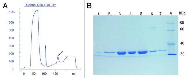
The mETX proteins with a 6× His tag on C-terminus were expressed in the E. coli BL21 (DE3) strain. The rETX and mETX proteins were expressed in soluble forms at 16°C after induction with 0.5 mM IPTG. These toxins were purified using a Ni2+-chelating affinity chromatography resin column. The induced conditions were optimized to provide high-level expression of mETX in a soluble form. Only mETXH106P has a low-level soluble expression. The soluble expression of mETXH106P reached 7.6% of the total protein concentration whereas mETXF199E could reach 24.1% (analyzed by BandScan software, Glyko). The concentrations of imidazole varied in the buffer used to elute the different mutant proteins. Only the affinity chromatographic profile of mETXF199E was shown (Fig. 1A). The molecular mass of the expressed proteins was approximately 34 kDa (Fig. 1B). The purified mutant proteins were analyzed using SDS-PAGE (Fig. 1B) and up to 98% purity was achieved according to BandScan software (GlykoPrep).

Figure 1. Affinity chromatographic profile and SDS-PAGE of mETX. (A) Purification of mETXF199E using a Ni2+-cheating HP column. The mETXF199E was eluted with buffer containing increasing concentrations of imidazole up to 500 mM (the arrow marking fraction). The purification data of other mETXs have not shown. (B) 12% SDS-PAGE analysis of purified rETX and its mutants. Lane 1-6, mETXH106P, mETXS111Y, mETXS111H, mETXF199H, mETXF199E, mETXS111YF199E; Lane 7, rETX; Lane M, ProteinRuler Ι protein marker (TransGen Biotech).
Antigenicity of the mETX
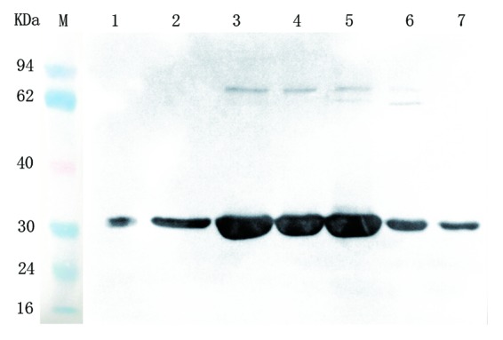
An indirect ELISA was used to detect the antigenicity of the toxin mutants. Compared with wild-type rETX, all of the mETX also showed high reactivity with the mouse monoclonal antibodies (mAb) against the recombinant ETX. (Table 1). Furthermore, an immunoblot assay showed that both mETXs and rETX were recognized by the mouse mAb against the rETX (Fig. 2). The results showed that the toxin mutants retained the same antigenicity as the rETX.
Table 1. Titration of mETXs compared with rETX.
| Mutation | Titration |
|---|---|
| H106P | 1:102 |
| S111H | 1:103 |
| S111Y | 1:103 |
| F199H | 1:103 |
| F199E | 1:103 |
| S111YF199E | 1:103 |
| rETX | 1:103 |

Figure 2. Immunoblot of purified rETX and its mutants with monoclonal mouse anti-rETX antibody detected using Super-ECL horseradish peroxidase. Lane 1-6, mETXH106P, mETXS111Y, mETXS111H, mETXF199H, mETXF199E, mETXS111YF199E; Lane 7, rETX; Lane 7, rETX. The pre-stained protein marker (TransGen Biotech) is indicated on the right side of the blot.
Activity of mETX in the cytotoxicity assay
The toxin mutants were assayed for cytotoxicity against the MDCK cells using the metabolic indicator MTS. The dose of mETX required to kill 50% of the MDCK cells (CT50) was calculated (Table 2). Statistical analysis revealed that four toxin mutants (mETXS111Y, mETXF199E, mETXH106P, mETXS111YF199E) had significant (P = 0.0094, 0.0002, 0.0023, 0.0159 respectively) decrease in cytotoxicity in MDCK cells compared with active rETX. The cytotoxicity of mETXS111Y in MDCK cells showed slight decrease compared with that of the rETX, meanwhile the mETXS111YF199E showed a higher cytotoxicity in MDCK cells than mETXF199E. So the mETXS111Y and mETXS111YF199E were excluded from further analyses. The CT50 of mETXF199E in the MDCK cells was approximately 97.06±5.79 μg/mL, which was about 2572 times of active rETX (37.73±12.55 ng/mL), indicating that the toxicity of mETXF199E was 2572-fold less than that of active rETX. The mETXH106P showed no cytotoxicity in the MDCK cells. Using published data,10,21 the ratio of CT50 to LD50 was calculated as 0.0056. The LD50 of the mETXF199E was calculated as 16.18±0.97 mg/kg, which is much higher than that of active rETX (65–110 ng/kg).2,3
Table 2. Cytotoxicity of mETX.
| Protein preparation | CT50(ng/mL)±SEM | P* |
|---|---|---|
| Wild type rETX | 37.73±12.55 | - |
| mETXH106P | NT | 0.0023 |
| mETXS111Y | 69.06±22.60 | 0.0094 |
| mETXS111H | 95.54±1.67 | 0.2435 |
| mETXF199E | 97060±5790 | 0.0002 |
| mETXF199H | 1280±220 | 0.1707 |
| mETXS111YF199E | 37090±1440 | 0.0159 |
NT, not toxic; *, each mETX compared with wild type rETX
Structures analysis of mETXs
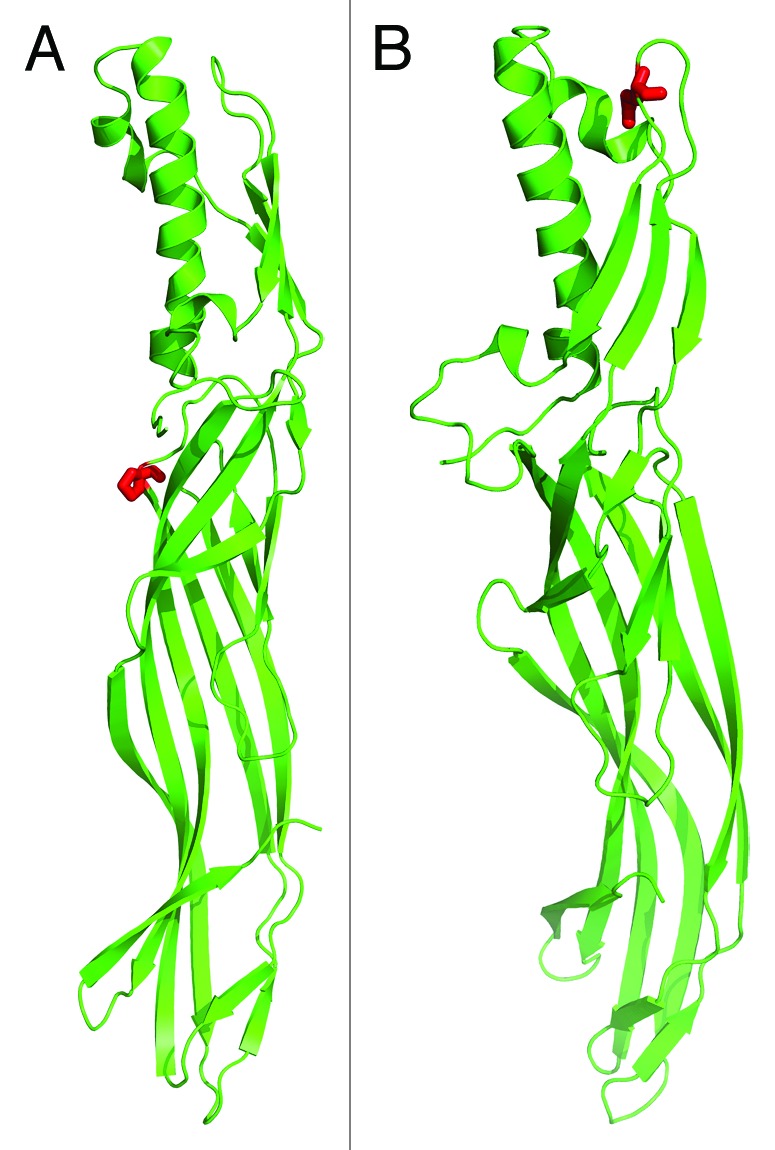
The three-dimensional structures of mETXH106P and mETXF199E were shown in cartoon. Pro106 and Glu199 were shown in rainbow sticks and colored in red (Fig. 3). The structures of mETXs didn’t show any obvious change compared with wild-type ETX according to the PyMOL Molecular Graphics Software.

Figure 3. The three-dimensional structures of mETXH106P (A) and mETXF199E (B). Pro106 and Glu199 were shown in rainbow sticks and colored in red.
Immunization of mice with mETXH106P and mETXF199E induces antibody response
The mice were immunized with mETXH106P and mETXF199E respectively, the ELISA results of the anti-mETX antibody titers in sera after each immunization are shown in Table 3. The data of the two groups (mETXH106P and mETXF199E) showed similar titers and has no significant differences, analyzed using the group t-test, the antibody titers increased following booster immunizations. After the third immunization, the mean IgG titer against the recombinant ETX of the two groups could reach 1:104. The sera antibody titer of the negative control group (immunized with PBS and adjuvant) was less than 1:10, which is obviously different from those of the test groups.
Table 3. The anti-mETX antibody titers of sera after each immunization.
| Group | Antibody titer* | ||
|---|---|---|---|
| Vaccination 1 | Vaccination 2 | Vaccination 3 | |
| mETXF199E | 1:102 | 1:103 | 1:104 |
| mETXH106P | 1:102 | 1:103 | 1:104 |
| PBS | 0 | 1:1 | 1:1 |
* The data shown is the arithmetic mean of the sera titer from three mice other than the geometric mean titers (GMTs), because the three mice picked randomly showed the same titers from a twice replicated experiment.
Protection in immunized mice against challenge with active rETX
Mice immunized with mETXH106P and mETXF199E were challenged with active rETX to evaluate the active protection effect. All of the mice in the group challenged up to 100 × LD50 dose survived. When the challenge dose increased to 500 × LD50 or 1000 × LD50 of ETX per mouse, all of the mice died (Table 4). Mice of the negative control group (immunized with PBS and adjuvant) did not survive when challenged with the 10× LD50 dose activated rETX. In addition, the body weight would have been an useful parameter to follow but was not recorded.
Table 4. Survival of mice challenged with active rETX.
| Groups | 10× LD50 | 100× LD50 | 500× LD50 | 1000× LD50 |
|---|---|---|---|---|
| alive/total | alive/total | alive/total | alive/total | |
| Mice vaccinated with mETXF199E | 3/3 | 3/3 | 0/3 | 0/3 |
| Mice vaccinated with mETXH106P | 3/3 | 3/3 | 0/3 | 0/3 |
| Mice vaccinated with PBS | 0/3 | - | - | - |
| Passive protection of mETXF199E | 3/3 | 0/3 | 0/3 | 0/3 |
| Passive protection of mETXH106P | 3/3 | 0/3 | 0/3 | 0/3 |
| Control for passive protection | 0/3 | - | - | - |
Passive protection of mice
The passive protection effect of the immunized sera for mice was also estimated. The survival results of the mice which were treated with the mixture of immunized sera and active rETX are shown in Table 4. The results suggested that the anti-mETXH106P or anti-mETXF199E antibodies could completely neutralize 10 × LD50 dose of activated recombinant ETX and there was no difference between the two groups.
Discussion
ETX, one of the most potent toxins just below the botulinum and tetanus toxins, is classified as a category B agent by the Centers for Disease Control and Prevention.1 It is necessary to develop a viable and safe vaccine against ETX for livestock and human. To date, no reports have examined the use of ETX toxoid vaccine in humans. A toxoid vaccine has been widely used over the past few decades and has been reported of variable immune responses and inflammatory responses following vaccination.9 Genetically detoxified toxins such as toxin mutants, which are low or non-toxic but retain their immunogenicity, are promising candidate vaccines for toxin proteins. Consequently, there will be greater applications in the future for genetically detoxified vaccines.
In this paper, the rETX (without 13 N-terminal and 23 C-terminal residues) that retains its natural biological activity and does not have to be activated by proteases, was used as the substitution of the natural ETX.20 Oliver et al. reported that rETX showed slightly reduced activity in the cytotoxicity experiment compared to native ETX, but overall, rETX can replace the natural toxin in biological activity assay.16 The rETX we used, which has a 6× His tag on the N-terminus, is convenient for purification. Meanwhile, the 6× His tag is very small and has no immunogenicity, so it is acceptable for use in laboratory research and even Phase 1 clinical trials.20,22 The His tag will be removed by protease if the candidate vaccine requires further research.
It has been reported that residues His106, Ser111 and Phe199 in ETX might influence the structure or bioactivity of the toxin.10,15,16 Oliver et al. constructed six mETXs (S111E, S111C, K117E, T130C, K117H, and P121H) and found that only Ser111Glu exhibited decreased cytotoxicity.16 In this study, we tried to replace the amino acid Ser111 with Tyr or His to elucidate whether it can show a significant difference in S111E cytotoxicity.16 However, we found that only mETXS111Y showed slightly decreased cytotoxicity and could not be considered as a candidate vaccine (Table 2). Phe199 was also predicted to be the center of receptor binding.14,15 Ivie et al. has already constructed the mETXF199E before, which lacked detectable cytotoxic activity, was supposed to be misfolded and excluded from further analyses.15 We obtained a different result in cytotoxicity of mETXF199E, because the highest concentration of mETXF199E they used (about 400 nM) is much less than what we used (about 8000 nM) in the experiment. In our study, the cytotoxicity assay revealed that mETXF199E had a large decrease in cytotoxicity compared with the wild-type activated rETX (about 2572-fold). In contrast, mETXF199H did not display significantly decreased cytotoxicity. This finding indicated that replacement with different amino acids could make biological activity vary. The H106P has been reported to be a non-toxic mutation, and we achieved the same result in this study.10 We also found that all the mETXs except mETXH106P showed high soluble expression. This maybe because the amino acid His106 is important for maintaining the structure of protein, which may change upon amino acid replacement. Compared with the wild-type ETX, the predicted three-dimensional structures of the mETXH106P and mETXF199E didn’t show any obvious change. This showed that reduction of the cytotoxicity of mETXH106P and mETXF199E is less likely due to the structure change. Furthermore, it contributes to the possibility that Phe199 is essential to the binding of ETX with cells.
We then used mETX as an immunogen to vaccinate BALB/c mice. The mETXH106P and mETXF199E groups showed the same protection of immunized mice against rETX (100 × LD50). The protection provided by mETXH106P was slightly different from that which described previously, which could protect against 1000× LD50 of activate recombinant wild-type ETX.10 This result maybe due to the fact that the rETX sequences were derived from different C. perfringens strains. Oyston et al.10 chosed strain NCTC8346 while we chosed strain NCTC8533 (single amino acid residues difference at 321 position, Tyr and Ser respectively). The different doses of ETX in challenge and varied methods of immunization such as adjuvant and immunizing dose could influence the titer and protection. Furthermore, the immunized sera provided effective passive protection when the mixture of immunized sera and 10 × LD50 of rETX were co-injected into mice. The two toxin mutants, mETXH106P and mETXF199E, have different advantages as candidate vaccines. The former showed non-toxic but low soluble expression, which increased the purification difficulty. The mETXF199E had high soluble expression but retained slight toxicity. It showed 2572-fold less cytotoxicity than that of rETX. Moreover, mETXF199E was comparatively safe, for the LD50 of the mETXF199E (16.18 ± 0.97 mg/kg) was much higher than that of active rETX (65–110 ng/kg). The tissue section of kidney did not show any pathological injury in the group of mice injected with mETXH106P and mETXF199E (the figure not shown), which also supported this point. Based on these results, mETXF199E could be more suitable than rest of the mETXs.
In this study, the F199E mutant of ETX was firstly reported as a candidate vaccine. Our results indicated that not only mETXH106P but mETXF199E also showed both strong immunogenicity and safety. The limitation of the mETXF199E is that it still has toxicity, so we plan to replace Phe199 with other amino acids to make toxin mutants with lower toxicity or even no toxicity in the future. The immunization protocol also should be optimized to achieve a more powerful protection and the stability of mETXF199E is going to be tested in the future work. Altogether, the mETXF199E as a promising candidate vaccine will have great potential applications in the future and should be investigated deeply.
Materials and Methods
Site-directed mutations of ETX in plasmid pTIG-trx
The pTIG-etx recombinant plasmid containing the truncated etx gene fragment (GenBank Accession no. M80837) with a 6 × His tag sequence at the 3’ end was constructed by Zhao et al.20 The nucleotide sequence of the rETX has been optimized according to the code usage of E. coli and was used as the template for site-directed mutagenesis. The oligonucleotide primer sequences used for the mutagenesis reaction are shown in Table 5. The six site-directed mutations (pTIG-mETXH106P, pTIG-mETXS111H, pTIG-mETXS111Y, pTIG-mETXF199H, pTIG-mETXF199E, pTIG-mETXS111YF199E) were generated using the QuikChange® Lightning Site-Directed Mutagenesis Kit (Stratagene).23 The mutant sequence was verified using nucleotide sequence analysis (Sangon).
Table 5. Primers used to produce the site-directed mutants of recombinant ε-toxin.
| Mutation | Primer |
|---|---|
| H106P | 5'-CGACCACCACCCCCACCGTGGGCAC 5'-GTGCCCACGGTGGGGGTGGTGGTCG |
| S111H | 5'-CCACACCGTGGGCACCCATATCCAGGCAACCGCTA 5'-TAGCGGTTGCCTGGATATGGGTGCCCACGGTGTGG |
| S111Y | 5'-CACCGTGGGCACCTATATCCAGGCAACCGC 5'-GCGGTTGCCTGGATATAGGTGCCCACGGTG |
| F199H | 5'-GTGAAATCCCGAGCTATCTGGCTCATCCGCGTGATGG 5'-CCATCACGCGGATGAGCCAGATAGCTCGGGATTTCAC |
| F199E | 5'-GGGTGAAATCCCGAGCTATCTGGCTGAGCCGCGTGATGGTT 5'-AACCATCACGCGGCTCAGCCAGATAGCTCGGGATTTCACCC |
Expression and purification of the rETX and its toxin mutants
The constructed pTIG-mETXs were transformed into competent E. coli BL21 (DE3) cells (TransGen) for expression. The expression of mETX was driven by a T7 promoter.
The E. coli BL21 (DE3) cells containing pTIG-mETX were grown in LB medium supplemented with 100 μg/mL ampicillin at 37°C until they reached an OD600 of 0.6–0.8. The production of rETX and its mutants was induced using isopropyl-β-D-thiogalactopyranoside (IPTG; Promega) in a final concentration of 0.5 mM and at 16°C overnight, about 16-20h. Meanwhile, we found induction in a final concentration of 0.3 mM IPTG and at the temperature of 30°C for 4–6 hours could make the mutant pTIG-mETXH106P be expressed efficiently in soluble status. The bacteria cultured in 1 L of LB medium were collected by centrifugation and washed with phosphate-buffered saline solution (PBS), and the precipitation was resuspended by buffer A (20 mM PB, 500 mM NaCl, 50 mM imidazole; pH 7.4) and lysed by sonication. The lysate was centrifuged and the supernatant then purified through a HiTrap Chelating high performance 5-mL column pre-packed with a precharged Ni2+ column (GE Healthcare). The proteins were eluted with buffer B (20 mM PB, 500 mM NaCl, 500 mM imidazole; pH 7.4). The purified proteins were analyzed using 12% sodium dodecyl sulfate-polyacrylamide gel electrophoresis (SDS-PAGE) gels. The highly purified proteins were dialyzed in 0.01 M PBS for 12 h and concentrated by a PEG20000 (Merck). The concentrations of the purified proteins were measured using a bicinchoninic acid assay kit (BCA; Pierce) featuring bovine serum albumin (BSA; Pierce) as a standard.
Antigenicity of the mETX
Enzyme-linked immunosorbent assay (ELISA)
ELISA was employed to identify the antigenicity of mETX. A 96-well plate (Corning) was coated with 10 μg/mL mETXs or rETX in 0.05 M carbonate-buffered saline solution (CBS), pH9.6, at a volume of 100 μL per well overnight at 4°C. After the plate was washed with PBST (0.01M PBS, 0.05% Tween-20) five times and blocked with 3% BSA in 0.01M PBS for 2 h, 100 μL 10-fold serial dilutions mouse anti-rETX monoclonal antibody (prepared by our laboratory) were added to the plate and incubated at 37°C for 1 h. The plate was then washed and 100 μL of horseradish peroxidase (HRP)-coupled goat anti-mouse IgG (Sigma) was added and incubated for 1 h. After the plate was washed with PBST five times, the substrate solution was added to each well and incubated at room temperature for 10 minutes and 2M H2SO4 was added to stop the reaction. The absorbance was measured at 450 nm using a microplate reader (Molecular Device). The values of OD450 greater than 2.1-fold negative control were considered as positive.
Western blotting.
After electrophoresis, the purified protein mETX were transferred from a SDS-PAGE gel to a nitrocellulose membrane (Amersham) using a transmembrane device (Amersham Biosciences). We then blocked the nonspecific sites on the membrane by incubating them in 0.01 M PBS containing 5% BSA for 1 h with shaking and incubated the blot in 0.01 M PBS with mouse anti-rETX monoclonal antibody (prepared by our laboratory) and 0.05% Tween-20 for 1 h at room temperature with shaking. Finally, we incubated the blot with HRP-coupled goat anti-mouse IgG at 37°C for 1 h, incubated the blot with SuperSignal Substrate Working Solution (Thermo) for 5 minutes, and exposure in AE-1000 cool CCD image analyzer (Beijing BGI-GBI Biotech Co., Ltd).
Cell culture and cytotoxicity assay
The rETX activity was measured by determining its effect on MDCK cells as described previously.10,20,24 Cytotoxicity was monitored using 3-(4, 5-dimethylthiazol-2-yl)-5-(3-carboxymethoxyphenyl)-2-(4-sulfophenyl)-2H-tetrazolium (MTS). The cytotoxicity assay of the MDCK cells (The Chinese Academy of Sciences) was performed using the CellTiter 96®AQueous One Solution Cell Proliferation Assay (Sigma). The MDCK cells were grown in 96-well plates with Dulbecco's modified Eagle medium (DMEM) supplemented with 10% fetal bovine serum. Cells were seeded at 1–2 × 10⁴ cells/well and cultured at 37°C in a 5% CO2 incubator for 24 h. The diluted ε-toxin and mETX were added to the cells and incubated at 37°C in a 5% CO2 incubator for 48 h, an equal volume of medium was added to the negative control and DMEM culture media was used as a blank. Aliquots of MTS (20 μL) were pipetted into each well of a 96-well assay plate containing 100 μL of culture medium. The plates were incubated in a 5% CO2 atmosphere at 37°C for 1–4 h. The OD at 490 nm was then measured using a SPECTRA MAX plus plate reader (Molecular Devices). In the pre-experiment, low concentrations of toxin with a 5-fold dilution were used to evaluate the range of CT50, and the mutants which didn’t have an obvious decrease in cytotoxicity were excluded from further analyses. The CT50 of the toxin mutants was calculated using the improved Karber’s method according to the formula below and compared with the CT50 of rETX, the results were assessed using the t-test.
CT50 = Log-1 [Xm - i × (∑p - 0.5) + i/4 × (1 - Pm - Pn)]
Xm, log value of the according concentration of maximum mortality; i, difference between the log value of the gradient concentrations; Pm, value of the maximum mortality; Pn, value of the minimum mortality.
Structural analysis
For generation of the three-dimensional structures of two mETXs (mETXF199E and mETXH106P), crystal structure of wild-type ETX was used as a starting model. The coordinate for the wild-type ETX was taken from the protein data bank (1UYJ.pdb). Molecular modelings were performed by MODELLER program in Discovery Studio 2.5 (Accelrys). Protein structure illustrations were generated with the PyMOL Molecular Graphics Software.
Vaccination of mice and challenge with rETX
In this test, low or non-toxic mutant ETX (mETXF199E and mETXH106P) was used to immunize six-week-old female BALB/c mice (purchased from Laboratory Animal Center, The Academy of Military Medical Sciences) that were randomly divided into two groups. Each group had 14 mice and two mice used as negative control were injected with 0.01M PBS (pH 7.4) mixed sufficiently with aluminum salt adjuvant (Pierce) at a ratio of 2:1 (v/v) for 30 minutes at 4°C. The purified mETX protein (10 μg in 150 μL mixture solution volume/mouse) in 0.01M PBS (pH 7.4) was mixed with adjuvant and administered via subcutaneous injection. The mice were given the same doses of antigen on days 17 and 38. One week after the last immunization, the mice were randomly divided into four subgroups, three mice for each group were challenged with 10, 100, 500, or 1000 × LD50 activate recombinant ETX by peritoneal injection, respectively. We observed the mice for 72 h and recorded their survival status. In addition, there are 14 mice immunized mETXF199E and mETXH106P, respectively, in the same way, used for passive protection. The animal studies were conducted in compliance with the Guide for the Care and Use of Laboratory Animals and the Association for Assessment and Accreditation of Laboratory Animal Care International.
Measurement of sera antibody titers
To detect the titers of anti-ETX antibody from blood serum, three mice were randomly chosen from each group and underwent bleeding from the caudal vein one week after each vaccination. The mice sera were then titrated using an ELISA. A 96-well plate was coated with 5 μg/mL rETX in 0.05M CBS, pH9.6, 100 μL in each well overnight at 4°C. The plate was washed with PBST and blocked with 3% BSA in 0.01M PBS. Then 100 μL 10-fold serial dilutions sera samples were added to the plate and incubated at 37°C for 1 h, the plate was washed and 100 μL of HRP-coupled goat anti-mouse IgG were added and incubated at 37°C for 1 h. After the plate was washed with PBST, the substrate solution was added to each well and incubated at room temperature for 10 minutes and 2M H2SO4 was added to stop the reaction. The absorbance was measured and the titer was calculated as described before.
Passive protection of mice against rETX challenge.
Sera samples were collected from the rest of 28 vaccinated mice and tested for the ability to protect mice again rETX challenge. The sera from two groups were mixed with the dose of 10, 100, 500, or 1000 × LD50 activated recombinant ETX (0.01M PBS) in an equal volume, respectively and incubated at 37°C for 30 minutes. 12 six-week-old female BALB/c mice were randomly divided into four subgroups and peritoneal injected with the mixed solution (500 μL per mouse). Another 3 mice were treated with the 10 × LD50 activated recombinant ETX mixed with an equal volume of non-immunized sera as control (500 μL per mouse). Observe the survival of mice for 72 h.
Disclosure of Potential Conflicts of Interest
No potential conflicts of interest were disclosed.
Acknowledgments
We thank Prof. Zhihu Zhao for providing the pTIG-trx vector and thank Dr. Junjie Yue for providing the molecular modelings of the three-dimensional structures of mETXs.
Note
In this paper, we used numbering system assigning 1 to the start of the mature toxin polypeptide.
Glossary
Abbreviations:
- His
histidine
- Pro
proline
- Ser
serine
- Tyr
tyrosine
- Phe
phenylalanine
- ETX
epsilon toxin
- mETX
mutant ETX
- E. coli
Escherichia coli
- LD50
50% lethal dose
- CT50
50% lethal dose of cells
- ELISA
enzyme-linked immunosorbent assay
- BCA
bicinchoninic acid assay
- MTS
3-(4,5-dimethylthiazol-2-yl)-5-(3-carboxymethoxyphenyl)-2-(4-sulfophenyl)-2H-tetrazolium
- SDS-PAGE
sodium dodecyl sulfate polyacrylamide gel electrophoresis
- TMB
tetramethyl benzidine
- PBS
phosphate-buffered saline solution
Footnotes
Previously published online: www.landesbioscience.com/journals/vaccines/article/25649
References
- 1.Popoff MR. Epsilon toxin: a fascinating pore-forming toxin. FEBS J. 2011;278:4602–15. doi: 10.1111/j.1742-4658.2011.08145.x. [DOI] [PubMed] [Google Scholar]
- 2.Minami J, Katayama S, Matsushita O, Matsushita C, Okabe A. Lambda-toxin of Clostridium perfringens activates the precursor of epsilon-toxin by releasing its N- and C-terminal peptides. Microbiol Immunol. 1997;41:527–35. doi: 10.1111/j.1348-0421.1997.tb01888.x. [DOI] [PubMed] [Google Scholar]
- 3.Rood JI. Virulence genes of Clostridium perfringens. Annu Rev Microbiol. 1998;52:333–60. doi: 10.1146/annurev.micro.52.1.333. [DOI] [PubMed] [Google Scholar]
- 4.Shortt SJ, Titball RW, Lindsay CD. An assessment of the in vitro toxicology of Clostridium perfringens type D epsilon-toxin in human and animal cells. Hum Exp Toxicol. 2000;19:108–16. doi: 10.1191/096032700678815710. [DOI] [PubMed] [Google Scholar]
- 5.Fernandez Miyakawa ME, Zabal O, Silberstein C. Clostridium perfringens epsilon toxin is cytotoxic for human renal tubular epithelial cells. Hum Exp Toxicol. 2011;30:275–82. doi: 10.1177/0960327110371700. [DOI] [PubMed] [Google Scholar]
- 6.Ivie SE, Fennessey CM, Sheng J, Rubin DH, McClain MS. Gene-trap mutagenesis identifies mammalian genes contributing to intoxication by Clostridium perfringens ε-toxin. PLoS One. 2011;6:e17787. doi: 10.1371/journal.pone.0017787. [DOI] [PMC free article] [PubMed] [Google Scholar]
- 7.Titball RW. Clostridium perfringens vaccines. Vaccine. 2009;27(Suppl 4):D44–7. doi: 10.1016/j.vaccine.2009.07.047. [DOI] [PubMed] [Google Scholar]
- 8.Uzal FA, Wong JP, Kelly WR, Priest J. Antibody response in goats vaccinated with liposome-adjuvanted Clostridium perfringens type D epsilon toxoid. Vet Res Commun. 1999;23:143–50. doi: 10.1023/A:1006206216220. [DOI] [PubMed] [Google Scholar]
- 9.Uzal FA, Bodero DA, Kelly WR, Nielsen K. Variability of serum antibody responses of goat kids to a commercial Clostridium perfringens epsilon toxoid vaccine. Vet Rec. 1998;143:472–4. doi: 10.1136/vr.143.17.472. [DOI] [PubMed] [Google Scholar]
- 10.Oyston PC, Payne DW, Havard HL, Williamson ED, Titball RW. Production of a non-toxic site-directed mutant of Clostridium perfringens epsilon-toxin which induces protective immunity in mice. Microbiology. 1998;144:333–41. doi: 10.1099/00221287-144-2-333. [DOI] [PubMed] [Google Scholar]
- 11.Lobato FC, Lima CG, Assis RA, Pires PS, Silva RO, Salvarani FM, et al. Potency against enterotoxemia of a recombinant Clostridium perfringens type D epsilon toxoid in ruminants. Vaccine. 2010;28:6125–7. doi: 10.1016/j.vaccine.2010.07.046. [DOI] [PubMed] [Google Scholar]
- 12.Mathur DD, Deshmukh S, Kaushik H, Garg LC. Functional and structural characterization of soluble recombinant epsilon toxin of Clostridium perfringens D, causative agent of enterotoxaemia. Appl Microbiol Biotechnol. 2010;88:877–84. doi: 10.1007/s00253-010-2785-y. [DOI] [PubMed] [Google Scholar]
- 13.Souza AM, Reis JK, Assis RA, Horta CC, Siqueira FF, Facchin S, et al. Molecular cloning and expression of epsilon toxin from Clostridium perfringens type D and tests of animal immunization. Genet Mol Res. 2010;9:266–76. doi: 10.4238/vol9-1gmr711. [DOI] [PubMed] [Google Scholar]
- 14.Cole AR, Gibert M, Popoff M, Moss DS, Titball RW, Basak AK. Clostridium perfringens epsilon-toxin shows structural similarity to the pore-forming toxin aerolysin. Nat Struct Mol Biol. 2004;11:797–8. doi: 10.1038/nsmb804. [DOI] [PubMed] [Google Scholar]
- 15.Ivie SE, McClain MS. Identification of amino acids important for binding of Clostridium perfringens epsilon toxin to host cells and to HAVCR1. Biochemistry. 2012;51:7588–95. doi: 10.1021/bi300690a. [DOI] [PMC free article] [PubMed] [Google Scholar]
- 16.Knapp O, Maier E, Benz R, Geny B, Popoff MR. Identification of the channel-forming domain of Clostridium perfringens Epsilon-toxin (ETX) Biochim Biophys Acta. 2009;1788:2584–93. doi: 10.1016/j.bbamem.2009.09.020. [DOI] [PubMed] [Google Scholar]
- 17.Pelish TM, McClain MS. Dominant-negative inhibitors of the Clostridium perfringens epsilon-toxin. J Biol Chem. 2009;284:29446–53. doi: 10.1074/jbc.M109.021782. [DOI] [PMC free article] [PubMed] [Google Scholar]
- 18.Worthington RW, Mülders MS. Physical changes in the epsilon prototoxin molecule of Clostridium perfringens during enzymatic activation. Infect Immun. 1977;18:549–51. doi: 10.1128/iai.18.2.549-551.1977. [DOI] [PMC free article] [PubMed] [Google Scholar]
- 19.Bhown AS, Habeerb AF. Structural studies on epsilon-prototoxin of Clostridium perfringens type D. Localization of the site of tryptic scission necessary for activation to epsilon-toxin. Biochem Biophys Res Commun. 1977;78:889–96. doi: 10.1016/0006-291X(77)90506-X. [DOI] [PubMed] [Google Scholar]
- 20.Zhao Y, Kang L, Gao S, Zhou Y, Su L, Xin W, et al. Expression and purification of functional Clostridium perfringens alpha and epsilon toxins in Escherichia coli. Protein Expr Purif. 2011;77:207–13. doi: 10.1016/j.pep.2011.02.001. [DOI] [PubMed] [Google Scholar]
- 21.Payne DW, Williamson ED, Havard H, Modi N, Brown J. Evaluation of a new cytotoxicity assay for Clostridium perfringens type D epsilon toxin. FEMS Microbiol Lett. 1994;116:161–7. doi: 10.1111/j.1574-6968.1994.tb06695.x. [DOI] [PubMed] [Google Scholar]
- 22.Han YH, Gao S, Xin WW, Kang L, Wang JL. A recombinant mutant abrin A chain expressed in Escherichia coli can be used as an effective vaccine candidate. Hum Vaccin. 2011;7:838–44. doi: 10.4161/hv.7.8.16258. [DOI] [PubMed] [Google Scholar]
- 23.Braman J, Papworth C, Greener A. Site-directed mutagenesis using double-stranded plasmid DNA templates. Methods Mol Biol. 1996;57:31–44. doi: 10.1385/0-89603-332-5:31. [DOI] [PubMed] [Google Scholar]
- 24.McClain MSCT, Cover TL. Functional analysis of neutralizing antibodies against Clostridium perfringens epsilon-toxin. Infect Immun. 2007;75:1785–93. doi: 10.1128/IAI.01643-06. [DOI] [PMC free article] [PubMed] [Google Scholar]


