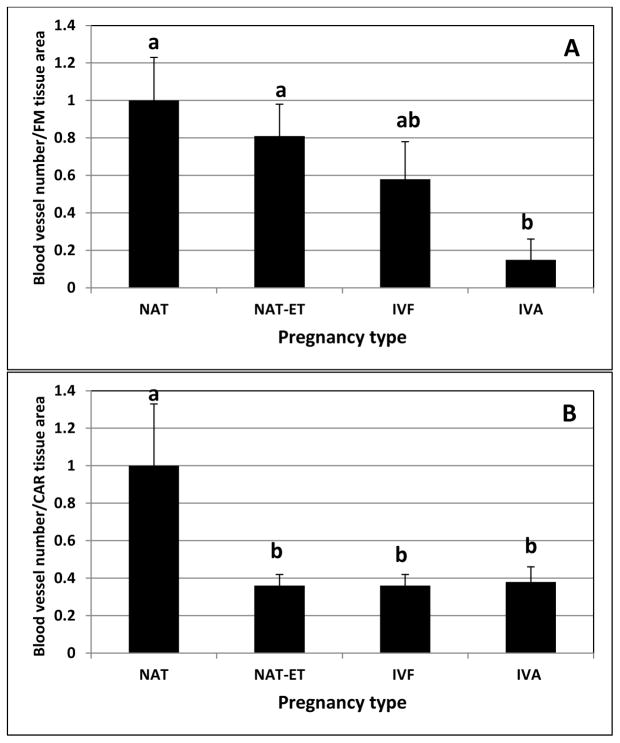
Fig. 3.
The density of blood vessels (based on SMCA staining) in FM (A) and CAR (B) from NAT, NAT-ET, IVF and IVA groups on Day 22 of pregnancy. Data are expressed as a fold-change compared to the NAT group, which was arbitrarily set as 1. In NAT, the density of blood vessels was 39.1±3.5/106 μm2 and 39.4±4.3/106 μm2 in FM and CAR, respectively. Means ± SEM with different superscript letters differ within tissue (a,bP < 0.01).

