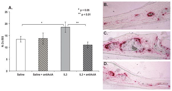
Figure 2.
Figure 2A: IL3 increases osteoclast numbers in vivo and AntiActA inhibits this effect in vivo. C57BL 9-week-old mice per group were injected intraperitoneally with saline (100 μl) or antiActA (1μg in 100μl PBS) for 7 days and injected subcutaneously over the calvaria with mIL-3 (1μg in 50μl PBS) or saline daily for 5 days under light isoflurane anesthesia. After 7 days, mice were sacrificed and calvaria fixed, decalcified, and sectioned in a coronal orientation posterior to the junction of the sagittal and coronal suture and stained for TRAP. TRAP+ osteoclasts were counted on the endosteal bone surfaces. IL-3 treatment resulted in significantly more TRAP+ cells on the endosteal surface of the calvaria as compared with saline treated controls (mean N.Oc/BS saline treated control 13.42, SD 1.18; supracalvarial IL-3 treatment 18.51, SD 2.14, p < 0.05). Animals treated with intraperitoneal anti-ActA in combination with IL-3 had a reduced number of TRAP+ cells as compared with IL-3 treated animals only, (mean N.Oc/BS supracalvarial IL-3 with intraperitoneal antiActA 11.01, SD 1.2, p<0.01).
Figure 2B - D: IL-3 increases TRAP+ OCL formation in vivo and intraperitoneal treatment with an ActA neutralizing antibody abrogates the osteoclastogenic effect of IL-3. Representative images of calvaria sections from mice treated with subcutaneous supracalvarial injections of saline (B) as compared with IL-3 (C) are shown. TRAP+ OCL stain red. Significantly more TRAP activity (red stained cells) is seen in calvarial sections from mice treated with IL-3 as compared with saline control. (D) Mice were treated with IL-3 in combination with intraperitonal antiActA, which significantly decreased the osteoclastogenic effect of IL-3. Images were captured at x20 magnification for four consecutive fields lateral to the sagittal suture. Reduced TRAP activity is seen in calvarial sections from mice treated with IL-3 in combination with anti-ActA as compared with IL-3 treatment alone.

