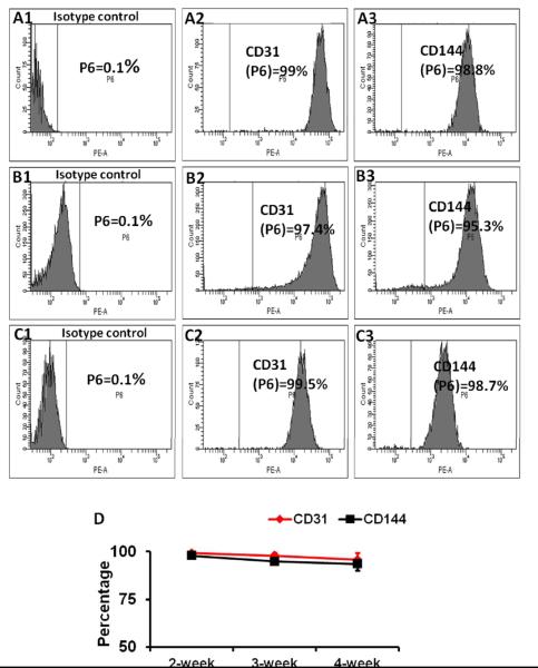
Figure 3. Differentiated hiPSC-ECs maintained EC characteristics for at least 4 weeks in vitro.
Differentiated hiPSC-ECs were purified to >95% CD31+ cells via flow cytometry, and maintained in medium supplemented with VEGF and SB431542 for 14 days (A1-A3), 21 days (B1-B3), and 28 days (C1-C3). The proportion of cells that expressed CD31 (A2, B2, C2) or CD144 (A3, B3, C3) were compared with isotype controls (A1, B1, and C1) and (D) presented as a function of time after purification.

