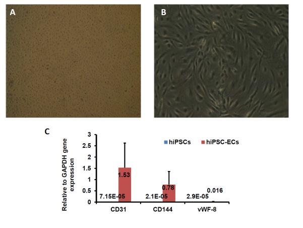
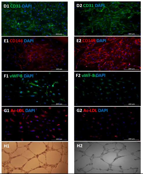
Figure 4. Characterization of differentiated hiPSC-ECs.
The morphology of the differentiated hiPSC-ECs was evaluated via images obtained at (A) 25X or (B) 100X magnification. (C) The gene expression levels of CD31, CD144, and vWF-8 mRNA were normalized to GAPDH, and presented as fold changes. Protein expression of CD31, CD144, and vWF-8 at week-1 (D1, E1, and F1) and week-4 (D2, E2, and F2) after isolation were evaluated via immunofluorescence; nuclei were counterstained with DAPI. The biological function of hiPSC-ECs was evaluated via Dil-ac-LDL uptake and the formation of tube-like structures on Matrigel at week-1 (G1 and H1) and week-4 (G2 and H2) after isolation; (Bar=200 μm, Panel E: magnification=200x).


