Abstract
Purpose
To examine the accuracy and predictive ability of B-scan ultrasonography in the post-repair assessment of an open globe injury.
Methods
In all, 965 open globe injuries treated at the Massachusetts Eye and Ear Infirmary between 1 January 2000 and 1 June 2010 were retrospectively reviewed. A total of 427 ultrasound reports on 210 patients were analyzed. Ultrasound reports were examined for the following characteristics: vitreous hemorrhage, vitreous tag, retinal tear, RD (including subcategories total RD, partial RD, closed funnel RD, open funnel RD, and chronic RD), vitreous traction, vitreous debris, serous choroidal detachment, hemorrhagic choroidal detachment, kissing choroidal detachment, dislocated crystalline lens, dislocated intraocular lens (IOL), disrupted crystalline lens, intraocular foreign body (IOFB), intraocular air, irregular posterior globe contour, disorganized posterior intraocular contents, posterior vitreous detachment, choroidal vs retinal detachment, vitreal membranes, and choroidal thickening. The main outcome measure was visual outcome at final follow-up.
Results
Among 427 B-scan reports, there were a total of 57 retinal detachments, 19 retinal tears, 18 vitreous traction, 59 serous choroidal detachments, 47 hemorrhagic choroidal detachments, and 10 kissing choroidal detachments. Of patients with multiple studies, 26% developed retinal detachments or retinal tears on subsequent scans. Ultrasound had 100% positive predictive value for diagnosing retinal detachment and IOFB. The diagnoses of retinal detachment, disorganized posterior contents, hemorrhagic choroidal detachment, kissing choroidal detachment, and irregular posterior contour were associated with worse visual acuity at final follow-up. Disorganized posterior contents correlated with particularly poor outcomes.
Conclusions
B-scan ultrasonography is a proven, cost-effective imaging modality in the management of an open globe injury. This tool can offer both diagnostic and prognostic information, useful for both surgical planning and further medical management.
Keywords: ultrasound, ultrasonography, open globe, ocular trauma
Introduction
One of the most important roles for B-scan ultrasonography remains diagnosis and follow-up of ocular trauma. Ultrasonography can function to diagnose intraocular pathology immediately following ocular trauma when no additional imaging is possible,1 but also during evaluation or follow-up if a posterior view is obscured. The seminal study in penetrating ocular trauma by Rubsamen et al2 demonstrated 100% sensitivity and specificity for preoperative ultrasound diagnosis of retinal detachments and intraocular foreign bodies (IOFBs) in 46 patients. In addition, the article revealed retinal detachment, subretinal hemorrhage, and hemorrhagic choroidal detachment on imaging to be poor prognostic factors for visual outcome. However, the study population was limited in size and restricted to preoperative use of B-scan ultrasound. More recently, ultrasound has had comparable results to CT scan in assessing ocular blast injuries in a military setting.3, 4
At our institution, B-scan is rarely used preoperatively in the setting of an open globe because of concerns over ocular pressure expulsing ocular contents in an open eye, increased artifacts in a smaller irregularly contoured globe, and ocular manipulation potentially increasing the risk of infection before surgical wound closing. Rather, CT scans are routinely used in the preoperative evaluation of the open globe patient as studied by Joseph et al.5 B-scan is a routine diagnostic tool after globe repair when developing a plan for further surgical rehabilitation. The purpose of this study is to examine B-scans in a large cohort of patients who have undergone surgical repair of open globe injuries to validate the accuracy of B-scan ultrasonography in diagnosing various traumatic pathologies and predict patient outcomes from these initial findings.
Materials and methods
A retrospective chart review was conducted on 965 open globe injuries at the Massachusetts Eye and Ear Infirmary between 1 January 2000 and 1 June 2010, as approved by the Massachusetts Eye and Ear Infirmary Institutional Review Board. An open globe injury was defined as a traumatic full-thickness break in the corneoscleral wall of the eye. B-scan ultrasonography was performed in cases of limited view to the posterior segment after surgical repair of the open globe injury. All available B-scan ultrasonography reports were examined and primary findings were entered into a computerized database with demographic and clinical data for review and analysis. Recorded ultrasound findings included: vitreous hemorrhage, vitreous tag, retinal tear, RD (including subcategories of total RD, partial RD, closed funnel RD, open funnel RD, and chronic RD), vitreous traction, vitreous debris, serous choroidal detachment, hemorrhagic choroidal detachment, kissing choroidal detachment, dislocated crystalline lens, dislocated intraocular lens (IOL), disrupted crystalline lens, IOFB, intraocular air, irregular posterior globe contour, disorganized posterior intraocular contents, posterior vitreous detachment, choroidal vs retinal detachment, vitreal membranes, and choroidal thickening. If the ultrasound was inconclusive for retinal pathology, the following categories were used: possible RD and possible retinal tear. The data included age, sex, information about the time and place of injury, mechanism of injury, initial examination, open globe repair surgical details including location of wound and surgical techniques, and follow-up examinations, and outcomes. The following data were entered for each surgery: zone of injury, canthotomy, uveal reposition, lensectomy, IOL insertion, IOFB removal, lid laceration repair, anterior chamber washout, zonular dialysis, disinsertion of rectus muscle, penetrating keratoplasty, cataract wound dehiscence, pars plana vitrectomy, anterior vitrectomy, Weck-cel vitrectomy, epiretinal membrane peel, scleral buckle, silicone oil, gas tamponade, endolaser therapy, cryotherapy, glaucoma operation (tube, shunt, or valve), or enucleation. The following data were entered for each clinic visit: corrected and uncorrected vision, intraocular pressure, corneal edema, cataract formation, choroidal detachment, endophthalmitis, glaucoma, hyphema, corneal neovascularization, corneal scar, phthisis, retinal detachment, retinal scar, and vitreous hemorrhage. If a specific data field was not available for patients, then they were excluded from that particular analysis. When possible, an ocular trauma score (OTS) was calculated based on the data available at presentation, as described by Kuhn et al.6 This scoring system helps determine severity of injury and predict visual outcome by assigning point values to initial visual acuity, afferent pupillary defect, endophthalmitis, retinal detachment, and mechanism of injury. The raw score ranges from 0 to 100, with 100 being the least traumatic.
Stepwise multiple linear regression was performed using SPSS version 17.0 software (SPSS, Inc., Chicago, IL, USA) to determine the predictive value of B-scan findings on final visual acuity. A P-value of <0.05 was considered statistically significant.
Results
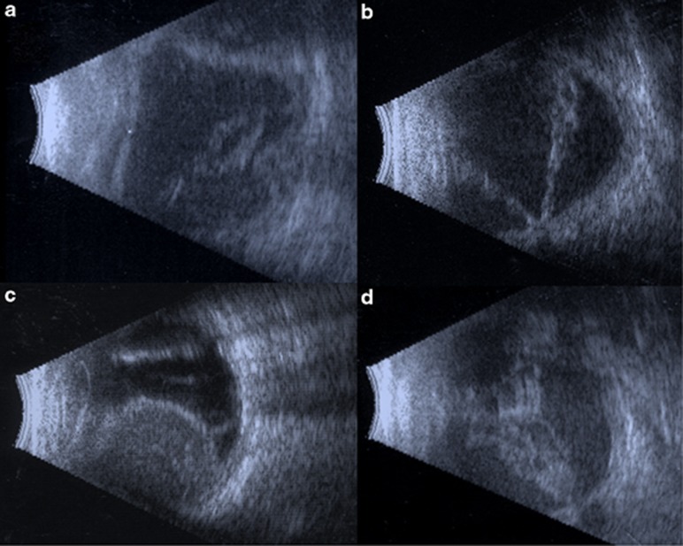
Of the 965 patients with open globe injuries at the Massachusetts Eye and Ear Infirmary between January 2000 and June 2010, 210 patients were found with media opacity limiting fundus examination who underwent B-scan ultrasonography at MEEI. The time of scan ranged from postoperative day 1 to 5 years after the initial injury, with a median of 9 days after the repair. Mean patient age was 43.4 years (range 1 to 94 years). The cohort was 73% male. A total of 427 B-scan reports were reviewed on these patients. Of the patients who received B-scan ultrasonography, 79 underwent subsequent retinal surgery at our institution. Of the 427 scans examined independently, there were the following findings (see Table 1): 57 retinal detachments (including 26 total RD, 4 open funnel RD, and 4 closed funnel RD), 19 retinal tears, 18 vitreous traction, 59 serous choroidal detachments, 47 hemorrhagic choroidal detachments, and 10 ‘kissing choroidal' detachments. When limiting the data set to a patient's initial B-scan, the diagnoses were as follows (see Table 2): 32 retinal detachments (14 total RD, 1 open funnel, and 1 closed funnel), 14 retinal tears, 15 vitreous traction, 47 serous choroidal detachments, 37 hemorrhagic choroidal detachments, and 7 ‘kissing choroidal' detachments. Figure 1 illustrates the ultrasound findings of four example cases. Retinal detachment could not be definitely evaluated in 39 scans (9.1%) because of traumatic changes in anatomy. Of the 210 total patients, 123 patients had multiple ultrasound reports. Of these 123 patients, 21 (17%) had identical results with subsequent scans and 102 (83%) had at least one diagnostic difference between the multiple scans. Most of these differences were minor findings. However, of 96 patients without a retinal tear or retinal detachment on initial ultrasound examination, 26% developed a retinal tear or retinal detachment on subsequent scans.
Table 1. Total B-scan findings.
| Variable | Number |
|---|---|
| Total B-scans | 427 |
| RD | 57 |
| Total RD | 26 |
| Open funnel RD | 4 |
| Closed funnel RD | 4 |
| Retinal tear | 19 |
| Serous choroidal detachment | 59 |
| Hemorrhagic choroidal detachments | 47 |
| Kissing choroidal detachment | 10 |
| Vitreous traction | 18 |
Abbreviation: RD, retinal detachment.
Table 2. B-scan findings on initial ultrasound.
| Variable | Number |
|---|---|
| Total B-scans | 210 |
| RD | 32 |
| Total RD | 14 |
| Open funnel RD | 1 |
| Closed funnel RD | 1 |
| Retinal tear | 14 |
| Serous choroidal detachment | 47 |
| Hemorrhagic choroidal detachments | 37 |
| Kissing choroidal detachment | 7 |
| Vitreous traction | 15 |
Abbreviation: RD, retinal detachment.
Figure 1.
Ultrasonography examples in ocular trauma. (a) A 71-year-old man with a ruptured globe in a motor vehicle accident. The wound involved zones II and III. B-scan performed 20 days after operation showed loose and membranous vitreous debris, flattening of the globe, and diffuse choroidal thickening with bulging of the optic nerve into the eye suggestive of hypotony. At 2 months after operation, the patient had light perception vision with phthisis. (b) A 14-year-old female who sustained a BB to the eye with retained intraocular FB. B-scan 24 days after the repair and removal of the BB showed a V-shaped membrane, nonmobile, diffusely thickened with cystic components, attaching at the optic nerve, suggestive of a total open funnel retinal detachment. This was verified and repaired 1 week later with PPV/retinectomy/laser and oil. She ultimately developed LP vision and pain and received a retrobulbar chlorpromazine injection for pain. (c) A 76-year-old man with a blunt rupture of his globe resulting in a ruptured extracapsular cataract extraction wound, limbal laceration, and uveal extrusion. B-scan 2 days after the repair showed large dome-shaped membranous elevations containing nonmobile moderate reflective debris with anterior attachment suggestive of non-kissing hemorrhagic choroidal detachments. An overlaying membrane off the base of the choroidal detachment is suggestive of a retinal detachment. After 6 weeks, B-scan showed shallow choroidal detachment, and total retinal detachment. He then underwent scleral buckle, lensectomy, PPV, laser and gas 6 weeks later. Final vision was LP. (d) A 57-year-old man with a blunt rupture resulting in a zone II and III scleral rupture. B-scan 5 weeks later showed diffusely thickened, nonmobile, V-shaped membrane with a point of attachment at the optic nerve, representing a total funnel retinal detachment. After 2 months, the patient had no light perception vision with phthisis.
There were 79 patients with available operative reports for subsequent retinal surgery. Of 16 patients with retinal detachment diagnosed on B-scan, all detachments were confirmed intraoperatively. Of the 3 patients diagnosed with IOFB on ultrasound, IOFB was found by the retina surgeon in all cases. Of the 19 patients with hemorrhagic choroidal detachment, the diagnosis was confirmed intraoperatively in 8 cases but not in the remaining 11 patients.
B-scan results were often predictive of operative outcome. Of the four patients found to have closed funnel RD on ultrasound, two of these detachments were found to be inoperable. Of the four patients with irregular posterior contour, one patient was found to have an inoperable RD. Of the four patients with kissing choroidal detachments, three patients were found to have retinal detachments during surgery. The report of ‘disorganized posterior contents' on B-scan predicted a poor prognosis. Of these 16 patients, 2 (13%) had no follow-up, 3 patients (19%) required secondary enucleation, 8 patients (50%) were found to be NLP at final follow-up, 3 patients (19%) were found to be LP at their last follow-up, and 3 patients (19%) underwent retinal surgery. Of the patients with surgical follow-up, all three patients were found to have total retinal detachment (one case was deemed inoperable and two patients were diagnosed with proliferative vitreoretinopathy).
We performed a linear regression analysis with primary outcome of LogMAR final visual acuity. The following B-scan findings correlated with worse final vision (adjusted R2=0.238; F=13.008; P<0.001; see Table 3): retinal detachment (β=0.258, P<0.001), disorganized posterior contents (β=0.260, P<0.001), hemorrhagic choroidal detachment (β=0.200, P=0.002), kissing choroidal detachment (β=0.160, P=0.013), and irregular posterior contour (β=0.165, P=0.014). There were no variables that were associated with improved visual acuity. The β-coefficient represents the slope of the regression line that correlates with the degree of visual acuity change for each variable studied.
Table 3. Linear regression model for B-scan findings predicting worse final visual outcome.
| B-scan finding | β | P-value |
|---|---|---|
| Retinal detachment | 0.258 | <0.001 |
| Disorganized posterior contents | 0.260 | <0.001 |
| Hemorrhagic choroidal detachment | 0.200 | 0.002 |
| Kissing choroidal detachment | 0.160 | 0.013 |
| Irregular posterior contour | 0.165 | 0.014 |
Discussion
Ultrasonography continues to prove itself as a relatively inexpensive and effective imaging modality. In this cohort of open globe injuries, hundreds of diagnoses were made that would have been impossible given the lack of view for a full ophthalmic examination. This platform was particularly useful for posterior segment pathology. The most common findings in open globe injury patients with limited view to the fundus were serous choroidal detachment, hemorrhagic choroidal detachment, retinal detachment, retinal tear, and vitreous traction.
In a trauma population, if a retinal detachment or intraocular foreign body is diagnosed on ultrasound, there is a high likelihood that these findings will be confirmed clinically. In fact, every IOFB and retinal detachment noted on ultrasound in our study was corroborated during vitreoretinal surgery. Thus, B-scan demonstrated a 100% positive predictive value for retinal detachment and IOFB. The imaging was also useful in diagnosing hemorrhagic choroidal detachment (42% positive predictive value). It is possible that this lower predictive value represents resolution of a hemorrhagic choroidal detachment between B-Scan and surgery. In addition, it is possible that pathology diagnosed as a hemorrhagic choroidal detachment was actually a serous choroidal detachment that resolved with infusion pressure during vitrectomy. However, ultrasound may not be a definitive imaging modality for differentiating serous from choroidal detachments. Thus, we recommend evaluating ultrasound findings in the full clinical context to maximize diagnostic yield.
Ocular contents are not static following severe trauma.7, 8, 9, 10 While a poor view to the fundus can take weeks to resolve without intervention, the degree of posterior disorganization may evolve. Oftentimes, B-scan can provide definitive diagnosis only after ocular contents settle, with a significant progression in diagnostic potential as time advances. For this reason, serial ultrasound imaging of a traumatized eye with media opacity can provide clinically useful information for planning management of complex pathology. As was seen in our data, a very high percentage of those patients who had serial scans had a change in interpretation of at least one finding, with one quarter developing retinal detachments or retinal tears diagnosed by ultrasound on follow-up examination.
Findings on ultrasonography can assist in prediction of visual outcomes. The diagnosis of retinal detachment, disorganized posterior contents, hemorrhagic choroidal detachment, and kissing choroidal detachment correlate with worse end vision. Perhaps the poorest prognostic factors are closed funnel retinal detachments, which are often inoperable, and disorganized posterior contents. The patients with disorganized contents typically have light perception or worse vision and frequently require secondary enucleation.
B-scan ultrasonography is an effective adjuvant imaging modality in the evaluation of ocular trauma patients. Although computed tomography is valuable in identifying foreign bodies, the lack of resolution for posterior pathology limits the clinical utility after the initial traumatic event. B-scan can provide more accurate diagnoses, as well as help to predict visual outcomes. The results of this study also validate the use of ultrasonography after open globe repair for planning of secondary surgical intervention.

Acknowledgments
Payment for lectures (to LH).
The authors declare no conflict of interest.
Footnotes
Author contributions
Design of the study (MTA, GY, and CMA); conduct of the study (MTA, GY, LH, and CMA); data collection (MTA, GY, LH, and CMA); management (MTA); analysis (MTA, GY, and CMA); interpretation of data (MTA, GY, LH, and CMA); and preparation, review, or approval of manuscript (MTA, GY, LH, and CMA).
References
- Sawyer MN. Ultrasound imaging of penetrating ocular trauma. J Emerg Med. 2009;36:181–182. doi: 10.1016/j.jemermed.2007.04.005. [DOI] [PubMed] [Google Scholar]
- Rubsamen PE, Cousins SW, Winward KE, Byrne SF. Diagnostic ultrasound and pars plana vitrectomy in penetrating ocular trauma. Ophthalmology. 1994;101:809–814. doi: 10.1016/s0161-6420(94)31254-1. [DOI] [PubMed] [Google Scholar]
- Ritchie JV, Horne ST, Perry J, Gay D. Ultrasound triage of ocular blast injury in the military emergency department. Mil Med. 2012;177:174–178. doi: 10.7205/milmed-d-11-00217. [DOI] [PubMed] [Google Scholar]
- Gay DA, Ritchie JV, Perry JN, Horne S. Ultrasound of penetrating ocular injury in a combat environment. Clin Radiol. 68:82–84. doi: 10.1016/j.crad.2012.05.015. [DOI] [PubMed] [Google Scholar]
- Joseph DP, Pieramici DJ, Beauchamp NJJr. Computed tomography in the diagnosis and prognosis of open-globe injuries. Ophthalmology. 2000;107:1899–1906. doi: 10.1016/s0161-6420(00)00335-3. [DOI] [PubMed] [Google Scholar]
- Kuhn F, Maisiak R, Mann L, Mester V, Morris R, Witherspoon CD.The Ocular Trauma Score (OTS) Ophthalmol Clin North Am 200215163–165.vi. [DOI] [PubMed] [Google Scholar]
- Andreoli MT, Andreoli CM. Surgical rehabilitation of the open globe injury patient. Am J Ophthalmol. 2012;153:856–860. doi: 10.1016/j.ajo.2011.10.013. [DOI] [PubMed] [Google Scholar]
- Salehi-Had H, Andreoli CM, Andreoli MT, Kloek CE, Mukai S. Visual outcomes of vitreoretinal surgery in eyes with severe open-globe injury presenting with no-light-perception vision. Graefes Arch Clin Exp Ophthalmol. 2009;247:477–483. doi: 10.1007/s00417-009-1035-4. [DOI] [PubMed] [Google Scholar]
- Savar A, Andreoli MT, Kloek CE, Andreoli CM. Enucleation for open globe injury. Am J Ophthalmol. 2009;147:595–600. e1. doi: 10.1016/j.ajo.2008.10.017. [DOI] [PubMed] [Google Scholar]
- Andreoli CM, Andreoli MT, Kloek CE, Ahuero AE, Vavvas D, Durand ML.Low rate of endophthalmitis in a large series of open globe injuries Am J Ophthalmol 2009147601–608.e2. [DOI] [PubMed] [Google Scholar]


