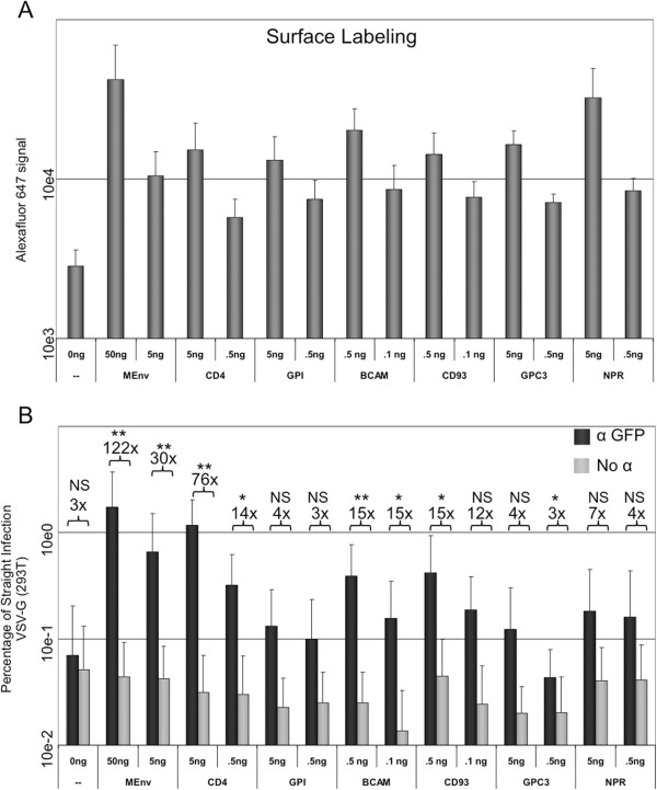
Figure 5.
Co-capture using cellular glycoproteins. Co-capture was performed on virus produced from cells transfected as before using VSV-G and the indicated amounts of GFP-tagged cellular proteins or YFP-tagged MLV Env. A) The surface expression of the indicated proteins in transfected cells was assayed by surface labeling GFP with an anti-GFP antibody conjugated to Alexa Fluor 647, which was detected by flow cytometry. B) The luciferase signal from the anti-GFP captured samples and the no-antibody captured samples normalized to the straight control is shown. The average fold increase of antibody/no antibody is shown for each pair. All graphs are the average of at least three independent experiments and standard deviations are shown. *p < 0.05; **p < 0.01; NS p > 0.05.

