Abstract
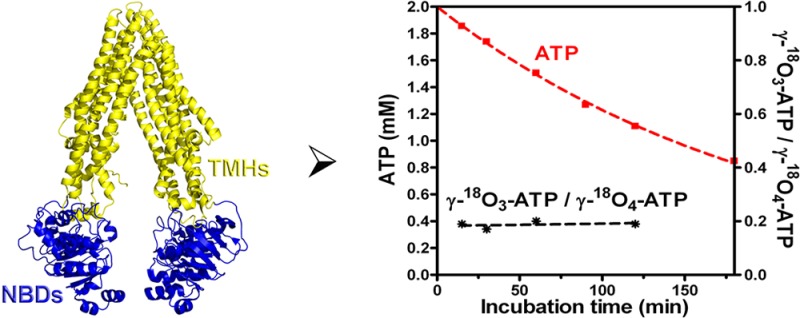
P-glycoprotein (P-gp) is a member of the ABC transporter family that confers drug resistance to many tumors by catalyzing their efflux, and it is a major component of drug–drug interactions. P-gp couples drug efflux with ATP hydrolysis by coordinating conformational changes in the drug binding sites with the hydrolysis of ATP and release of ADP. To understand the relative rates of the chemical step for hydrolysis and the conformational changes that follow it, we exploited isotope exchange methods to determine the extent to which the ATP hydrolysis step is reversible. With γ18O4-labeled ATP, no positional isotope exchange is detectable at the bridging β-phosphorus–O−γ-phosphorus bond. Furthermore, the phosphate derived from hydrolysis includes a constant ratio of three 18O/two 18O/one 18O that reflects the isotopic composition of the starting ATP in multiple experiments. Thus, H2O-exchange with HPO42– (Pi) was negligible, suggesting that a [P-gp·ADP·Pi] is not long-lived. This further demonstrates that the hydrolysis is essentially irreversible in the active site. These mechanistic details of ATP hydrolysis are consistent with a very fast conformational change immediately following, or concomitant with, hydrolysis of the γ-phosphate linkage that ensures a high commitment to catalysis in both drug-free and drug-bound states.
The ATP-binding cassette (ABC) transporters comprise a large family of transmembrane ATP-dependent efflux pumps that are best described by their shared “ATP-switch” mode of action.1 In humans, the isoform ABCB1, or P-glycoprotein, plays a significant role in cellular drug resistance in tumors in which it is overexpressed, and it contributes to drug–drug interactions due to its high level expression in hepatic, renal, and intestinal tissue.2−6 As a result, there is significant interest in designing inhibitors of P-gp that could be used to modulate drug efflux, particularly in the central nervous system,7 and efforts to develop inhibitors could be facilitated by further understanding of the role of substrate–nucleotide binding and concomitant structural changes in transmembrane domains (TMDs) and nucleotide binding domains (NBDs) during the ATP catalytic cycle.
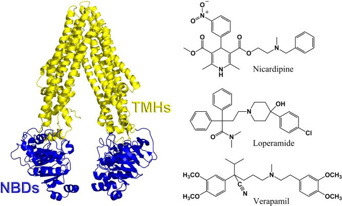
On the basis of structural models of murine and Caenorhabditis elegans proteins,8,9 the human P-gp likely consists of a dimer of two TMDs with six transmembrane helices (TMHs) that form a hydrophobic and promiscuous drug binding site, or sites with access to the plasma membrane inner leaflet (Figure 1). These sites are coupled functionally to two NBDs on the cytosolic side of the membrane that catalyze the hydrolysis of ATP. On the basis of the structural models, it has been suggested that the NBDs are brought into close proximity upon binding nucleotide,10,11 but the magnitude of functionally important conformational changes remains unknown. All three steps in the NBD cycle (ATP binding, hydrolysis, and release of products) are associated with release of energy coupled to some form of conformational change in either the TMDs or NBDs. Although mechanistic models differ in their details depending on the particular ABC transporter, the available data indicate that ATP hydrolysis alternates between the two NBDs, and the hydrolysis, or dissociation of ADP, is used to drive distant conformational changes in the transmembrane helices, to allow drugs to be released to the extracellular surface and to “reset” the conformational state of the protein.12−15
Figure 1.
Left: Ribbon structure of murine P-gp (pdb: 3G5U) in the inward-facing nucleotide free state. The NBDs (blue) move into close proximity and the TMHs (yellow) rearrange upon nucleotide binding. Right: Chemical structures of three P-gp substrates studied in this work.
P-gp binds a remarkably wide range of drugs or probe ligands that differentially stimulate or inhibit the ATPase activity at saturating concentrations.16 In fact, several distinct binding sites have been proposed within the transmembrane helices, which may communicate allosterically despite their distinct selectivities.2 These binding sites include residues on helices 4 and 5 and 10 and 11, and, taken together, they exhibit impressive promiscuity.17 On the basis of many biochemical data, it is likely that the large binding site within the TMDs includes subsites with overlapping but distinct substrate preferences, and this could result in multiple drug translocation pathways.18,19
Despite major progress in our understanding of the human P-gp mechanism, including the availability of X-ray structures of closely related homologues,8,9 the molecular details of several aspects of the P-gp reaction cycle remain uncertain. For example, P-gp exhibits a basal ATP hydrolysis even in the absence of substrates or drug, but no physiological purpose is known for this activity. In addition, different substrates bind in different regions of the large promiscuous binding site, so it is challenging to understand how drugs bound at different sites can communicate with the NBDs to stimulate ATP hydrolysis. Although significant evidence indicates that the D-loops of the NBDs and helices 6 and 12 of the TMDs are important in the interdomain communication, it remains unknown how different substrates utilize a common mechanism or whether there are drug-dependent differences in the coupling between these domains.20−22
P-gp poses an interesting example of ATP hydrolysis because it is widely proposed that release of ADP is rate limiting in the catalytic cycle,21,23,24 which implies a significant population of the [P-gp·ADP·Pi] or [P-gp·ADP] complex at steady state, where Pi is used throughout this manuscript to abbreviate HPO42–. In addition, there is a vast body of evidence that indicates that the enzyme undergoes significant conformational change immediately after ATP hydrolysis and prior to ADP release.15,22,26 The majority of data that support the posthydrolysis conformational change exploit a nonphysiologic “trapped state” with VO43– ion replacing the Pi to form a pseudoirreversible complex. On the basis of the resulting mechanistic models, it is reasonable to expect that, if the release of ADP is sufficiently slow or incompletely coupled to formation of the posthydrolysis conformation, then the highly populated [P-gp·ADP·Pi] complex could regenerate ATP at a measurable rate, as observed for other classes of ATPases.27,28 On the other hand, if a sufficiently fast conformational change preceded ADP release, then no regeneration of ATP would be possible, and the hydrolysis step would be completely coupled to the subsequent conformational change. In effect, the relative rates of hydrolysis and conformational change are not well understood, yet they determine the commitment to catalysis once ATP is hydrolyzed.
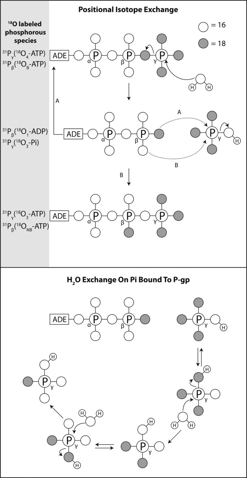
Because many ATPases demonstrate substantial reversibility of the hydrolytic step, without full commitment to catalysis, particularly in the absence of one or more of their cosubstrates, we considered the possibility that P-gp could include a reversible component in the ATP hydrolysis. For other ATPases, such reversibility has been documented by NMR-based approaches as well as mass spectrometry which both rely on the rearrangement or “isomerization” of 18O in labeled ATP and is referred to as positional isotope exchange (PIX).27−32 Therefore, we performed NMR-based PIX experiments aimed to quantify the relative rate of regeneration of ATP from hydrolytic products ADP and Pi at the active sites of the NBDs. With PIX experiments using γ-18O4-ATP, it is possible to monitor the relocation of 18O label in the bridging position of the γ-phosphate of ATP into the nonbridging position on the β-phosphate, as a measure of reformation of the ATP from ADP and 16O-/18O-containing free phosphate ion (Figure 2). The extent of PIX that occurs is essentially a measure of the forward flux to the new conformation associated with the [P-gp·ADP·Pi] complex versus backward flux to the [P-gp·ATP] complex. PIX would not be observed if the new conformation that is populated immediately after hydrolysis differed significantly in the ATP binding sites. In addition, if the ATP hydrolysis of P-gp was reversible, then different [P-gp·ADP·Pi·drug] complexes would potentially yield varying amounts of PIX. In effect, a change in reversibility, if present, would provide a direct measure of the impact of different substrates or inhibitors on linking conformational changes with ATP hydrolysis.
Figure 2.
Top: Schematic of PIX. Open and shaded circles represent 16O and 18O, respectively. Starting with γ-18O4-ATP with 18O at each of the γ-phosphorus positions, hydrolysis followed by isomerization and reformation of ATP (pathway B) yields 18O in the nonbridging (NB) versus the initial bridging (B) position of the β-phosphorus. Bottom: Schematic of H2O exchange with Pi in the [P-gp·ADP·Pi] complex. Attack of H2O on the Pi bound to P-gp leads to exchange of the 18O from the Pi with 16O.
A second measure of reaction dynamics for ATP hydrolysis has been exploited for many well-studied enzymes, wherein there is incorporation of additional water-borne oxygen atoms into the enzyme-bound Pi prior to release. If the [enzyme·ADP·Pi] complex is sufficiently long-lived, then additional rounds of H2O attack at the Pi lead to further incorporation of oxygen from H2O. We analyzed both PIX and H2O exchange with Pi with human P-gp in liposomes. We report that no detectable PIX is observed in either the basal ATPase activity of P-gp or in the presence of the probe substrates nicardipine, verapamil, or loperamide, or the inhibitor cyclosporin A. Furthermore, none of the Pi in the [P-gp·ADP·Pi·drug] complex exchanges oxygen with water in any of the complexes studied. Thus, these data further support the model wherein there is fast conformational change immediately post hydrolysis, which allows for fast Pi release and a high commitment to catalysis in the NBDs.
Materials and Methods
Materials
n-Dodecyl-β-d-maltopyranoside (DDM) was purchased from Affymetrix. His60 nickel affinity resin was from Clontech. Escherichia coli total lipid extract was purchased from Avanti Polar Lipids. C219 mouse IgG1 monoclonal antibody for immunoblots was from Covance. Goat antimouse IgG (H + L) secondary antibody, DyLight 800 conjugate was from Thermo Scientific. Commercial P-gp supersomes were purchased from BD Biosciences.
Adenosine 5′-triphosphate sodium salt, γ-18O4-ATP, with a per-site basis isotopic enrichment of at least 94% was purchased from Cambridge Isotopes.
Insect cell lines and expression medium were from Invitrogen. Cholesterol, chloroform, and all other reagents were from Sigma-Aldrich.
Baculovirus Expression of Human P-Glycoprotein (P-gp)
The strains, expression constructs, and growth conditions for expression of human P-gp in insects cells have been described previously.33
Preparation of Supersomes or Microsomes
Throughout the manuscript the term “supersome” refers to microsomes obtained from insect cells that overexpress P-gp, and “microsome” refers to those obtained from “control” insect cells not overexpressing P-gp. Trichoplusia ni (T.ni) cells expressing P-gp were harvested by centrifugation and then resuspended in ice-cold hypotonic homogenization buffer (5 mM Tris, 5 mM TCEP, 40 μM leupeptin, 2 μM pepstatin A, 4 mM benzamidine, 200 μM PMSF, pH 7.4) at a ratio of 5 mL of buffer per gram wet-weight of cells and incubated for 45 min on ice. All further steps were carried out at 4 °C or on ice. The mixture was lysed with a minimum of 10 strokes using a Potter-S homogenizer and then preclarified by spinning at 500g for 10 min. The supernatant was centrifuged at 200000g for 60 min. The supersome or microsome pellet was resuspended in a minimum of storage buffer (50 mM Tris, 250 mM sucrose, 20% w/v glycerol, 5 mM TCEP, 40 μM leupeptin, 2 μM pepstatin A, 4 mM benzamidine, 200 μM PMSF, pH 7.4) and stored at −80 °C until further use. Aliquots were removed prior to freezing to determine total membrane protein concentration and test for drug-stimulated ATPase activity.
Colorimetric Determination of Drug-Stimulated ATPase Activity
Basal versus drug-stimulated ATPase reactions were carried out with various concentrations of ATP at 37 °C and quenched with 10 mM EDTA. Liberated phosphate was quantified by colorimetric assay (34) using phosphate standards and commercial P-gp-supersomes (BD Biosciences) as a positive control.
Solubilization and Purification of P-gp
Frozen supersomes were thawed quickly and then resuspended in solubilization buffer (2% w/v DDM, 50 mM Tris, 300 mM NaCl, 1.5 mM MgSO4, 20 mM imidazole, 0.4% w/v E. coli lipids, 20% w/v glycerol, 5 mM TCEP, 40 μM leupeptin, 2 μM pepstatin A, 4 mM benzamidine, 200 μM PMSF, pH 7.4). The mixture was homogenized by passing through a narrow 25-gauge syringe and then incubated at 4 °C with mixing for 1 h. The mixture was clarified by centrifugation at 10000g for 30 min at 15 °C and the supernatant transferred to pre-equilibrated His60 resin. The protein was batch adsorbed to the resin over 4 h at 4 °C and then washed extensively with equilibration buffer (0.1% w/v DDM, 50 mM Tris, 300 mM NaCl, 1.5 mM MgSO4, 20 mM imidazole, 0.4% w/v E. coli lipids, 20% w/v glycerol, 5 mM TCEP, 40 μM leupeptin, 2 μM pepstatin A, 4 mM benzamidine, 200 μM PMSF, pH 7.4). Next, the resin was washed with buffer containing 200 mM imidazole without protease inhibitors. Fractions containing pure P-gp were collected by eluting three times with 2 bed volumes each of 500 mM imidazole and lowering the pH from 7.4 to 6.8.
Preparation of P-gp Liposomes
Purified P-gp was incorporated into preformed liposomes as follows. E. coli lipids/cholesterol 4:1 films were resuspended in 50 mM Tris, 150 mM NaCl, pH 7.4 using a bath sonicator. The opaque mixture was subjected to several freeze–thaw cycles in liquid nitrogen prior to passing through a mini-extruder (Avanti Polar lipids) fitted with a 200 nm polycarbonate (PC) membrane. Detergent -saturated liposomes were used for P-gp-reconstitution, and the Rsat value was determined by titrating a sample of preformed liposomes with detergent and measuring the change in absorbance at 540 nm. When preparing detergent-saturated liposomes for mixing with purified P-gp, the Rsat value was adjusted to compensate for detergent and lipid present in purified P-gp sample. Upon mixing, the P-gp/detergent/liposome sample was incubated for 1 h with mixing. Detergent was removed by incubating the mixture with prewashed Amberlite-XAD beads that were prewashed with methanol and resuspended in buffer for a minimum of 2 h at 25 °C, and the mixture was passed several times through a 200 nm PC membrane using an extruder. The resulting P-gp-liposome prep was stable at −80 °C with no significant loss in ATPase activity.
Determination of Total Phosphorus in Phospholipids and Protein Concentration
Phospholipids were quantified based on the colorimetric assay recommended by Avanti Polar Lipids, Inc., using phosphate standards. Protein concentration was determined using trichloroacetic acid (TCA)/acetone protein precipitation, followed by Bio-Rad DC protein assay.
NMR Spectroscopy
All NMR experiments were performed at 25 °C on a 499.73 MHz Agilent DD2 spectrometer equipped with a 5-mm AutoX Dual Broadband, z-axis pulsed-field gradient probe head.
Solutions of 31 nM liposomes-reconstituted P-gp (200 nm E. coli lipids/cholesterol liposomes) were incubated at 37 °C with 2 mM γ-18O4-ATP, 50 mM Tris·HCl, 15 mM NH4Cl, 5 mM MgSO4, 2.5 mM EGTA, 0.02 wt % NaN3, 2 mM TCEP, pH 7.4 in the absence (basal activity) or in presence (stimulated activity) of 50 μM nicardipine, loperamide or verapamil (from 10 mM stock solutions in dimethyl sulfoxide, Sigma-Aldrich). Inhibition of P-gp was studied with final concentrations of cyclosporin A of 20 μM, by dilution from a stock solution in DMSO, to yield a final DMSO concentration of 1%. Aliquots were taken at variable times, quenched by addition of 20 mM EDTA, pH adjusted to approximately 8.5 with 5 M NaOH, and spun at 14 000 rpm for 5 min to remove insoluble material. The supernatant was transferred to a clean tube and D2O added to a final concentration of 10% v/v. The final cosolvent content was <1% v/v. Adding EDTA quenched ATPase activity and sequestered all divalent cations to minimize the exchange broadening. A high pH value was also employed to minimize broadening due to protons exchange.35
PIX positive control samples were prepared as above but using commercial human P-gp-supersomes (28.2 μg/mL total protein, BD Gentest) or sf9 insect cell-microsomes (28.2 μg/mL total protein, BD Supersomes) and adding 2.5 mM EGTA to inhibit Ca2+-dependent ATPases. Alternatively, to inhibit other major ATPases other than P-gp including Na+/K+ ATPases and F1, H+/K+ ATPases, 1 mM ouabain (Sigma-Aldrich), 8 mM sodium azide (Sigma-Aldrich), and 50 μM mellitin were used.
31P NMR spectra (at 202.29 MHz) were acquired at a resolution of 16k complex points in the time domain with 2048 accumulations each (sw = 7062.1 Hz, d1 = 1.5 s). Data were zero filled to 32k points, and no apodization function was applied before Fourier transformation. Spectra were referenced to Phosphoric acid 85 wt % in H2O (Sigma-Aldrich).
All data were processed and analyzed using MNova 8.1 processing software (Mestrelab Research, Santiago de Compostela, Spain). All NMR experiments were performed at least four times on at least two different preparations of P-gp supersomes or P-gp liposomes.
Results
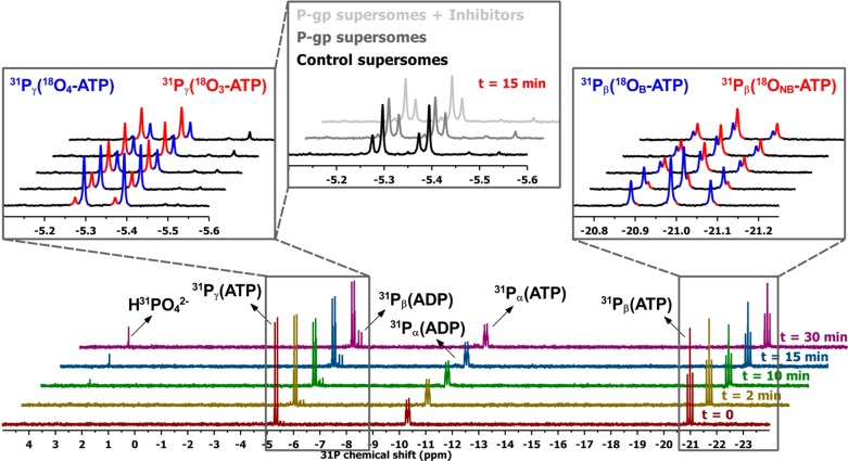
To establish a PIX assay, “positive control” supersomes known to contain multiple ATPases were incubated for varying times with labeled ATP, where each oxygen atom attached to the γ-phosphorus was 18O. The aim of these experiments was to demonstrate the feasibility of observing PIX with membrane ATPases in our hands, rather than to quantify PIX for any specific ATPase, and to determine if PIX due to P-gp could be detected in this environment. The 31P nuclei of ATP were monitored by NMR; it is well established that the chemical shift of 31P is sensitive to the oxygen isotope that is attached to it, and whether the oxygen isotope is in a position that bridges the γ and β phosphorus atoms or in a nonbridging position.29 Specifically, the substitution of an 18O for an 16O induces a ∼0.02 ppm (about 4 Hz at 202.93 MHz) downfield shift in the 31P NMR resonance per oxygen substituted, while the effect of 18O exchange from the βγ-bridge position to a β-nonbridge position on the 31Pβ resonance is a less pronounced upfield shift (∼0.01 ppm or about 2 Hz at 202.93 MHz). This enables PIX at the γ-phosphoryl group to be easily detected by inspection of the 31P NMR spectrum. In fact, the 18O exchange from the βγ-bridge position to a β-nonbridge position results in a decrease in the signal of 31Pγ with four bound 18O and an increase in the signal for the 31Pγ with three 18O atoms (Figure 3).
Figure 3.
Positional isotope exchange with commercial human P-gp supersomes. Total proteins (28.2 μg/mL) were incubated with 2 mM γ-18O4-ATP, 50 mM Tris·HCl, 15 mM NH4Cl, 5 mM MgSO4, 2.5 mM EGTA, 0.02 wt % NaN3, 2 mM TCEP, pH 7.4 in the presence of 50 μM nicardipine. For the samples with inhibitors (gray spectra top middle panel), conditions were as described in Materials and Methods. 31P NMR spectra were recorded at the times indicated. In the left-hand inset, the 31Pγ signals of ATP bearing four or three 18O are presented in blue and red, respectively. The 31Pβ signals from ADP clearly demonstrate that ADP is formed concomitant with the changes in isotopic composition of the 31Pγ signal (red vs blue). In the right-hand inset, the 31Pβ signals originating from ATP with 18O at the bridging position (18OB) or at the nonbridging position (18ONB) are shown in blue and red, respectively.
After incubation of supersomes with 18O-labeled ATP, the Mg2+ in the assay buffer was chelated with EDTA to eliminate the line broadening that results from rapid dissociation and association of Mg2+ to the ATP. For a series of incubations, the NMR spectra of the ATP were then collected at varying times. For supersomes with expressed P-gp, clear NMR signals from ADP were observed and increased in intensity with increasing time of incubation (Figure 3). In addition, the appearance of 18O in the nonbridging position of the β-phosphorus was readily apparent. PIX was easily detected in commercial supersomes that contain overexpressed P-gp. Interestingly, for several preparations of microsomes and P-gp supersomes, those containing overexpressed P-gp consistently demonstrated a clear increase in PIX compared to control supersomes (Figure 3). This suggested that P-gp was contributing significantly to the observed PIX. In order to determine whether other highly abundant ATPases contributed to the PIX, experiments were performed in the presence of 2.5 mM EGTA, to inhibit Ca2+-dependent ATPases, and 1 mM oubain, 50 μM mellitin and 8 mM sodium azide, to inhibit Na+/K+ ATPases and F1 H+/K+ ATPases. These inhibitors had no detectable effect on the observed PIX, suggesting the possibility that P-gp contributes significantly in the P-gp supersomes. Similarly, however, the addition of nicardipine at concentrations that stimulate purified P-gp had no effect on the overall ATPase activity or PIX of P-gp supersomes, indicating that P-gp is not a dominant source of ATPase activity or PIX in these membranes. Results shown in Figure 3 are representative. The degree of PIX in P-gp supersomes as measured by the ratio γ-18O3-ATP/γ-18O4-ATP varied less than 10% between preparations and between experiments with the same preparation.
Apparently, multiple ATPases are active in these membranes and contribute to PIX, such that inhibition or stimulation of one or a few ATPases does not yield a detectable change.
After 30 min, 31.8% of the residual (nonhydrolyzed) ATP was γ-18O4-ATP, 63.1% was present as γ-18O3-ATP, 5.2% as γ-18O2-ATP, and γ-18O-ATP was not detectable. Therefore, ∼63% of the total residual ATP was exchanged after 30 min. At t0, γ-18O4-ATP was ∼85.4%, and γ-18O3-ATP was ∼14.6%. PIX clearly occurs with these P-gp supersomes, and the potential contribution of P-gp is discussed further below. Most importantly, these experiments demonstrated that PIX is observable in our hands.
Having established that we could observe PIX in a sample with our experimental protocol using the “positive control”, we examined purified P-gp reconstituted in liposomes. The purified protein was judged to be >90% pure based on gel electrophoresis (Supporting Information). In preparing P-gp reconstituted liposomes using purified P-gp expressed in insect cells, it was observed that using preformed liposomes to which detergent had been added to the saturation point (Rsat) increased the yield and activity of the preparation. The addition of TCEP as a reducing agent was also critical for maintaining stability in P-gp liposome preparations and activity in PIX experiments. These P-gp liposomes exhibited a 2.8–4.0-fold increase in ATPase activity with nicardipine compared to basal hydrolysis in the absence of drug, depending on the liposome preparation used.
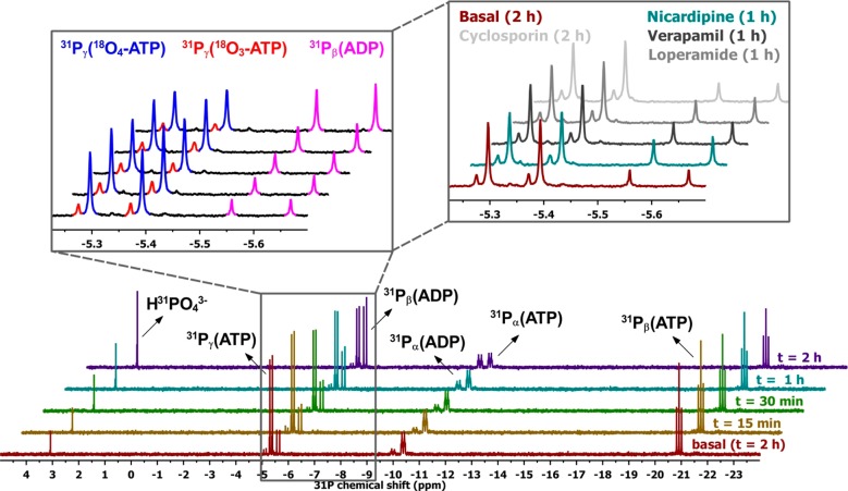
In contrast to the P-gp supersomes, purified P-gp in liposomes exhibited no detectable PIX. Using 18O-labeled ATP as above, the data for drug-free P-gp or in the presence of either 50 μM nicardipine, 50 μM loperamide or 50 μM verapamil or 20 μM cyclosporin A demonstrate no 18O in the nonbridging position at any point during the time course of ATP hydrolysis (Figure 4). This is a remarkable result, inasmuch as many two-substrate enzymes that use ATP exhibit detectable reversibility in the hydrolysis.36,37 Notably, however, some ATPases yield no PIX.31
Figure 4.
Absence of positional isotope exchange with purified liposome-reconstituted P-gp. 31 nM P-gp was incubated with 2 mM γ-18O4-ATP, 50 mM Tris·HCl, 15 mM NH4Cl, 5 mM MgSO4, 2.5 mM EGTA, 0.02 wt % NaN3, 2 mM TCEP, pH 7.4 in the presence of 50 μM nicardipine. The specific activity for the Nicardipine-stimulated P-gp was 1660 nmol of Pi/min/mg protein, at 2 mM ATP concentration, 37 °C. The reactions were conducted as described in the text, and 31P NMR spectra were recorded at the times indicated. The incubation was also conducted in the presence of 50 μM loperamide or 50 μM verapamil, or 20 μM cyclosporin A (right-hand side inset).
Exchange of Oxygen from H2O into Phosphate Ion
As a second probe of the lifetime of the [P-gp·ADP·Pi] complex, we monitored the incorporation of 16O from water into the Pi resulting from ATP hydrolysis. For some ATPases, further “attack” of water at Pi results in incorporation of multiple oxygen atoms from H2O in addition to the one incorporated as a result of attack on the initial γ-phosphate of ATP. This occurs on the enzyme, but not in bulk solution, when active site features “activate” water as a nucleophile or Pi as an electrophile via specific or general acid or base catalysis.29,30 For each round of H2O reaction with the Pi, an additional 16O atom is incorporated (Figure 2). Thus, if no exchange occurs, the Pi will contain the single 16O from the original ATP hydrolytic reaction. In the presence of P-gp liposomes, the fraction of Pi containing either two or three 16O atoms reflected the isotopic composition of the starting ATP, indicating no incorporation of 16O resulting from a second or third attack of H2O at this phosphorus, after the initial attack on ATP. Clearly, the Pi formed in the active site is highly protected from any “activated” H2O, or it is rapidly released into bulk solution where the reaction is undetectably slow.
The combined results for the P-gp supersomes and P-gp liposomes are shown in Figure 5, which shows the time-dependent changes in the isotopically labeled species. Specifically, the concentration of ATP with three 18O atoms on the γ-phosphorus (γ-18O3-ATP), resulting from a single round of PIX, and the ATP with two 18O atoms on the γ-phosphorus (γ-18O2-ATP), resulting from two rounds of PIX, increase at the expense of starting γ-18O4-ATP. The concentrations of species with two or three 18O atoms in the ATP do change as ATP is depleted with P-gp in liposomes, as expected (Figure 4). The data for the concentration of ATP in the liposome experiment (Figure 5B) fit well to a first-order decay even at the longest time point of the experiments. This, combined with direct measurement of ATPase activity via the colorimetric assay in P-gp liposome samples incubated for times comparable to NMR experiments, indicates that no significant loss of enzyme activity occurred during the experiment. The first-order decrease in [ATP] (Figure 5B) reflects the non-steady-state nature of these experiments in which substrate depletion is significant by the end of experiments. It is useful to emphasize that, although the ATP concentration is changing in this experimental design, the ratio of γ-18O3-ATP/γ-18O4-ATP is a valid indicator of PIX at low extent of ATP hydrolysis, unless there were a kinetic isotope effect favoring hydrolysis of the γ-18O3-ATP. Such heavy atom isotope effects would be negligible in these experiments, and no significant kinetic selection for hydrolysis of initially reformed γ-18O3-ATP is expected. Even if reversibly formed γ-18O3-ATP were hydrolyzed preferentially, the ratio γ-18O3-ATP/γ-18O4-ATP would be expected to increase, as it does with supersomes, where it changes from a value of ∼0.2 to 2.0 in 30 min (Figure 5A black curve). The ratio does not change with P-gp liposomes (black curve in Figure 5B). As with the experiments with supersomes, the results with P-gp liposomes in Figures 4 and 5 are representative; three preparations of P-gp liposomes demonstrated no PIX at comparable levels of ATPase activity.
Figure 5.
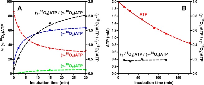
Rates of positional isotope exchange or ATP hydrolysis. (A) P-gp supersomes. The fractions of the exchanging species γ-18O4-ATP in red, γ-18O3-ATP in blue, and γ-18O2-ATP in green as a function of the incubation time are shown. Also, the time dependence of the ratio γ-18O3-ATP/γ-18O4-ATP is presented (black line). (B) Purified liposome-reconstituted P-gp. The hydrolysis rate (red line) and the time dependence of the ratio γ-18O3-ATP/γ-18O4-ATP (black line) are shown. The reactions were conducted as described in the text, and 31P NMR spectra were recorded at the times indicated. ATP concentrations and the PIX rates were determined from the areas of the 31P NMR signals of the γ-phosphorus atom of ATP corresponding to the γ-18O4-ATP, γ-18O3-ATP, and γ-18O2-ATP moieties.
Discussion
Despite significant progress in understanding the mechanistic details of P-gp, few data report on the details of ATP hydrolysis and functionally important conformational changes in the protein that gate the release of spent nucleotide or drugs. The “vanadate-trapped” P-gp has provided a valuable model for the posthydrolysis [P-gp·ADP·Pi] state, and it is clear that this state is conformationally distinct from others throughout the reaction cycle.38−40 However, little is known about the reaction dynamics for the ATP hydrolysis step and the relative rates of chemical steps vs the conformational changes that follow. Although PIX studies are well established probes of ATP hydrolases, none have been reported for P-gp. The PIX and H2O exchange experiments reported here are consistent with a few possible scenarios.
First, we discuss the results of the experiments with supersomes, for which some uncertainty exists regarding the contribution of P-gp to PIX. The observation that PIX occurs to a greater extent at equivalent degrees of ATP hydrolysis for the P-gp supersomes vs “normal” sf9 cell microsomes suggests that P-gp is either contributing to the PIX or its overexpression results in a change in levels of other ATPases that catalyze PIX. To determine whether other ATPases catalyze the PIX in these samples, incubations were performed with established inhibitors of Ca2+-ATPases, Na+/K+-ATPases, and the F1 H+/K+ ATPase. None of these inhibitors resulted in a decrease in PIX at any time point we examined, supporting the possibility that P-gp is responsible. However, it is extremely difficult to completely isolate PIX due to any specific enzyme in crude supersomes, so although these experiments are suggestive, they do not prove that P-gp in supersomes catalyzes PIX. Further experiments with mutants with altered ATPase activity may be useful to determine whether the P-gp contributes.
In contrast, the results with the P-gp liposomes are clear: no PIX occurs in this system. If the PIX in supersomes includes a contribution from P-gp, this difference obviously requires consideration. Possibly, the lipid environment of supersomes vs our liposomes could be sufficiently different to alter the catalytic properties of P-gp. Alternatively, other constituents of supersomes, including sterols or other proteins, could interact with P-gp and alter its properties. As above, further work is required to explore these possibilities. However, given the definitive lack of PIX in P-gp liposomes, we discuss the implications of those results.
One possibility is that there is fast conformational change immediately after the initial hydrolysis step that prevents reversible formation of ATP, as summarized in Figure 6. In this case, conformational rearrangement to a state that is chemically incompetent for either reformation of ATP or for attack of water on the product Pi is significantly faster than either of these chemical processes. A second possibility is that Pi dissociates very rapidly, and this causes a subsequent conformational change that disfavors Pi rebinding and ATP synthesis. In either case, rapid changes in structure or ligand occupancy disfavor the reverse reaction.
Figure 6.

Schematized mechanism for conformational changes coupled to ATP hydrolysis in P-gp. A portion of the entire catalytic cycle is shown starting from the drug and nucleotide-bound state. After hydrolysis of one ATP, the conformational change and release of Pi are fast, preventing reformation of ATP or exchange of bound HPO42– with H2O. The conformational change is unlikely to include full dissociation of the NBD dimers but rather is fast with minor structural rearrangement. This conformational change is sufficient to ensure a high commitment to catalysis for ATP hydrolysis.
Regarding the role of conformational change in P-gp catalysis, there are currently two competing models proposed to explain the events involved in drug efflux by P-gp that differ in the timing of drug/substrate release with ATP binding or hydrolysis. The first uses ATP binding and NBD dimerization as the pumping mechanism.41,42 Interaction of drug/substrate in the active site causes a conformational change in the NBDs that enhances ATP binding and initiates dimerization of the NBDs. This dimerization then results in another conformational change propagated to the TMDs, which open and expose the drug to the extracellular space. This TMD conformational change reduces affinity for the substrate allowing for its release. The next step in this model involves ATP hydrolysis to separate the NBD dimer and release Pi and ADP. In the other model, binding of ATP and substrate occur followed by hydrolysis of ATP at one NBD, which causes a conformational change that lowers affinity for the drug/substrate thereby releasing it.43 Release of Pi and ADP is followed by hydrolysis of ATP at the second NBD resulting in a conformational change that resets the enzyme. In addition to the uncertainty concerning the timing of ATP hydrolysis relative to drug release, the magnitude of the conformational changes that take place during the catalytic cycle is debated. The crystal structures of the nucleotide-free murine P-gp or its homologue from C. elegans suggest that the inward-facing conformation to which drugs bind has the NBDs very far apart with no inter-NBD contact.8,9 However, cross-linking studies in which the NBDs are tethered suggest that their full dissociation from one another is not required for P-gp function.44
Although the evidence for a significant posthydrolysis conformational change is abundant, it remains unknown whether the conformations observed in the crystal structures are physiologically relevant. In fact, recent MD simulations based on crystal structures of NBD domains from the Sav1866 homologue suggest a detailed mechanism in which more localized conformational changes are sufficient to allosterically communicate with the drug binding sites and release ADP.17 In those studies, the ATP/ATP structure (ATP bound in each of the two sites at the NBD–NBD interface) was used as a starting point, and one ATP was removed to yield an ATP/apo NBD dimer. The highly conserved D-loop of the ATP-bound site was observed to undergo a conformational switch to allow hydrogen bonding between a conserved glutamate with a water molecule, which also hydrogen bonded to the backbone of the other D-loop of the other NBD. This oriented the water molecule for in-line displacement of ADP from the γ-phosphate of ATP. The rearrangement of the D-loop occurs with movement of other elements, which are thought to communicate with the TMHs, suggesting a mechanism for communication between the local reaction trajectory and the drug binding sites. However, for the ATP/ADP structure, with one ATP and one ADP bound, this conformational change to favor ATP hydrolysis did not take place. Thus, the MD simulations suggest that large scale conformational changes, to fully dissociated NBDs observed in the crystal structures, are not required to release ADP or drug. Such a full dissociation of the NBDs from the original nucleotide-bound NBD dimer conformation would be expected to be slow due to the need to disrupt many inter-NBD contacts. In turn, a slow dissociation would likely occur with a low commitment to catalysis by the NBDs with significant reversibility, in contrast to our observations here. In addition, other water molecules did not rearrange to provide additional nucleophilic waters in the vicinity. Thus, the PIX and H2O exchange data support these MD simulations, requiring only local conformational changes to disrupt the ATP hydrolysis machinery, which happen fast relative to chemical steps. Notably, our experiments report only on the aggregate ATP hydrolysis at both sites. However, the data do indicate that neither ATP hydrolysis event is reversible nor yields a long-lived [P-gp·ADP·Pi] complex. If either ATP site yielded PIX or H2O exchange, it would be observed. Figure 6 schematically demonstrates the relative rates of conformational change and chemical steps that are consistent with, but not proven by, the PIX results.
A second possible interpretation of the PIX and H2O exchange results is that ADP and Pi are bound so rigidly that no rearrangement of oxygen ligands on phosphorus is possible. In principle, if all atoms were sufficiently immobilized with respect to their orientation on the phosphorus atoms, then the 18O oxygen atom of initially formed β-phosphate of ADP that was initially the bridging oxygen of γ-phosphate of ATP could attack the bound Pi and release the 16O atom derived from solvent, thus reforming γ-18O4-ATP. This would yield no PIX despite reversible ATP hydrolysis. Similarly for the water exchange with Pi, it is possible that the Pi could be rigidly held so that even if the water exchanged with it, it could be displaced upon reversible reformation of the fully labeled 18O4-ATP. A few observations suggest that these formal possibilities are unlikely. Rapid pseudo rotation of phosphorus ligands in hydrolysis reactions is common, wherein oxygen atoms readily exchange between positions in pentavalent phosphorus en route to hydrolysis products. Also, ADP and Pi have low binding affinities for P-gp and therefore would be expected to be able to “tumble” to some degree and allow for rearrangement of the oxygen atoms. For these reasons, we propose the lack of PIX or H2O exchange in the liposome experiments represents irreversibility, or very low reversibility, of ATP hydrolysis, rather than rigidly held hydrolysis products.
To the extent that this is the case, it is interesting that PIX and H2O exchange with Pi are absent in both the drug-free P-gp and with several different drugs bound. A hallmark of P-gp is its extraordinary promiscuity wherein it couples ATP hydrolysis with transport of a remarkable range of structurally unrelated substrates. Presumably, different drugs lead to different conformations of the TMHs, and this prompted the expectation that different drugs could promote PIX or H2O exchange to different extents. However, the results indicate that P-gp is able to ensure a high commitment to catalysis for each ATPase half reaction for the drug-free state as well as with the structurally distinct drugs we examined. This is particularly interesting in comparison to ATPases that demonstrate PIX or H2O exchange with Pi at appreciable rates.25 Typically, PIX is maximal when a cosubstrate is not present.28 Such enzymes appear “perched” to optimize rates of ATP hydrolysis when cosubstrate is added but exhibit a high degree of reversible ATP hydrolysis, possibly, to minimize wasteful expenditure of ATP. That is, they are “perched” to optimize rates of ATP hydrolysis in the presence of cosubstrate and to minimize wasteful hydrolysis in their absence. To the extent that the vast array of drug substrates recognized by P-gp represent “cosubstrates”, measurable PIX would be expected in the absence of any drug. However, the basal ATPase activity exhibited no PIX or H2O exchange. Unlike other ATPases, P-gp does not minimize or reduce wasteful ATP consumption with a reversible reaction manifold with drug-dependent increase in the commitment to catalysis. This further amplifies the enigmatic role, if any, of the basal ATPase activity of P-gp.
Acknowledgments
PIX experiments were performed at the University of Washington Analytical Biopharmacy Core, which is supported by the Washington State Life Sciences Discovery Fund and the Center for the Intracellular Delivery of Biologics.
Glossary
Abbreviations
- EGTA
ethylene glycol tetraacetic acid
- EDTA
ethylene diamintetraacetic acid
- NBDs
nucleotide binding domains
- TCEP
tris(2-carboxyethyl)phosphine
- PC membrane
polycarbonate membrane
- PIX
positional isotope exchange
- PMSF
phenylmethanesulfonyl fluoride
- TMDs
transmembrane domains
Supporting Information Available
Results for gel electrophoresis with purified P-gp. This material is available free of charge via the Internet at http://pubs.acs.org.
Author Contributions
# M.S. and M.A. contributed equally to this work.
The authors declare no competing financial interest.
This work was supported by NIH GM098457 (W.M.A.) and by the Department of Medicinal Chemistry.
Funding Statement
National Institutes of Health, United States
Supplementary Material
References
- Jones P. M.; O’Mara M. L.; George A. M. (2009) ABC transporters: a riddle wrapped in a mystery inside an enigma. Trends Biochem. Sci. 34, 520–531. [DOI] [PubMed] [Google Scholar]
- Lugo M. R.; Sharom F. J. (2005) Interaction of LDS-751 and rhodamine 123 with P-glycoprotein: evidence for simultaneous binding of both drugs. Biochemistry 44, 14020–14029. [DOI] [PubMed] [Google Scholar]
- Gillet J. P.; Gottesman M. M. (2010) Mechanisms of multidrug resistance in cancer. Methods Mol. Biol. 596, 47–76. [DOI] [PubMed] [Google Scholar]
- Gottesman M. M. (2002) Mechanisms of cancer drug resistance. Annu. Rev. Med. 53, 615–627. [DOI] [PubMed] [Google Scholar]
- Juliano R. (1976) Drug-resistant mutants of Chinese hamster ovary cells possess an altered cell surface carbohydrate component. J. Supramol. Struct. 4, 521–526. [DOI] [PubMed] [Google Scholar]
- Sharom F. J. (2011) The P-glycoprotein multidrug transporter. Essays Biochem. 50, 161–178. [DOI] [PubMed] [Google Scholar]
- Jeynes B.; Provias J. (2011) An investigation into the role of P-glycoprotein in Alzheimer’s disease lesion pathogenesis. Neurosci. Lett. 487, 389–393. [DOI] [PubMed] [Google Scholar]
- Aller S. G.; Yu J.; Ward A.; Weng Y.; Chittaboina S.; Zhuo R.; Harrell P. M.; Trinh Y. T.; Zhang Q.; Urbatsch I. L.; Chang G. (2009) Structure of P-glycoprotein reveals a molecular basis for poly-specific drug binding. Science 323, 1718–1722. [DOI] [PMC free article] [PubMed] [Google Scholar]
- Jin M. S.; Oldham M. L.; Zhang Q.; Chen J. (2012) Crystal structure of the multidrug transporter P-glycoprotein from Caenorhabditis elegans. Nature 490, 566–569. [DOI] [PMC free article] [PubMed] [Google Scholar]
- Sonveaux N.; Shapiro A. B.; Goormaghtigh E.; Ling V.; Ruysschaert J. M. (1996) Secondary and tertiary structure changes of reconstituted P-glycoprotein. A Fourier transform attenuated total reflection infrared spectroscopy analysis. J. Biol. Chem. 271, 24617–24624. [DOI] [PubMed] [Google Scholar]
- Qu Q.; Russell P. L.; Sharom F. J. (2003) Stoichiometry and affinity of nucleotide binding to P-glycoprotein during the catalytic cycle. Biochemistry 42, 1170–1177. [DOI] [PubMed] [Google Scholar]
- Callaghan R.; Ford R. C.; Kerr I. D. (2006) The translocation mechanism of P-glycoprotein. FEBS Lett. 580, 1056–1063. [DOI] [PubMed] [Google Scholar]
- George A. M.; Jones P. M. (2012) Perspectives on the structure-function of ABC transporters: the Switch and Constant Contact models. Prog. Biophys. Mol. Biol. 109, 95–107. [DOI] [PubMed] [Google Scholar]
- Sauna Z. E.; Smith M. M.; Muller M.; Kerr K. M.; Ambudkar S. V. (2001) The mechanism of action of multidrug-resistance-linked P-glycoprotein. J. Bioenerg. Biomembr. 33, 481–491. [DOI] [PubMed] [Google Scholar]
- Urbatsch I. L.; Tyndall G. A.; Tombline G.; Senior A. E. (2003) P-glycoprotein catalytic mechanism: studies of the ADP-vanadate inhibited state. J. Biol. Chem. 278, 23171–23179. [DOI] [PubMed] [Google Scholar]
- Polli J. W.; Wring S. A.; Humphreys J. E.; Huang L.; Morgan J. B.; Webster L. O.; Serabjit-Singh C. S. (2001) Rational use of in vitro P-glycoprotein assays in drug discovery. J. Pharmacol. Exp. Ther. 299, 620–628. [PubMed] [Google Scholar]
- Jones P. M.; George A. M. (2012) Role of the D-loops in allosteric control of ATP hydrolysis in an ABC transporter. J. Phys. Chem. A 116, 3004–3013. [DOI] [PubMed] [Google Scholar]
- Parveen Z.; Stockner T.; Bentele C.; Pferschy S.; Kraupp M.; Freissmuth M.; Ecker G. F.; Chiba P. (2011) Molecular dissection of dual pseudosymmetric solute translocation pathways in human P-glycoprotein. Mol. Pharmacol. 79, 443–452. [DOI] [PMC free article] [PubMed] [Google Scholar]
- Rautio J.; Humphreys J. E.; Webster L. O.; Balakrishnan A.; Keogh J. P.; Kunta J. R.; Serabjit-Singh C. J.; Polli J. W. (2006) In vitro p-glycoprotein inhibition assays for assessment of clinical drug interaction potential of new drug candidates: a recommendation for probe substrates. Drug Metab. Dispos. 34, 786–792. [DOI] [PubMed] [Google Scholar]
- Crowley E.; O’Mara M. L.; Kerr I. D.; Callaghan R. (2010) Transmembrane helix 12 plays a pivotal role in coupling energy provision and drug binding in ABCB1. FEBS J. 277, 3974–3985. [DOI] [PubMed] [Google Scholar]
- Kerr K. M.; Sauna Z. E.; Ambudkar S. V. (2001) Correlation between steady-state ATP hydrolysis and vanadate-induced ADP trapping in Human P-glycoprotein. Evidence for ADP release as the rate-limiting step in the catalytic cycle and its modulation by substrates. J. Biol. Chem. 276, 8657–8664. [DOI] [PubMed] [Google Scholar]
- Storm J.; O’Mara M. L.; Crowley E. H.; Peall J.; Tieleman D. P.; Kerr I. D.; Callaghan R. (2007) Residue G346 in transmembrane segment six is involved in inter-domain communication in P-glycoprotein. Biochemistry 46, 9899–9910. [DOI] [PubMed] [Google Scholar]
- Syberg F.; Suveyzdis Y.; Kotting C.; Gerwert K.; Hofmann E. (2012) Time-resolved Fourier transform infrared spectroscopy of the nucleotide-binding domain from the ATP-binding Cassette transporter MsbA: ATP hydrolysis is the rate-limiting step in the catalytic cycle. J. Biol. Chem. 287, 23923–23931. [DOI] [PMC free article] [PubMed] [Google Scholar]
- Verhalen B.; Ernst S.; Borsch M.; Wilkens S. (2012) Dynamic ligand-induced conformational rearrangements in P-glycoprotein as probed by fluorescence resonance energy transfer spectroscopy. J. Biol. Chem. 287, 1112–1127. [DOI] [PMC free article] [PubMed] [Google Scholar]
- Bagshaw C. R.; Trentham D. R.; Wolcott R. G.; Boyer P. D. (1975) Oxygen exchange in the gamma-phosphoryl group of protein-bound ATP during Mg2+-dependent adenosine triphosphatase activity of myosin. Proc. Natl. Acad. Sci. U. S. A. 72, 2592–2596. [DOI] [PMC free article] [PubMed] [Google Scholar]
- Ramachandra M.; Ambudkar S. V.; Chen D.; Hrycyna C. A.; Dey S.; Gottesman M. M.; Pastan I. (1998) Human P-glycoprotein exhibits reduced affinity for substrates during a catalytic transition state. Biochemistry 37, 5010–5019. [DOI] [PubMed] [Google Scholar]
- Fan F.; Williams H. J.; Boyer J. G.; Graham T. L.; Zhao H.; Lehr R.; Qi H.; Schwartz B.; Raushel F. M.; Meek T. D. (2012) On the catalytic mechanism of human ATP citrate lyase. Biochemistry 51, 5198–5211. [DOI] [PubMed] [Google Scholar]
- von der Saal W.; Crysler C. S.; Villafranca J. J. (1985) Positional isotope exchange and kinetic experiments with Escherichia coli guanosine-5′-monophosphate synthetase. Biochemistry 24, 5343–5350. [DOI] [PubMed] [Google Scholar]
- Cohn M.; Hu A. (1978) Isotopic (18O) shift in 31P nuclear magnetic resonance applied to a study of enzyme-catalyzed phosphate--phosphate exchange and phosphate (oxygen)--water exchange reactions. Proc. Natl. Acad. Sci. U. S. A. 75, 200–203. [DOI] [PMC free article] [PubMed] [Google Scholar]
- Hackney D. D.; Boyer P. D. (1978) Evaluation of the partitioning of bound inorganic phosphate during medium and intermediate phosphate in equilibrium water oxygen exchange reactions of yeast inorganic pyrophosphatase. Proc. Natl. Acad. Sci. U. S. A. 75, 3133–3137. [DOI] [PMC free article] [PubMed] [Google Scholar]
- Thomas J.; Fishovitz J.; Lee I. (2010) Utilization of positional isotope exchange experiments to evaluate reversibility of ATP hydrolysis catalyzed by Escherichia coli Lon protease. Biochem. Cell Biol. 88, 119–128. [DOI] [PubMed] [Google Scholar]
- Williams L.; Fan F.; Blanchard J. S.; Raushel F. M. (2008) Positional isotope exchange analysis of the Mycobacterium smegmatis cysteine ligase (MshC). Biochemistry 47, 4843–4850. [DOI] [PubMed] [Google Scholar]
- Ritchie T. K.; Grinkova Y. V.; Bayburt T. H.; Denisov I. G.; Zolnerciks J. K.; Atkins W. M.; Sligar S. G. (2009) Chapter 11 - Reconstitution of membrane proteins in phospholipid bilayer nanodiscs. Methods Enzymol. 464, 211–231. [DOI] [PMC free article] [PubMed] [Google Scholar]
- Chifflet S.; Torriglia A.; Chiesa R.; Tolosa S. (1988) A method for the determination of inorganic phosphate in the presence of labile organic phosphate and high concentrations of protein: application to lens ATPases. Anal. Biochem. 168, 1–4. [DOI] [PubMed] [Google Scholar]
- Raushel F. M.; Villafranca J. J. (1988) Positional isotope exchange. Crit. Rev. Biochem. 23, 1–26. [DOI] [PubMed] [Google Scholar]
- Midelfort C. F.; Rose I. A. (1976) A stereochemical method for detection of ATP terminal phosphate transfer in enzymatic reactions. Glutamine synthetase. J. Biol. Chem. 251, 5881–5887. [PubMed] [Google Scholar]
- Raushel F. M.; Villafranca J. J. (1980) P-31 Nuclear Magnetic-Resonance Application to Positional Isotope Exchange-Reactions Catalyzed by Escherichia coli Carbamoyl-Phosphate Synthetase - Analysis of Forward and Reverse Enzymatic-Reactions. Biochemistry 19, 3170–3174. [DOI] [PubMed] [Google Scholar]
- Urbatsch I. L.; Sankaran B.; Weber J.; Senior A. E. (1995) P-glycoprotein is stably inhibited by vanadate-induced trapping of nucleotide at a single catalytic site. J. Biol. Chem. 270, 19383–19390. [DOI] [PubMed] [Google Scholar]
- Rothnie A.; Storm J.; Campbell J.; Linton K. J.; Kerr I. D.; Callaghan R. (2004) The topography of transmembrane segment six is altered during the catalytic cycle of P-glycoprotein. J. Biol. Chem. 279, 34913–34921. [DOI] [PubMed] [Google Scholar]
- Ritchie T. K.; Kwon H.; Atkins W. M. (2011) Conformational analysis of human ATP-binding cassette transporter ABCB1 in lipid nanodiscs and inhibition by the antibodies MRK16 and UIC2. J. Biol. Chem. 286, 39489–39496. [DOI] [PMC free article] [PubMed] [Google Scholar]
- Higgins C. F.; Linton K. J. (2004) The ATP switch model for ABC transporters. Nat. Struct. Mol. Biol. 11, 918–926. [DOI] [PubMed] [Google Scholar]
- Linton K. J.; Higgins C. F. (2007) Structure and function of ABC transporters: the ATP switch provides flexible control. Pflugers Arch. 453, 555–567. [DOI] [PubMed] [Google Scholar]
- Ambudkar S. V.; Kim I. W.; Sauna Z. E. (2006) The power of the pump: mechanisms of action of P-glycoprotein (ABCB1). Eur. J. Pharm. Sc. 27, 392–400. [DOI] [PubMed] [Google Scholar]
- Verhalen B.; Wilkens S. (2011) P-glycoprotein retains drug-stimulated ATPase activity upon covalent linkage of the two nucleotide binding domains at their C-terminal ends. J. Biol. Chem. 286, 10476–10482. [DOI] [PMC free article] [PubMed] [Google Scholar]
Associated Data
This section collects any data citations, data availability statements, or supplementary materials included in this article.


