
Figure 7.
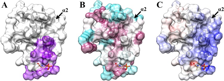
Transparent surface representation and ribbon diagram of DNAJA1-JD bound with O-phospho-l-serine with (A) the proposed inhibition site based on the TIM14–TIM16 interaction (purple), (B) the highly conserved (magenta) and poorly conserved (cyan) residues from Consurf, and (C) the positively charged surface (blue) and negatively charged surface (red) from Delphi. Helix α2, which is potentially an important component of the DnaJ–DnaK interaction site and the TIM16-like inhibitory binding site, is labeled.