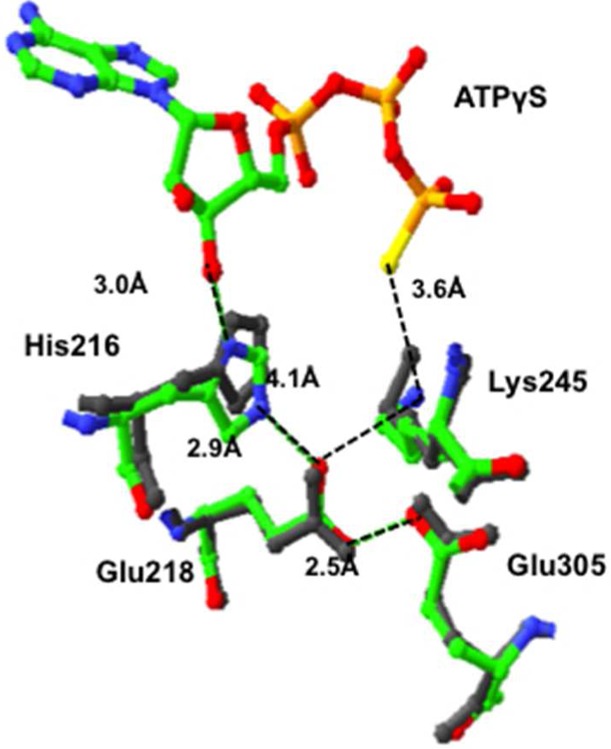
Figure 5.

Molecular model showing the position of His216 relative to Glu218 and Glu305 in the BC domain of RePC in a subunit with ATPγS bound (colored residues, Protein Data Bank entry 2QF7) and in a subunit of RePC without nucleotide bound (gray residues, Protein Data Bank entry 3TW7). Distances between atoms are indicated by the black dotted lines and are in units of angstroms. Distances are measured between atoms in the structure of RePC with ATPγS bound (Protein Data Bank entry 2QF7).
