Abstract
Background
This study is a report on the long-term analysis of acute and late toxicities for patients with localized prostate cancer treated with hypofractionated helical tomotherapy.
Methods
From January 2008 through August 2013, 70 patients with localized prostate cancer were treated definitively with hypofractionated helical tomotherapy. The helical tomotherapy was designed to deliver 75 Gy in 2.5 Gy per fraction to the prostate gland, 63 Gy in 2.1 Gy per fraction to the seminal vesicles, and 54 Gy in 1.8 Gy per fraction to the pelvic lymph nodes. Incidence rates and predictive factors for radiation toxicities were analyzed retrospectively.
Results
The incidences of grades 0, 1, and 2 acute gastrointestinal (GI) toxicity were 51.4%, 42.9%, and 5.7%, and those of acute genitourinary (GU) toxicity were 7.1%, 64.3%, and 28.6%, respectively. The maximum dose of rectum and bladder V40 and V50 were significant predictive factors for acute GI and GU toxicity. The cutoff value of rectum maximum dose and bladder V40 and V50 by receiver-operating characteristic curves analysis were 76.5 Gy, 17.3%, and 10.2%, respectively. The incidences of grades 0, 1, and 2 late GI toxicity were 82.0%, 14.0%, and 4.0%, and those of late GU toxicity were 18.0%, 56.0%, and 26.0%, respectively. Rectum V70 and bladder V70 and V75 were significant predictive factors for late GI and GU toxicity. The cutoff value of rectum V70 and bladder V70 and V75 by receiver-operating characteristic curves analysis was 2.8%, 2.8%, and 1.0%, respectively.
Conclusion
Hypofractionated helical tomotherapy using a schedule of 75 Gy at 2.5 Gy per fraction had favorable acute and late toxicity rates and no serious complication, such as grade 3 or worse toxicity. To minimize radiation toxicities, constraining the rectum maximum dose to less than 76.5 Gy, rectum V70 to less than 2.8%, bladder V40 to less than 17.3%, bladder V50 to less than 10.2%, bladder V70 to less than 2.8%, and bladder V75 to less than 1.0% would be necessary.
Keywords: prostate cancer, helical tomotherapy, hypofractionated radiotherapy, radiation toxicity, predictive factor
Introduction
In recent years, hypofractionated radiotherapy (RT) has been integrated into standard regimens in many treatment centers. Because the presumed α/β ratio in prostate cancer is close to 1.5, a hypofractionated RT regime provides a radiobiological benefit over conventional fractionated RT.1 In addition, because several clinical studies have demonstrated a clear radiation dose response, reflected as an improved biochemical failure-free survival rate observed with a dose-escalated RT schedule in localized prostate cancer, a hypofractionated RT regime could offer a more convenient way to treat prostate cancer.2–4 However, the potential toxicity that could develop in normal organs surrounding the prostate causes some concern when using a hypofractionated RT regime.5
Hypofractionated helical tomotherapy using a fractionation schedule of 75 Gy at 2.5 Gy per fraction for localized prostate cancer began at our institution in January 2008. Preliminary observations of the acute and late toxicities have been reported and showed that hypofractionated helical tomotherapy is well tolerated with a favorable toxicity rate.6 This study reports the long-term analysis of acute and late toxicities for patients with localized prostate cancer treated with hypofractionated helical tomotherapy.
Materials and methods
Eligibility criteria for this study were histologically confirmed adenocarcinoma of the prostate, completion of definitive hypofractionated helical tomotherapy with or without androgen deprivation therapy, no previous pelvic RT, no evidence of pelvic lymph node involvement or distant metastasis, no other concomitant malignant disease, no history of inflammatory bowel disease, no previous major pelvic surgery, Eastern Cooperative Oncology Group performance status of 2 or lower, and a follow-up period of more than 6 months after helical tomotherapy. At our institution, 70 prostate cancer patients met the eligibility criteria from January 2008 through August 2013 and were enrolled in this study. The institutional review board of Kyung Hee University Medical Center approved the retrospective review and analysis of patient data for this study.
The initial evaluation for all 70 patients included determination of the American Joint Committee on Cancer, 7th edition, clinical stage; risk group according to the D’Amico risk classification; pretreatment prostate-specific antigen levels; biopsy Gleason score; percentage of positive prostate biopsies; pelvic magnetic resonance imaging (MRI); and bone scan. Additional workup with transrectal ultrasonography and abdomen computed tomography (CT) was obtained according to physician preference.
Radiotherapy
All patients underwent CT simulation in the supine position after immobilization with a posterior vacuum bag and an anterior vacuum-sealed cover sheet. A rectal balloon catheter was also inserted into the rectum and inflated with 60–80 cc of air, and all patients were instructed to drink 400–600 cc water 1 hour before simulation and before every treatment session. A planning CT scan of the pelvis was obtained at 5 mm intervals from above the iliac crest through the midfemur.
The simulation CT data were transferred to Hi·Art Planning Station (TomoTherapy Inc., Madison, WI, USA) for inverse planning. Clinical target volume (CTV) 1 was the prostate gland. The seminal vesicles were CTV2 and were contoured in patients with seminal vesicle invasion on the pelvic MRI or in patients with a proposed risk of seminal vesicle involvement greater than 15%, as calculated by Roach’s formula.7 Pelvic lymph nodes including common iliac, internal and external iliac, obturator, and presacral lymph nodes were defined as CTV3 and contoured in patients with seminal vesicle invasion on pelvic MRI and a proposed risk of occult lymph nodal involvement higher than 15%, as calculated by Roach’s formula.7 CTV1 and CTV2 were expanded in three dimensions with a 1 cm margin to obtain planning target volumes (PTVs) 1 and 2, except at the prostate-rectal interface, where a 0.5 cm margin was used. PTV3 was defined by a 0.5 cm expansion of CTV3. Rectum, bladder, and femoral heads were contoured as organs at risk.
The prescription dose was 75 Gy in 30 fractions (2.5 Gy per fraction) for PTV1. PTV2 and PTV3 simultaneously received 63 Gy (2.1 Gy per fraction) and 54 Gy (1.8 Gy per fraction), respectively. Assuming an α/β ratio of 1.5 for prostate cancer, 75 Gy in 30 fractions can be considered to be similar to 86 Gy in 43 fractions. Each treatment plan was evaluated with a cumulative dose-volume histogram. In general, plans were considered acceptable if the PTV was covered by 95% of isodose curves and doses to normal organs were limited in their tolerances. The limits used for normal organs were no more than 25% and 5% of bladder and 20% and 5% of rectum to receive greater than 50 and 70 Gy (V50 <25% and V70 <5% for bladder, V50 <20% and V70 <5% for rectum), with a maximum dose level of 75 Gy. Planning objectives were prioritized to give the greatest importance to covering the PTV, with trying to keep radiation doses in normal structures as low as possible.
RT was performed using a TomoTherapy (TomoTherapy Inc.). Triangulation marks were used to make sure the patient did not roll and to quickly position the patient in the correct location. Before each treatment, a 3.5 megavoltage fan beam CT image was acquired, using a CT detector mounted on a ring gantry and matched to the planning CT image. If necessary, the patient position was corrected. Some patients received 6 months of androgen-deprivation therapy consisting of a daily oral administration of 50 mg bicalutamide and a 4 week interval of subcutaneous injection of a luteinizing hormone-releasing hormone analogue at the physician’s discretion. RT started 8 weeks after the first injection of luteinizing hormone-releasing hormone analogue.
Toxicity evaluation
Before the start of RT, pretreatment gastrointestinal (GI) and genitourinary (GU) symptoms were assessed according to the Common Terminology Criteria for Adverse Events, version 4.0. Patients were monitored weekly during RT and then at 2 month intervals for 1 year and at 3–4 month intervals thereafter. At each visit, GI and GU toxicities were prospectively scored by the radiation oncologist, according to Radiation Therapy Oncology Group (RTOG) criteria. Acute toxicity was defined as experiencing toxicity during or within 6 months of RT. Late toxicity was defined as any toxicity occurring or persisting 6 months or more after RT. The worst toxicity grade scored at any time was considered the final grade of toxicity.
Statistical analysis
The Kaplan–Meier method was used to compute the actuarial incidence of toxicity. The correlation of the development of toxicity with potential predictive factors was determined using the log-rank test. Parameters evaluated as potential predictive factors for toxicity were age, RT target, androgen-deprivation therapy, pretreatment symptoms, and several dosimetric parameters. For multivariate analysis, the Cox proportional regression hazard model was used. Elapsed time was calculated from the date of RT start to the date of maximum toxicity recognition or final follow-up visit. Receiver-operating characteristic (ROC) curves were generated to define the cutoff values for significant parameters. For all analyses, a P-value <0.05 was considered statistically significant. All analyses were performed using SPSS version 18.0 (IBM Corporation, Armonk, NY, USA).
Results
Patient characteristics
All patients completed the planned RT without any interruption. All patients tolerated the rectal inflation procedure well throughout the simulation and treatment course. Five patients did not tolerate the bladder-filling procedure during the treatment course, and these patients were instructed to drink 100–200 cc water 1 hour before treatment. Characteristics of all patients are summarized in Table 1. The median follow-up duration was 3.0 years (range, 0.7–5.8 years). Four patients (5.7%) had grade 1 pretreatment GI symptoms such as abdominal distension, anal pain, and fecal incontinence. Fifty-two (74.3%) and 5 (7.1%) patients had grade 1 and 2 pretreatment GU symptoms, respectively, such as urinary frequency, urgency, dysuria, urinary retention, and hematuria.
Table 1.
Patient characteristics (n=70)
| Characteristics | n (%) |
|---|---|
| Age, years, median (range) | 72.3 (54.2–82.1) |
| Pre-RT PSA, ng/mL, median (range) | 13.5 (3.2–235.6) |
| Gleason score | |
| 3+3 | 27 (38.6%) |
| 3+4 | 13 (18.6%) |
| 4+3 | 10 (14.2%) |
| 4+4 | 7 (10.0%) |
| 4+5 | 8 (11.4%) |
| 5+4 | 4 (5.7%) |
| 5+5 | 1 (1.5%) |
| Percentage of positive prostate biopsies, %, median (range) | 42.3 (8.3–100.0) |
| T stage | |
| 1c | 19 (27.1%) |
| 2a | 1 (1.6%) |
| 2b | 12 (17.1%) |
| 2c | 5 (7.1%) |
| 3a | 14 (20.0%) |
| 3b | 19 (27.1%) |
| Risk group | |
| Low | 10 (14.3%) |
| Intermediate | 14 (20.0%) |
| High | 46 (65.7%) |
| RT target | |
| PG | 39 (55.7%) |
| PG + SV | 22 (31.4%) |
| PG + SV + PLN | 9 (12.9%) |
| Androgen-deprivation therapy | |
| Yes | 16 (22.9%) |
| No | 54 (77.1%) |
| Pretreatment gastrointestinal symptoms | |
| Grade 0 | 66 (94.3%) |
| Grade 1 | 4 (5.7%) |
| Pretreatment genitourinary symptoms | |
| Grade 0 | 13 (18.6%) |
| Grade 1 | 52 (74.3%) |
| Grade 2 | 5 (7.1%) |
Abbreviations: RT, radiotherapy; PSA, prostate-specific antigen; PG, prostate gland; SV, seminal vesicles; PLN, pelvic lymph nodes.
Radiotherapy dosimetric data
The radiation dose-volume relationship for PTV and normal structures was analyzed and summarized in Table 2. In all patients, the PTV was covered by the 95% isodose curve. The dose-volume limits of normal structures were met in almost all patients, but the maximum dose limits of the normal structures were not reached and were put in to try to keep maximum doses in normal structures as low as possible.
Table 2.
Dosimetric data of hypofractionated helical tomotherapy for whole patients (n=70)
| Dosimetric parameters | Median (range) |
|---|---|
| PTV1 | |
| Mean dose, Gy | 75.7 (75.4–78.6) |
| Maximum dose, Gy | 77.7 (77.2–81.5) |
| Minimum dose, Gy | 73.1 (73.6–75.6) |
| PTV2 | |
| Mean dose, Gy | 67.7 (63.3–70.5) |
| Maximum dose, Gy | 76.0 (68.8–77.8) |
| Minimum dose, Gy | 61.4 (52.3–64.4) |
| PTV3 | |
| Mean dose, Gy | 57.7 (54.8–61.7) |
| Maximum dose, Gy | 69.6 (60.4–79.2) |
| Minimum dose, Gy | 50.4 (46.2–53.1) |
| Rectum | |
| V5, % | 93.8 (65.0–100.0) |
| V10, % | 87.3 (58.0–100.0) |
| V20, % | 74.5 (42.3–98.0) |
| V30, % | 48.6 (24.5–93.2) |
| V40, % | 27.0 (15.0–64.1) |
| V50, % | 15.0 (8.2–28.2) |
| V60, % | 7.9 (4.6–14.3) |
| V70, % | 3.0 (1.0–8.7) |
| V75, % | 1.0 (0.1–5.6) |
| Maximum dose, Gy | 76.5 (75.6–79.7) |
| Bladder | |
| V5, % | 82.2 (18.0–100.0) |
| V10, % | 71.5 (9.3–100.0) |
| V20, % | 57.5 (10.7–99.5) |
| V30, % | 40.5 (7.2–82.5) |
| V40, % | 24.5 (3.0–50.0) |
| V50, % | 14.3 (1.4–40.0) |
| V60, % | 8.0 (0.6–29.1) |
| V70, % | 3.0 (0.1–16.4) |
| V75, % | 1.1 (0–10.5) |
| Maximum dose, Gy | 76.7 (74.9–80.6) |
Abbreviation: PTV, planning target volume.
Acute toxicity and predictive factors
The maximum RTOG acute GI toxicity scores were 0 in 36 patients (51.4%), 1 in 30 patients (42.9%), and 2 in 4 patients (5.7%). There was no grade 3 or worse acute GI toxicity, and all acute GI toxicities developed within 10 weeks after RT. The 25-week actuarial incidence rate of grade 1 or 2 acute GI toxicity was 48.6%. The most common acute GI toxicity was anal pain, and many patients experienced several acute GI toxicities simultaneously. All acute GI toxicities are summarized in Table 3.
Table 3.
Acute gastrointestinal toxicities (n=70)
| Maximum grade and specific toxicities | n (%) |
|---|---|
| Grade 0 | 36 (51.4) |
| Grade 1 | 30 (42.9) |
| Anal pain | 25 (35.7) |
| Rectal bleeding | 5 (7.1) |
| Diarrhea | 2 (2.9) |
| Constipation | 2 (2.9) |
| Fecal incontinence | 2 (2.9) |
| Grade 2 | 4 (5.7) |
| Anal pain | 3 (4.3) |
| Diarrhea | 1 (1.4) |
| Fecal incontinence | 1 (1.4) |
The maximum RTOG acute GU toxicity scores were 0 in 5 patients (7.1%), 1 in 45 patients (64.3%), and 2 in 20 patients (28.6%). There was no grade 3 or worse acute GU toxicity; however, almost all patients experienced acute GU toxicity. All acute GU toxicities developed within 10 weeks after RT, and the 25 week actuarial incidence rate of grade 1 or 2 acute GU toxicity was 92.9%. Urinary frequency developed in all patients who experienced acute GU toxicity, and many patients experienced several acute GU toxicities simultaneously. All acute GU toxicities are summarized in Table 4.
Table 4.
Acute genitourinary toxicities (n=70)
| Maximum grade and specific toxicities | n (%) |
|---|---|
| Grade 0 | 5 (7.1) |
| Grade 1 | 45 (64.3) |
| Urinary frequency | 45 (64.3) |
| Urinary urgency | 11 (15.7) |
| Dysuria | 8 (11.4) |
| Urinary retention | 5 (7.1) |
| Urinary incontinence | 4 (5.7) |
| Grade 2 | 20 (28.6) |
| Urinary frequency | 20 (28.6) |
| Urinary urgency | 6 (8.6) |
| Dysuria | 3 (4.3) |
| Urinary incontinence | 1 (1.4) |
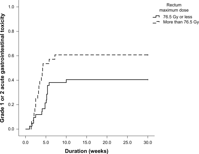
Predictive factors for grade 1 or 2 acute GI toxicity were analyzed. Only rectum maximum dose was significantly associated with grade 1 or 2 acute GI toxicity on univariate analysis (P=0.042). On multivariate analysis, rectum maximum dose remained a significant predictive factor (hazard ratio =1.980; 95% confidence interval, 1.008–3.891; P=0.047; Table 5 and Figure 1). The cutoff value of rectum maximum dose by ROC curves analysis was 76.5 Gy (sensitivity, 61.8%; specificity, 69.4%). Because of the low incidence of grade 2 acute GI toxicity, we did not attempt to find predictive factors for grade 2 acute GI toxicity.
Table 5.
Analysis of predictive factors for grade 1 or 2 acute gastrointestinal toxicity
| Variables | 25-week actuarial rate of grade 1 or 2 acute GI toxicity, % |
P-value
|
|
|---|---|---|---|
| Univariate analysis | Multivariate analysis | ||
| Age, years, ≤70 versus >70 | 58.3 versus 43.5 | 0.352 | 0.385 |
| RT target, PG versus PG + SV versus PG + SV + PLN | 46.2 versus 45.5 versus 66.7 | 0.474 | 0.499 |
| Androgen-deprivation therapy, yes versus no | 25.0 versus 55.6 | 0.097 | 0.096 |
| Pretreatment GI symptoms, yes versus no | 50.0 versus 25.0 | 0.416 | 0.394 |
| Rectum | |||
| V5 ≤93% versus >93% | 38.2 versus 58.3 | 0.138 | 0.232 |
| V10 ≤87% versus >87% | 41.9 versus 53.8 | 0.419 | 0.498 |
| V20 ≤75% versus >75% | 38.5 versus 61.3 | 0.105 | 0.222 |
| V30 ≤48% versus >48% | 41.2 versus 55.6 | 0.502 | 0.418 |
| V40 ≤27% versus >27% | 40.5 versus 57.6 | 0.275 | 0.391 |
| V50 ≤15% versus >15% | 45.0 versus 53.3 | 0.700 | 0.757 |
| V60 ≤8% versus >8% | 47.2 versus 50.0 | 0.996 | 0.969 |
| V70 ≤3% versus >3% | 45.7 versus 51.4 | 0.705 | 0.967 |
| V75 ≤1% versus >1% | 40.0 versus 57.1 | 0.184 | 0.450 |
| Max dose ≤76.5 Gy versus >76.5 Gy | 40.5 versus 60.7 | 0.042 | 0.047 |
Abbreviations: RT, radiotherapy; PG, prostate gland; SV, seminal vesicles; PLN, pelvic lymph nodes; GI, gastrointestinal; Max dose, maximum dose.
Figure 1.
Incidence of grade 1 or 2 acute gastrointestinal toxicity according to rectum maximum dose. The 25-week actuarial rate of grade 1 or 2 acute gastrointestinal toxicity was 40.5% if the maximum dose was 76.5 Gy or lower and 60.7% if the maximum dose was higher than 76.5 Gy (P=0.042 on univariate and P=0.047 on multivariate analysis).
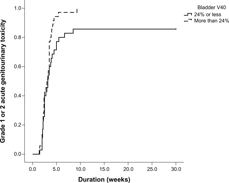
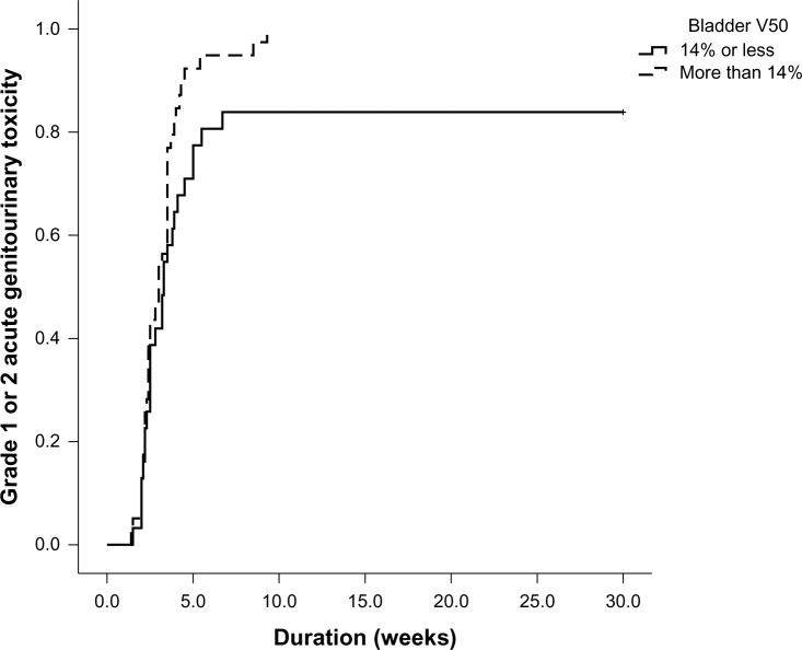
Predictive factors for grade 1 or 2 acute GU toxicity were analyzed. RT target (P=0.022), bladder V40 (P=0.038), and bladder V50 (P=0.040) were significantly associated with grade 1 or 2 acute GU toxicity on univariate analysis. On multivariate analysis, bladder V40 (hazard ratio =1.801; 95% confidence interval, 1.065–3.145; P=0.025) and bladder V50 (hazard ratio =1.814; 95% confidence interval, 1.087–3.027; P=0.021) remained significant predictive factors for grade 1 or 2 acute GU toxicity (Table 6, Figures 2 and 3). The cutoff values of bladder V40 and V50, by ROC curves analysis, were 17.3% (sensitivity, 76.9%; specificity, 80.0%) and 10.2% (sensitivity, 70.8%; specificity, 100%), respectively.
Table 6.
Analysis of predictive factors for grade 1 or 2 acute genitourinary toxicity
| Variables | 25-week actuarial rate of grade 1 or 2 acute GU toxicity, % |
P-value
|
|
|---|---|---|---|
| Univariate analysis | Multivariate analysis | ||
| Age, years, ≤70 versus >70 | 86.5 versus 95.0 | 0.867 | 0.532 |
| RT target, PG versus PG + SV versus PG + SV + PLN | 87.2 versus 100 versus 100 | 0.022 | 0.103 |
| Androgen-deprivation therapy, yes versus no | 87.8 versus 94.4 | 0.913 | 0.695 |
| Pretreatment GU symptoms, yes versus no | 94.7 versus 84.6 | 0.087 | 0.099 |
| Bladder | |||
| V5 ≤82% versus >82% | 85.7 versus 100 | 0.231 | 0.328 |
| V10 ≤71% versus >71% | 85.7 versus 100 | 0.318 | 0.345 |
| V20 ≤57% versus >57% | 85.7 versus 100 | 0.251 | 0.332 |
| V30 ≤40% versus >40% | 85.7 versus 100 | 0.084 | 0.056 |
| V40 ≤24% versus >24% | 85.7 versus 100 | 0.038 | 0.025 |
| V50 ≤14% versus >14% | 83.9 versus 100 | 0.040 | 0.021 |
| V60 ≤8% versus >8% | 86.8 versus 100 | 0.118 | 0.112 |
| V70 ≤3% versus >3% | 89.7 versus 96.8 | 0.573 | 0.381 |
| V75 ≤1% versus >1% | 90.9 versus 94.6 | 0.845 | 0.694 |
| Max dose ≤76.7 Gy versus >76.7 Gy | 89.7 versus 96.8 | 0.254 | 0.500 |
Abbreviations: RT, radiotherapy; PG, prostate gland; SV, seminal vesicles; PLN, pelvic lymph nodes; GU, genitourinary; Max dose, maximum dose.
Figure 2.
Incidence of grade 1 or 2 acute genitourinary toxicity according to bladder V40. The 25-week actuarial rate of grade 1 or 2 acute genitourinary toxicity was 85.7% if V40 was 24% or lower and 100% if V40 was higher than 24% (P=0.038 on univariate and P=0.025 on multivariate analysis).
Figure 3.
Incidence of grade 1 or 2 acute genitourinary toxicity according to bladder V50. The 25-week actuarial rate of grade 1 or 2 acute genitourinary toxicity was 83.9% if V50 was 14% or lower and 100% if V50 was higher than 14% (P=0.040 on univariate and P=0.021 on multivariate analysis).
Predictive factors for grade 2 acute GU toxicity were also analyzed. Pretreatment GU symptoms were significantly associated with grade 2 acute GU toxicity on univariate analysis (P=0.018). However, on multivariate analysis, no parameters were significantly associated with grade 2 acute GU toxicity (Table 7).
Table 7.
Analysis of predictive factors for grade 2 acute genitourinary toxicity
| Variables | 25-week actuarial rate of grade 2 acute GU toxicity, % |
P-value
|
|
|---|---|---|---|
| Univariate analysis | Multivariate analysis | ||
| Age, years, ≤70 versus >70 | 29.2 versus 28.3 | 0.863 | 0.911 |
| RT target, PG versus PG + SV versus PG + SV + PLN | 25.6 versus 22.7 versus 55.6 | 0.102 | 0.171 |
| Androgen-deprivation therapy, yes versus no | 31.2 versus 27.8 | 0.733 | 0.879 |
| Pretreatment GU symptoms, yes versus no | 35.1 versus 0 | 0.018 | 0.147 |
| Bladder | |||
| V5 ≤82% versus >82% | 22.9 versus 34.3 | 0.339 | 0.605 |
| V10 ≤71% versus >71% | 22.9 versus 34.3 | 0.339 | 0.408 |
| V20 ≤57% versus >57% | 22.9 versus 34.3 | 0.334 | 0.479 |
| V30 ≤40% versus >40% | 22.9 versus 34.3 | 0.318 | 0.568 |
| V40 ≤24% versus >24% | 22.9 versus 34.3 | 0.299 | 0.796 |
| V50 ≤14% versus >14% | 19.4 versus 35.9 | 0.140 | 0.555 |
| V60 ≤8% versus >8% | 21.1 versus 37.5 | 0.131 | 0.404 |
| V70 ≤3% versus >3% | 25.6 versus 32.3 | 0.544 | 0.951 |
| V75 ≤1% versus >1% | 24.2 versus 32.4 | 0.517 | 0.803 |
| Max dose ≤76.7 Gy versus >76.7 Gy | 20.5 versus 38.7 | 0.109 | 0.262 |
Abbreviations: RT, radiotherapy; PG, prostate gland; SV, seminal vesicles; PLN, pelvic lymph nodes; GU, genitourinary; Max dose, maximum dose.
Late toxicity and predictive factors
Of all patients, 50 were followed-up for more than 2 years. The median follow-up duration of these 50 patients was 3.9 years (range, 2.0–5.8 years). Thirty-two (64.0%) and 9 (18.0%) patients had high- and intermediate-risk disease, respectively, and the other 9 patients (18.0%) had low-risk disease. Only 4 patients of these patients received androgen-deprivation therapy. The RT target was the prostate gland in 24 patients (48.0%), prostate gland and seminal vesicles in 18 patients (36.0%), and prostate gland, seminal vesicles, and pelvic lymph nodes in 8 patients (16.0%). Three patients (6.0%) had grade 1 pretreatment GI symptoms, and 36 (72.0%) and 3 (6.0%) patients had grade 1 and 2 pretreatment GU symptoms, respectively. Late toxicities were evaluated in these 50 patients.
The maximum RTOG late GI toxicity scores were 0 in 41 patients (82.0%), 1 in 7 patients (14.0%), and 2 in 2 patients (4.0%). There was no grade 3 or worse late GI toxicity, and the incidence of late GI toxicity seemed to reach a plateau at 24 months after treatment. The 24-month actuarial incidence rate of grade 1 or 2 late GI toxicity was 18.0%. Most common late GI toxicity was rectal bleeding. Two patients experienced grade 2 rectal bleeding, which was successfully treated with argon plasma coagulation. Four patients experienced grade 1 rectal bleeding, which stopped spontaneously without treatment. All late GI toxicities are summarized in Table 8.
Table 8.
Late gastrointestinal toxicities in patients followed-up for more than 2 years (n=50)
| Maximum grade and specific toxicities | n (%) |
|---|---|
| Grade 0 | 41 (82.0) |
| Grade 1 | 7 (14.0) |
| Rectal bleeding | 4 (8.0) |
| Proctitis | 4 (8.0) |
| Rectal ulcer | 2 (4.0) |
| Fecal incontinence | 1 (2.0) |
| Grade 2 | 2 (4.0) |
| Rectal bleeding | 2 (4.0) |
| Proctitis | 2 (4.0) |
| Rectal ulcer | 1 (2.0) |
| Rectal telangiectasia | 1 (2.0) |
The maximum RTOG late GU toxicity scores were 0 in 9 patients (18.0%), 1 in 28 patients (56.0%), and 2 in 13 patients (26.0%). There was no grade 3 or worse late GU toxicity. All late GU toxicities developed within 23.5 months after RT, and incidence of late GU toxicity seemed to plateau at 23.5 months after treatment. The 24-months actuarial incidence rate of grade 1 or 2 late GU toxicity was 82.0%. The most common late GU toxicity was urinary frequency, which developed in all patients who experienced late GU toxicity. All late GU toxicities are summarized in Table 9.
Table 9.
Late genitourinary toxicities in patients followed-up for more than 2 years (n=50)
| Maximum grade and specific toxicities | n (%) |
|---|---|
| Grade 0 | 9 (18.0) |
| Grade 1 | 28 (56.0) |
| Urinary frequency | 28 (56.0) |
| Urinary urgency | 7 (14.0) |
| Urinary incontinence | 5 (10.0) |
| Dysuria | 2 (4.0) |
| Urinary retention | 1 (2.0) |
| Grade 2 | 13 (26.0) |
| Urinary frequency | 13 (26.0) |
| Urinary incontinence | 8 (16.0) |
| Urinary urgency | 3 (6.0) |
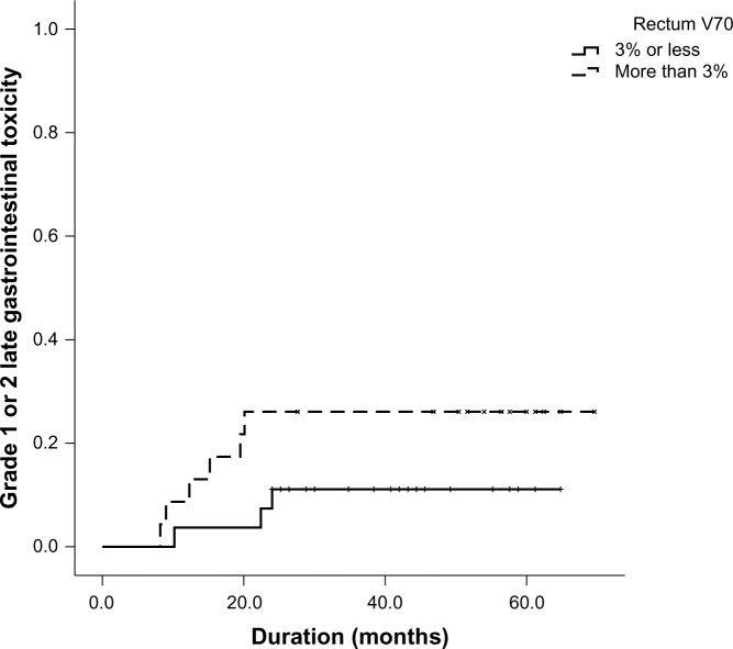
Predictive factors for grade 1 or 2 late GI toxicity were analyzed. Age (P=0.018) and pretreatment GI symptoms (P=0.023) were significantly associated with grade 1 or 2 late GI toxicity on univariate analysis. However, on multivariate analysis, age and pretreatment GI symptoms were not significantly associated with grade 1 or 2 late GI toxicity, and rectum V70 was a significant predictive factor (hazard ratio, 6.472; 95% confidence interval, 0.559–74.905; P=0.032; Table 10 and Figure 4). The cutoff value of rectum V70, by ROC curves analysis, was 2.8% (sensitivity, 85.6%; specificity, 71.2%). Because of the low incidence of grade 2 late GI toxicity, no attempt was made to find predictive factors for grade 2 late GI toxicity.
Table 10.
Analysis of predictive factors for grade 1 or 2 late gastrointestinal toxicity
| Variables | 24-month actuarial rate of grade 1 or 2 late GI toxicity, % |
P-value
|
|
|---|---|---|---|
| Univariate analysis | Multivariate analysis | ||
| Age, years, ≤70 versus >70 | 0 versus 30.0 | 0.018 | 0.717 |
| RT target, PG versus PG + SV versus PG + SV + PLN | 0 versus 11.1 versus 29.2 | 0.143 | 0.159 |
| Androgen deprivation therapy, yes versus no | 19.6 versus 0 | 0.354 | 0.931 |
| Pretreatment GI symptoms, yes versus no | 66.7 versus 14.9 | 0.023 | 0.869 |
| Rectum | |||
| V5 ≤95% versus >95% | 8.7 versus 25.9 | 0.120 | 0.973 |
| V10 ≤90% versus >90% | 8.7 versus 25.9 | 0.120 | 0.983 |
| V20 ≤79% versus >79% | 8.0 versus 28.0 | 0.064 | 0.915 |
| V30 ≤50% versus >50% | 16.7 versus 19.2 | 0.863 | 0.393 |
| V40 ≤27% versus >27% | 13.6 versus 21.4 | 0.454 | 0.829 |
| V50 ≤15% versus >15% | 16.0 versus 20.0 | 0.682 | 0.610 |
| V60 ≤8% versus >8% | 13.8 versus 23.8 | 0.341 | 0.078 |
| V70 ≤3% versus >3% | 11.1 versus 26.1 | 0.149 | 0.032 |
| V75 ≤1% versus >1% | 17.4 versus 18.5 | 0.932 | 0.135 |
| Max dose ≤76.7 Gy versus >76.7 Gy | 16.0 versus 20.0 | 0.793 | 0.483 |
Abbreviations: RT, radiotherapy; PG, prostate gland; SV, seminal vesicles; PLN, pelvic lymph nodes; GI, gastrointestinal; Max dose, maximum dose.
Figure 4.
Incidence of grade 1 or 2 late gastrointestinal toxicity according to rectum V70. The 24-month actuarial rate of grade 1 or 2 late gastrointestinal toxicity was 11.1% if V70 was 3% or lower and 26.1% if V70 was higher than 3% (P=0.149 on univariate and P=0.032 on multivariate analysis).
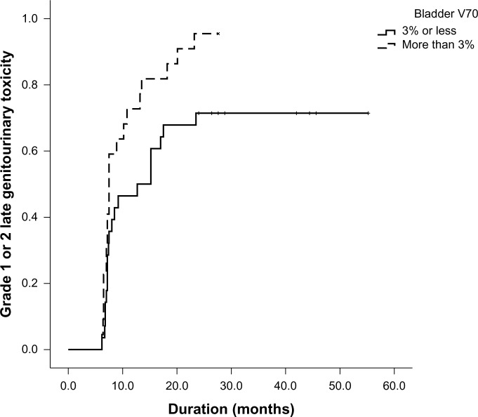
Predictive factors for grade 1 or 2 late GU toxicity were analyzed. Bladder V60 (P=0.036) and V70 (P=0.024) were significantly associated with grade 1 or 2 late GU toxicity on univariate analysis. On multivariate analysis, bladder V70 (hazard ratio, 1.992; 95% confidence interval, 1.068–3.718; P=0.030) remained a significant predictive factor (Table 11 and Figure 5). The cutoff value of bladder V70, by ROC curves analysis, was 2.8% (sensitivity, 68.3%; specificity, 77.8%).
Table 11.
Analysis of predictive factors for grade 1 or 2 late genitourinary toxicity
| Variables | 24-month actuarial rate of grade 1 or 2 late GU toxicity, % |
P-value
|
|
|---|---|---|---|
| Univariate analysis | Multivariate analysis | ||
| Age, years, ≤70 versus >70 | 80.0 versus 83.3 | 0.642 | 0.836 |
| RT target, PG versus PG + SV versus PG + SV + PLN | 75.0 versus 83.3 versus 100 | 0.428 | 0.230 |
| Androgen-deprivation therapy, yes versus no | 100 versus 80.4 | 0.107 | 0.117 |
| Pretreatment GU symptoms, yes versus no | 87.2 versus 63.6 | 0.092 | 0.099 |
| Bladder | |||
| V5 ≤85% versus >85% | 73.1 versus 91.7 | 0.071 | 0.078 |
| V10 ≤75% versus >75% | 73.1 versus 91.7 | 0.100 | 0.108 |
| V20 ≤61% versus >61% | 72.0 versus 92.0 | 0.076 | 0.083 |
| V30 ≤44% versus >44% | 76.0 versus 88.0 | 0.334 | 0.346 |
| V40 ≤27% versus >27% | 76.9 versus 87.5 | 0.327 | 0.339 |
| V50 ≤15% versus >15% | 73.1 versus 91.7 | 0.081 | 0.089 |
| V60 ≤8% versus >8% | 74.1 versus 91.3 | 0.036 | 0.051 |
| V70 ≤3% versus >3% | 71.4 versus 95.5 | 0.024 | 0.030 |
| V75 ≤1% versus >1% | 73.1 versus 91.7 | 0.111 | 0.120 |
| Max dose ≤76.9 Gy versus >76.9 Gy | 72.0 versus 92.0 | 0.054 | 0.060 |
Abbreviations: RT, radiotherapy; PG, prostate gland; SV, seminal vesicles; PLN, pelvic lymph nodes; GU, genitourinary; Max dose, maximum dose.
Figure 5.
Incidence of grade 1 or 2 late genitourinary toxicity according to bladder V70. The 24-month actuarial rate of grade 1 or 2 late genitourinary toxicity was 71.4% if V70 was 3% or lower and 95.5% if V70 was higher than 3% (P=0.024 on univariate and P=0.030 on multivariate analysis).
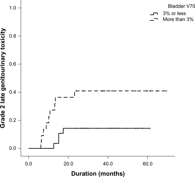
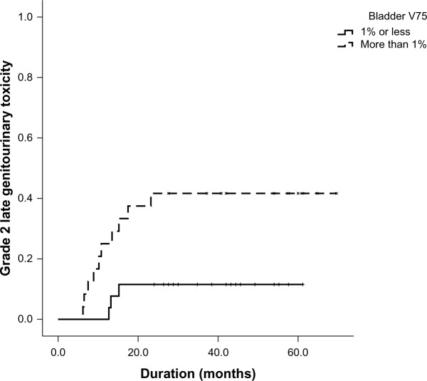
Predictive factors for grade 2 late GU toxicity were also analyzed. Pretreatment GU symptoms (P=0.036), bladder V60 (P=0.034), bladder V70 (P=0.021), and bladder V75 (P=0.013) were significantly associated with grade 2 late GU toxicity on univariate analysis. On multivariate analysis, bladder V70 (hazard ratio, 4.001; 95% confidence interval, 1.165–17.364; P=0.034) and bladder V75 (hazard ratio, 4.417; 95% confidence interval, 1.214–16.066; P=0.024) remained significant predictive factors for grade 2 late GU toxicity (Table 12, Figures 6 and 7). The cutoff values of bladder V70 and V75, by ROC curves analysis, were 2.8% (sensitivity, 76.9%; specificity, 66.8%) and 1.0% (sensitivity, 84.3%; specificity, 62.2%), respectively.
Table 12.
Analysis of predictive factors for grade 2 late genitourinary toxicity
| Variables | 24-month actuarial rate of grade 2 late GU toxicity, % |
P-value
|
|
|---|---|---|---|
| Univariate analysis | Multivariate analysis | ||
| Age, years, ≤70 versus >70 | 20.0 versus 35.0 | 0.214 | 0.162 |
| RT target, PG versus PG + SV versus PG + SV + PLN | 16.7 versus 29.2 versus 37.5 | 0.533 | 0.991 |
| Androgen-deprivation therapy, yes versus no | 50 versus 23.9 | 0.182 | 0.181 |
| Pretreatment GU symptoms, yes versus no | 33.3 versus 0 | 0.036 | 0.093 |
| Bladder | |||
| V5 ≤85% versus >85% | 23.1 versus 29.2 | 0.630 | 0.704 |
| V10 ≤75% versus >75% | 23.1 versus 29.2 | 0.630 | 0.894 |
| V20 ≤61% versus >61% | 20.0 versus 32.0 | 0.315 | 0.703 |
| V30 ≤44% versus >44% | 20.0 versus 32.0 | 0.315 | 0.703 |
| V40 ≤27% versus >27% | 19.2 versus 33.3 | 0.212 | 0.489 |
| V50 ≤15% versus >15% | 19.2 versus 33.3 | 0.212 | 0.810 |
| V60 ≤8% versus >8% | 14.8 versus 39.1 | 0.034 | 0.254 |
| V70 ≤3% versus >3% | 14.3 versus 40.9 | 0.021 | 0.034 |
| V75 ≤1% versus >1% | 11.5 versus 41.7 | 0.013 | 0.024 |
| Max dose ≤76.9 Gy versus >76.9 Gy | 20.0 versus 30.0 | 0.259 | 0.667 |
Abbreviations: RT, radiotherapy; PG, prostate gland; SV, seminal vesicles; PLN, pelvic lymph nodes; GU, genitourinary; Max dose, maximum dose.
Figure 6.
Incidence of grade 2 late genitourinary toxicity according to bladder V70. The 24-month actuarial rate of grade 2 late genitourinary toxicity was 14.3% if V70 was 3% or lower and 40.9% if V70 was higher than 3% (P=0.021 on univariate and P=0.034 on multivariate analysis).
Figure 7.
Incidence of grade 2 late genitourinary toxicity according to bladder V75. The 24-month actuarial rate of grade 2 late genitourinary toxicity was 11.5% if V75 was 1% or lower and 41.7% if V75 was higher than 1% (P=0.013 on univariate and P=0.024 on multivariate analysis).
Discussion
The results of our previous study, which analyzed radiation toxicities in 22 prostate cancer patients treated with helical tomotherapy using a fractionation schedule of 75 Gy at 2.5 Gy per fraction, showed a favorable toxicity rate.6 That study, however, had several limitations. The sample size was too small, and the follow-up period was not sufficiently long. As a consequence, we had difficulty making a firm conclusion. The current study analyzed the acute and late toxicities of all patients who underwent the same therapeutic regimen at our institution between January 2008 and August 2013. Because of the larger sample size and longer follow-up period, this study provided the opportunity to confirm the outcomes of our previous study.
Several studies have shown improved biochemical failure-free survival with dose-escalated RT schedules in localized prostate cancer.2–4,8 Thus, greater equivalent doses should be delivered using hypofractionated RT schedules. At the Cleveland Clinic Foundation, Kupelian et al showed that 70 Gy at 2.5 Gy per fraction was given safely to patients with localized prostate cancer, using intensity-modulated radiotherapy (IMRT).9,10 Therefore, it would be possible to give more total dose than 70 Gy at 2.5 Gy per fraction without serious acute or late toxicities. Our hypofractionated helical tomotherapy schedule (total 75 Gy, 2.5 Gy per fraction) was designed to give a higher biological equivalent dose than that of the Cleveland Clinic Foundation, with an acceptable risk of serious GI and GU complications. Assuming an α/β ratio of 1.5 for prostate cancer, the total equivalent dose of our fractionation schedule would be 86 Gy if delivered at 2 Gy per fraction. The total equivalent doses of various hypofractionated RT schedules for prostate cancer from other studies ranged from 76 Gy to 82 Gy if delivered at 2 Gy per fraction.9,11–16 The total equivalent dose of our fractionation schedule is higher than that of other studies. Because of the higher equivalent dose of our fractionation schedule, the accuracy of the daily patient setup was crucial. To minimize organ motion secondary to bladder or rectal filling, we inserted a rectal balloon catheter into the rectum and inflated it with 60–80 cc air, and all patients were instructed to drink 400–600 cc water 1 hour before every treatment session. Several studies have reported that rectal balloon and bladder filling can decrease daily motion variation.17–20
The reported incidence rate of GI and GU toxicities after hypofractionated RT for prostate cancer has varied widely.9,11–14,21–24 The reported rates of grade 0, 1, 2, and 3 acute GI toxicities ranged from 30%–66% to 22%–60%, 10%–35%, and 0%–3.5%, respectively, and those of acute GU toxicities ranged from 10%–33% to 43%–70%, 7%–40%, and 0%–8%, respectively. Inconsistencies in reported rates may be attributed to subjectivity in the scoring criteria for radiation toxicities. Because most scoring criteria for radiation toxicities are based on evaluation by the treating physician, inter- and intraobserver variation may be present. Other possible reasons for inconsistencies in the reported rates of radiation toxicities include various RT techniques and definitions of target volume, different indications for adjuvant hormonal therapy, and heterogeneous pretreatment symptoms of patient populations. In our study, there was no grade 3 or worse acute toxicity. The incidences of grade 0, 1, and 2 acute GI toxicity were 51.4%, 42.9%, and 5.7%, and those of acute GU toxicity were 7.1%, 64.3%, and 28.6%, respectively. Compared with previous reports, our study generally showed comparable toxicity rates; however, the rate of grade 2 acute GI toxicity was markedly lower (10%–35% versus 5.7%). In contrast, the reported rates of grade 0, 1, 2, and 3 late GI toxicities ranged from 66%–93% to 2%–27%, 3%–17%, and 0%–7%, and those of late GU toxicities ranged from 46%–90% to 4%–43%, 0%–17%, and 0%–4%, respectively. In our study, the incidences of grade 0, 1, and 2 late GI toxicity were 82.0%, 14.0%, and 4.0%, and those of late GU toxicity were 18.0%, 56.0%, and 26.0%, respectively. Our study showed comparable or favorable late GI toxicity rates compared with previous reports. However, our study showed unfavorable late GU toxicity rates. In our study, the rate of grade 0 late GU toxicity was lower (46%–90% versus 18%) and the rates of grade 1 (4%–43% versus 56%) and 2 (0%–17% versus 26%) late GU toxicities were higher than previous reports. However, all patients tolerated the treatment course and follow-up period well throughout, and our study showed no grade 3 or worse late GU toxicity. Therefore, taken altogether, the late GU toxicity rates of this study seemed to be acceptable (Table 13).
Table 13.
Comparison of reported toxicity rates between previous studies and our study
| Grade | Previous studies | Our study |
|---|---|---|
| Acute gastrointestinal toxicity rate, % | ||
| 0 | 30–66 | 51.4 |
| 1 | 22–60 | 42.9 |
| 2 | 10–35 | 5.7 |
| 3 | 0–3.5 | 0 |
| Acute genitourinary toxicity rate, % | ||
| 0 | 10–33 | 7.1 |
| 1 | 43–70 | 64.3 |
| 2 | 7–40 | 28.6 |
| 3 | 0–8 | 0 |
| Late gastrointestinal toxicity rate, % | ||
| 0 | 66–93 | 82.0 |
| 1 | 2–27 | 14.0 |
| 2 | 3–17 | 4.0 |
| 3 | 0–7 | 0 |
| Late genitourinary toxicity rate, % | ||
| 0 | 46–90 | 18.0 |
| 1 | 4–43 | 56.0 |
| 2 | 0–17 | 26.0 |
| 3 | 0–4 | 0 |
To our knowledge, no published study has analyzed predictive factors for radiation toxicities after helical tomotherapy in prostate cancer. Kupelian et al21 reported that rectum V70 was a significant predicting factor for grade 2 or 3 late rectal toxicity after linac-based IMRT, and Huang et al25 reported that rectum V60, V70, and V78 were significantly associated with grade 2 or 3 late rectal toxicity after three-dimensional conformal RT. However, because a large volume of normal tissue is exposed to low-dose radiation during helical tomotherapy, predictive factors for radiation toxicity after helical tomotherapy may differ from those after 3-dimensional conformal RT or linac-based IMRT in prostate cancer. In our study, several parameters were evaluated as potential predictive factors for acute GI and GU and for late GI and GU toxicities. Among the several parameters, rectum maximum dose (cutoff value, 76.5 Gy) was a significant predictive factor for grade 1 or 2 acute GI toxicity. In addition, bladder V40 (cutoff value, 17.3%) and V50 (cutoff value, 10.2%) for grade 1 or 2 acute GU toxicity, rectum V70 (cutoff value, 2.8%) for grade 1 or 2 late GI toxicity, bladder V70 (cutoff value, 2.8%) for grade 1 or 2 late GU toxicity, and bladder V70 (cutoff value, 2.8%) and V75 (cutoff value, 1.0%) were significant predictive factors for grade 2 late GU toxicity. Therefore, we propose constraining the rectum maximum dose to less than 76.5 Gy, rectum V70 to less than 2.8%, bladder V40 to less than 17.3%, bladder V50 to less than 10.2%, bladder V70 to less than 2.8%, and bladder V75 to less than 1.0% to reduce the development of radiation toxicity after helical tomotherapy in prostate cancer.
There were some limitations in this study. First, this study was retrospective and may have inherent bias. However, all toxicities were scored prospectively, and therefore, selection and reporting bias were limited. Second, although the sample size of this study was larger than our previous study, it was still small. Third, the patient characteristics were heterogeneous. Fourth, because of incomplete patient medical records, we could not analyze the incidence of radiation-induced erectile dysfunction. Despite these limitations, if additional studies show favorable tumor control and survival rates in prostate cancer patients treated with our fractionation regimen, hypofractionated helical tomotherapy using a fractionation schedule of 75 Gy at 2.5 Gy per fraction could be a safe and effective way of dose escalation in treating localized prostate cancer.
Conclusion
With a long follow-up period, hypofractionated helical tomotherapy using a fractionation schedule of 75 Gy at 2.5 Gy per fraction showed favorable acute and late toxicity rates and no serious complications, such as grade 3 or worse toxicity. To minimize radiation toxicities, rectum maximum dose, rectum V70, and bladder V40, V50, V70, and V75 should be kept as low as possible. In addition, further studies are necessary to analyze the tumor control and survival rates in prostate cancer patients treated with hypofractionated helical tomotherapy, using a fractionation schedule of 75 Gy at 2.5 Gy per fraction.
Author contributions
All authors have made substantive contributions to the article and assume full responsibility for its content. Also, all authors fulfilled the International Committee of Medical Journal Editors requirements for authorship.
Disclosure
The authors report no conflicts of interest in this work.
References
- 1.Fowler JF. The radiobiology of prostate cancer including new aspects of fractionated radiotherapy. Acta Oncol. 2005;44(3):265–276. doi: 10.1080/02841860410002824. [DOI] [PubMed] [Google Scholar]
- 2.Lyons JA, Kupelian PA, Mohan DS, Reddy CA, Klein EA. Importance of high radiation doses (72 Gy or greater) in the treatment of stage T1–T3 adenocarcinoma of the prostate. Urology. 2000;55(1):85–90. doi: 10.1016/s0090-4295(99)00380-5. [DOI] [PubMed] [Google Scholar]
- 3.Kupelian PA, Mohan DS, Lyons J, Klein EA, Reddy CA. Higher than standard radiation doses (≥72 Gy) with or without androgen deprivation in the treatment of localized prostate cancer. Int J Radiat Oncol Biol Phys. 2000;46(3):567–574. doi: 10.1016/s0360-3016(99)00455-1. [DOI] [PubMed] [Google Scholar]
- 4.Kupelian PA, Buchsbaum JC, Reddy CA, Klein EA. Radiation dose response in patients with favorable localized prostate cancer (Stage T1–T2, biopsy Gleason ≤6, and pretreatment prostate-specific antigen ≤10) Int J Radiat Oncol Biol Phys. 2001;50(3):621–625. doi: 10.1016/s0360-3016(01)01466-3. [DOI] [PubMed] [Google Scholar]
- 5.Brenner DJ, Hall EJ. Fractionation and protraction for radiotherapy of prostate carcinoma. Int J Radiat Oncol Biol Phys. 1999;43(5):1095–1101. doi: 10.1016/s0360-3016(98)00438-6. [DOI] [PubMed] [Google Scholar]
- 6.Kong M, Hong SE, Choi J, Chang S-G. Hypofractionated helical intensity-modulated radiotherapy (75 Gy at 2.5 Gy/fraction) for intermediate- and high-risk prostate cancer: Assessment of toxicity. J Radiother Pract. 2012;11:145–154. [Google Scholar]
- 7.Roach M, 3rd, Marquez C, Yuo HS, et al. Predicting the risk of lymph node involvement using the pre-treatment prostate specific antigen and Gleason score in men with clinically localized prostate cancer. Int J Radiat Oncol Biol Phys. 1994;28(1):33–37. doi: 10.1016/0360-3016(94)90138-4. [DOI] [PubMed] [Google Scholar]
- 8.Peeters ST, Heemsbergen WD, Koper PC, et al. Dose-response in radiotherapy for localized prostate cancer: results of the Dutch multicenter randomized phase III trial comparing 68 Gy of radiotherapy with 78 Gy. J Clin Oncol. 2006;24(13):1990–1996. doi: 10.1200/JCO.2005.05.2530. [DOI] [PubMed] [Google Scholar]
- 9.Kupelian PA, Willoughby TR, Reddy CA, Klein EA, Mahadevan A. Hypofractionated intensity-modulated radiotherapy (70 Gy at 2.5 Gy per fraction) for localized prostate cancer: Cleveland Clinic experience. Int J Radiat Oncol Biol Phys. 2007;68(5):1424–1430. doi: 10.1016/j.ijrobp.2007.01.067. [DOI] [PubMed] [Google Scholar]
- 10.Kupelian PA, Thakkar VV, Khuntia D, Reddy CA, Klein EA, Mahadevan A. Hypofractionated intensity-modulated radiotherapy (70 gy at 2.5 Gy per fraction) for localized prostate cancer: long-term outcomes. Int J Radiat Oncol Biol Phys. 2005;63(5):1463–1468. doi: 10.1016/j.ijrobp.2005.05.054. [DOI] [PubMed] [Google Scholar]
- 11.Coote JH, Wylie JP, Cowan RA, Logue JP, Swindell R, Livsey JE. Hypofractionated intensity-modulated radiotherapy for carcinoma of the prostate: analysis of toxicity. Int J Radiat Oncol Biol Phys. 2009;74(4):1121–1127. doi: 10.1016/j.ijrobp.2008.09.032. [DOI] [PubMed] [Google Scholar]
- 12.Arcangeli G, Fowler J, Gomellini S, et al. Acute and late toxicity in a randomized trial of conventional versus hypofractionated three-dimensional conformal radiotherapy for prostate cancer. Int J Radiat Oncol Biol Phys. 2011;79(4):1013–1021. doi: 10.1016/j.ijrobp.2009.12.045. [DOI] [PubMed] [Google Scholar]
- 13.Martin JM, Rosewall T, Bayley A, et al. Phase II trial of hypofractionated image-guided intensity-modulated radiotherapy for localized prostate adenocarcinoma. Int J Radiat Oncol Biol Phys. 2007;69(4):1084–1089. doi: 10.1016/j.ijrobp.2007.04.049. [DOI] [PubMed] [Google Scholar]
- 14.Arcangeli G, Saracino B, Gomellini S, et al. A prospective phase III randomized trial of hypofractionation versus conventional fractionation in patients with high-risk prostate cancer. Int J Radiat Oncol Biol Phys. 2010;78(1):11–18. doi: 10.1016/j.ijrobp.2009.07.1691. [DOI] [PubMed] [Google Scholar]
- 15.Chen LN, Suy S, Uhm S, et al. Stereotactic body radiation therapy (SBRT) for clinically localized prostate cancer: the Georgetown University experience. Radiat Oncol. 2013;8:58. doi: 10.1186/1748-717X-8-58. [DOI] [PMC free article] [PubMed] [Google Scholar]
- 16.Arcangeli S, Strigari L, Gomellini S, et al. Updated results and patterns of failure in a randomized hypofractionation trial for high-risk prostate cancer. Int J Radiat Oncol Biol Phys. 2012;84(5):1172–1178. doi: 10.1016/j.ijrobp.2012.02.049. [DOI] [PubMed] [Google Scholar]
- 17.McGary JE, Teh BS, Butler EB, Grant W., 3rd Prostate immobilization using a rectal balloon. J Appl Clin Med Phys. 2002;3(1):6–11. doi: 10.1120/jacmp.v3i1.2590. [DOI] [PMC free article] [PubMed] [Google Scholar]
- 18.Wachter S, Gerstner N, Dorner D, et al. The influence of a rectal balloon tube as internal immobilization device on variations of volumes and dose-volume histograms during treatment course of conformal radiotherapy for prostate cancer. Int J Radiat Oncol Biol Phys. 2002;52(1):91–100. doi: 10.1016/s0360-3016(01)01821-1. [DOI] [PubMed] [Google Scholar]
- 19.O’Doherty UM, McNair HA, Norman AR, et al. Variability of bladder filling in patients receiving radical radiotherapy to the prostate. Radiother Oncol. 2006;79(3):335–340. doi: 10.1016/j.radonc.2006.05.007. [DOI] [PubMed] [Google Scholar]
- 20.Jain S, Loblaw DA, Morton GC, et al. The effect of radiation technique and bladder filling on the acute toxicity of pelvic radiotherapy for localized high risk prostate cancer. Radiother Oncol. 2012;105(2):193–197. doi: 10.1016/j.radonc.2012.09.020. [DOI] [PubMed] [Google Scholar]
- 21.Kupelian PA, Reddy CA, Carlson TP, Altsman KA, Willoughby TR. Preliminary observations on biochemical relapse-free survival rates after short-course intensity-modulated radiotherapy (70 Gy at 2.5 Gy/fraction) for localized prostate cancer. Int J Radiat Oncol Biol Phys. 2002;53(4):904–912. doi: 10.1016/s0360-3016(02)02836-5. [DOI] [PubMed] [Google Scholar]
- 22.McCammon R, Rusthoven KE, Kavanagh B, Newell S, Newman F, Raben D. Toxicity assessment of pelvic intensity-modulated radiotherapy with hypofractionated simultaneous integrated boost to prostate for intermediate- and high-risk prostate cancer. Int J Radiat Oncol Biol Phys. 2009;75(2):413–420. doi: 10.1016/j.ijrobp.2008.10.050. [DOI] [PubMed] [Google Scholar]
- 23.Pollack A, Hanlon AL, Horwitz EM, et al. Dosimetry and preliminary acute toxicity in the first 100 men treated for prostate cancer on a randomized hypofractionation dose escalation trial. Int J Radiat Oncol Biol Phys. 2006;64(2):518–526. doi: 10.1016/j.ijrobp.2005.07.970. [DOI] [PMC free article] [PubMed] [Google Scholar]
- 24.Joo JH, Kim YJ, Kim YS, et al. Whole pelvic intensity-modulated radiotherapy for high-risk prostate cancer: a preliminary report. Radiat Oncol J. 2013;31(4):199–205. doi: 10.3857/roj.2013.31.4.199. [DOI] [PMC free article] [PubMed] [Google Scholar]
- 25.Huang EH, Pollack A, Levy L, et al. Late rectal toxicity: dose-volume effects of conformal radiotherapy for prostate cancer. Int J Radiat Oncol Biol Phys. 2002;54(5):1314–1321. doi: 10.1016/s0360-3016(02)03742-2. [DOI] [PubMed] [Google Scholar]


