Abstract
Exserohilum is a saprophytic fungal pathogen responsible for a wide spectrum of infections in humans. It causes life-threatening acute invasive infections in the immunocompromised individuals, particularly those having haematological disorders. We report a proven case of chronic invasive rhinosinusitis with orbital involvement by Exserohilum rostratum in an immunocompetent child. The patient responded well to endoscopic sinus surgery followed by oral itraconazole. An aggressive surgical approach is required for improving the outcome of patients with invasive infections. A microbiological diagnosis may help in deciding the systemic antifungal agent in fungal rhinosinusitis.
Background
Exserohilum is a saprophytic, dematiaceous fungus found in soil and plants. The genus includes three species known to cause human infections, namely Exserohilum rostratum, Exserohilum longirostratum and Exserohilum mcginnisii. The clinical manifestations may vary from benign cutaneous involvement to fulminant invasive disease associated with high mortality.1 Recently, it has caused an outbreak of spinal and paraspinal infections following contaminated injections of methylprednisolone acetate in the USA.2 Most cases of invasive disease are known to occur in immunocompromised patients, particularly those having haematological disorders. We report a unique case of chronic invasive fungal rhinosinusitis in an immunocompetent child from north India.
Case presentation
An 11-year-old girl, a resident of Uttar Pradesh, North India, presented with bilateral nasal stuffiness and nasal discharge since 12 months and left-sided proptosis since 8 months. The patient was a known case of bronchial asthma and recurrent allergic fungal sinusitis (AFS) since she was 2 years old. She had undergone endoscopic surgery 6 years previously. Nasal discharge was more on the left side, variable in quantity, thick in consistency, non-foul smelling and occasionally bloodstained. Left-sided proptosis was not associated with any visual disturbance or restriction of eye movements. General physical and systemic examinations of the patient were normal. Local examination was normal except for left cheek erythema and left axial proptosis.
CT of paranasal sinuses revealed the involvement of the left maxillary, frontal, sphenoid and ethmoidal sinuses along with a breach in the lamina papyracea. There was bony erosion of the medial wall of the left maxillary sinus. On the basis of CT findings, a diagnosis of left-sided pansinusitis with intraorbital extension was made and the patient underwent left-sided functional endoscopic sinus surgery (FESS). Preoperatively, all the routine haematological and biochemical parameters were within normal limits. Intraoperatively, multiple pale polypi were seen in lateral and medial to middle turbinate. The polypi were excised and the ethmoid, sphenoid sinus, spheno-ethmoidal recess and frontonasal duct area were cleared of disease. Dehiscence was seen in the lamina papyracea and the periorbita was visible. Excised tissue samples were sent for histopathological and microbiological examinations. The postoperative course was uneventful and the patient was subsequently discharged.
Investigations
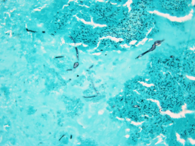
Histopathological examination by H&E staining of the ethmoidal polyp and other excised tissues revealed allergic mucin and chronic granulation tissue with areas of necrosis without any evidence of granuloma formation. Periodic acid-Schiff (PAS) with diastase and Gomori methanamine-silver (GMS) stainings revealed fungal elements within the necrotic material (figure 1).
Figure 1.

Gomori methenamine-silver staining of the tissue showing fungal hyphae, magnification ×400.
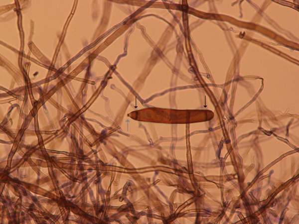
On microbial examination, the potassium hydroxide mount of the tissue revealed hyaline septate hyphae. The sample was cultured on Sabouraud's dextrose agar media on which rapidly growing black-coloured colonies having a velvet-like surface were noted within a week. The reverse of the culture tubes showed a black-coloured pigment suggesting a dematiaceous fungus. On lactophenol cotton blue mount, the isolate showed brown-coloured septate hyphae with sympodial conidiophores. The conidia were straight or slightly curved, elongated and cylindrical in shape, having predominantly 7–9 septa with a characteristically protruding, darkly pigmented hilum. Septa at both ends of the conidia were characteristically darker than other septa (figure 2). The fungus was identified as E rostratum on the basis of these morphological features.
Figure 2.

Typical conidium of Exserohilum rostratum showing a protruding hilum (grey arrow) and characteristically darker septa at both ends (black arrows), magnification ×400.
Treatment
A diagnosis of chronic invasive fungal rhinosinusitis was made after considering the clinical, histopathological and microbiological findings. The patient was subsequently given oral itraconazole and advised regular follow-up.
Outcome and follow-up
The patient is coming for regular follow-up without any evidence of disease.
Discussion
Exserohilum species are being increasingly recognised as human pathogens causing infections accidentally by either traumatic implantations or inhalation of spores.1 Recently, the fungus has come to attention because it caused a large outbreak of spinal and paraspinal infections in the USA due to contaminated injections of methylprednisolone acetate, resulting in high morbidity and mortality.2 Prior to this outbreak, the fungus was reported mainly from areas with hot climates such as southern states of the USA, Israel and India.2 It has never been reported from the Indian subcontinent as a causative agent for fungal rhinosinusitis. Until now, only six cases have been reported from the Indian subcontinent, five of them involving the cornea3 and one involving the skin.4
We report a unique case of chronic invasive fungal rhinosinusitis by E rostratum in an immunocompetent patient of the paediatric age group. Apart from the outbreak in the USA, this fungus has been implicated in 10 cases of acute invasive rhinosinusitis and 9 cases of allergic fungal sinusitis until now. All cases presenting as acute invasive rhinosinusitis were reported in patients having haematological disorders, mainly aplastic anaemia and haematological malignancies. No case of invasive rhinosinusitis has been reported by this fungus in immunocompetent individuals.1
In clinical laboratories, the genera Exserohilum need to be differentiated morphologically from other dematiaceous fungi such as Alternaria, Helminthosporium, Bipolaris and Drechslera. The important differentiating microscopic features are conidial shape, the presence or absence of a protruding hilum and the manner of origination of the germ tube from the basal cell. Exserohilum species have elongated, cylindrical conidia showing the characteristic protruding hilum from which an originating germ tube follows the long axis of the conidium. E rostratum is further differentiated microscopically from E longistratum and E mcginnisii on the basis of the presence of characteristic darkly pigmented septa at both ends of the conidium and typically having 7–9 septa in a single conidium.5
There is no definite optimal duration and choice of therapy for sinonasal diseases due to Exserohilum species. Most acute invasive cases have responded well to aggressive surgical debridement followed by systemic antifungal agents. Of the antifungals available, azoles seem to have better activity than amphotericin B1; however, strains resistant to echinocandins have been noted.6 Unfortunately, most patients succumb due to the underlying immunosuppression or other infections.7 Most cases of AFS respond well to surgical modalities without the institution of systemic antifungals;1 however, chronic invasive forms of fungal rhinosinusitis are usually treated by surgical debridement followed by the institution of systemic antifungals in immunocompromised8 as well as immunocompetent patients.9 This case adds to the growing body of literature on the spectrum of invasive sinonasal disease due to Exserohilum species. To conclude, Exserohilum species are being recognised as emerging fungal pathogens associated with high morbidity and mortality. A microbiological diagnosis may help in deciding the choice of antifungal agents, thereby improving the outcome of patients.
Learning points.
Exserohilum is an emerging fungal pathogen causing a wide spectrum of infections, ranging from benign involvement in immunocompetent individuals to life-threatening invasive disease in immunocompromised patients.
Invasive rhinosinusitis in the immunocompetent patient is exceedingly rare.
Microbiological diagnosis is essential to establish the fungal aetiology which may help to decide the choice of antifungal agent.
Treatment of invasive disease due to Exserohilum species requires aggressive surgical approach followed by systemic antifungals, preferably azoles.
Footnotes
Contributors: AG drafted the manuscript and did the microbiology work. IX was responsible for microbiological diagnosis and revising the manuscript. SCS is the treating physician responsible for patient management and revision of the manuscript. SM is responsible for histopathological diagnosis and editing of the manuscript.
Competing interests: None.
Patient consent: Obtained.
Provenance and peer review: Not commissioned; externally peer reviewed.
References
- 1.Adler A, Yaniv I, Samra Z, et al. Exserohilum: an emerging human pathogen. Eur J Clin Microbiol Infect Dis 2006;25:247–53 [DOI] [PubMed] [Google Scholar]
- 2.Centers for Disease Control and Prevention (CDC). Spinal and Exserohilum paraspinal infections associated with contaminated methylprednisolone acetate injections—Michigan, 2012–13. MMWR Morb Mortal Wkly Rep 2013; 62:377–81 [PMC free article] [PubMed] [Google Scholar]
- 3.Joseph NM, Kumar MA, Stephen S, et al. Keratomycosis caused by Exserohilum rostratum. Indian J Pathol Microbiol 2012;55:248–9 [DOI] [PubMed] [Google Scholar]
- 4.Agarwal A, Sing SM. A case of cutaneous phaeohyphomycosis caused by Exserohilum rostratum: in vitro sensitivity and review of literature. Mycopathologia 1995;131:9–12 [DOI] [PubMed] [Google Scholar]
- 5.McGinnis MR, Rinaldi MG, Winn RE. Emerging agents of phaeohyphomycosis: pathogenic species of Bipolaris and Exserohilum. J Clin Microbiol 1986;24:250–9 [DOI] [PMC free article] [PubMed] [Google Scholar]
- 6.da Cunha KC, Sutton DA, Gene J, et al. Molecular identification and In vitro response to antifungal drugs of clinical isolates of Exserohilum. Antimicrob Agents Chemother 2012;56:4951–4 [DOI] [PMC free article] [PubMed] [Google Scholar]
- 7.Derber C, Elam K, Bearman G. Invasive sinonasal disease due to dematiaceous fungi in immunocompromised individuals: case report and review of literature. Int J Infect Dis 2010;14S:e329–32 [DOI] [PubMed] [Google Scholar]
- 8.Soler ZM, Sclosser RJ. The role of fungi in diseases of the nose and sinuses. Am J Rhinol Allergy 2012;26:351–8 [DOI] [PMC free article] [PubMed] [Google Scholar]
- 9.Alrajhi AA, Enani M, Mahasin Z, et al. Chronic invasive aspergillosis of the paranasal sinuses in immunocompetent hosts from Saudi Arabia. Am J Trop Med Hyg 2001;65:83–6 [DOI] [PubMed] [Google Scholar]


