Abstract
Background
Hypertension is the most prevalent and modifiable risk factor for atrial fibrillation. The pressure overload in the left atrium induces pathophysiological changes leading to alterations in contractile function and electrical properties.
Objective
In this study our aim was to assess left atrial function in hypertensive patients to determine the association between left atrial function with paroxysmal atrial fibrillation (PAF).
Method
We studied 57 hypertensive patients (age: 53±4 years; left ventricular ejection fraction: 76±6.7%), including 30 consecutive patients with PAF and 30 age-matched control subjects. Left atrial (LA) volumes were measured using the modified Simpson's biplane method. Three types of LA volume were determined: maximal LA(LAVmax), preatrial contraction LA(LAVpreA) and minimal LA volume(LAVmin). LA emptying functions were calculated. LA total emptying volume = LAVmax−LAVmin and the LA total EF = (LAVmax-LAVmin )/LAVmax, LA passive emptying volume = LAVmax− LAVpreA and the LA passive EF = (LAVmax-LAVpreA)/LAVmax, LA active emptying volume = LAVpreA−LAVmin and LA active EF = (LAVpreA-LAVmin )/LAVpreA.
Results
The hypertensive period is longer in hypertensive group with PAF. LAVmax significantly increased in hypertensive group with PAF when compared to hypertensive group without PAF (p=0.010). LAAEF was significantly decreased in hypertensive group with PAF as compared to hypertensive group without PAF (p=0.020). A' was decreased in the hypertensive group with PAF when compared to those without PAF (p = 0.044).
Conclusion
Increased LA volume and impaired LA active emptying function was associated with PAF in untreated hypertensive patients. Longer hypertensive period is associated with PAF.
Keywords: Hypertension; Atrial function, left; Atrial fibrillation / physiopathology
Introduction
Atrial fibrillation (AF) is one of the most common arrhythmias in the clinical setting and associated with several systemic conditions. Hypertension is the most prevalent and modifiable risk factor for AF1,2. High blood pressure (BP) increases left ventricle (LV) end-diastolic pressure and induce LV diastolic dysfunction, which subsequently increases the left atrial (LA) pressure and causes stress on LA walls3,4. The pressure overload in the left atrium induces pathophysiological changes, which causes structural and functional remodeling. These changes alter LA electrophysiological features and increase atrial ectopic activity, culminating in the onset of paroxysmal atrial fibrillation (PAF) attacks. Increased morbidity and mortality of AF require the prediction of this arrhythmia in the early stages of the disease.
The LA chamber is not a simple transport space, but also has a dynamic structure. LA function includes LA expansion during LV systole (reservoir phase), passive (conduit phase) and active (booster or contractile phase) left atrial emptying during early and late LV diastole5-9. Two-dimensional and tissue Doppler imaging at different phases of the cardiac cycle have been successfully used in LA volume measurement and various LA functions10,11. LA dimension and LA volume have been reported as predictors of AF12-14. Furthermore, LA function may deteriorate before LA enlargement and rather than LA volumes, LA functions should be assessed for prediction of paroxysmal atrial fibrillation (PAF)15,16.
In this study our aim was to investigate the LA volume and LA functions in hypertensive subjects that were not adequately treated and to assess whether LA functions should be a useful parameter for estimation of PAF in hypertensive patients.
Methods
The study population consisted of two main groups; patients with increased BP and a normotensive control group. The normotensive control group (Group 1) included 30 adult patients with structurally normal hearts. The group with increased BP comprised 57 patients attending cardiology outpatient clinics for myocardial ischemia and hypertensive assessment. All patients had clinical systolic blood pressure (SBP) ≥140 mmHg and/or diastolic blood pressure ≥ 90 mmHg, measured on 3 different occasions by the attending physician. After the subject had rested in the sitting position for at least 5 minutes, blood pressure was measured twice with a standard mercury sphygmomanometer (a large cuff was used when necessary). The secondary causes of increased blood pressure (BP) were ruled out by laboratory, radiological and other clinical examinations. Patients' coronary angiography results showed normal coronaries; no narrowing or occlusion was detected in the coronary arteries. All patients were in sinus rhythm at the time of the basic assessment, which was confirmed by a 12-lead electrocardiogram. A total of 52% of 57 patients had an at least one episode of paroxysmal atrial fibrillation, which comprised group 3. The 27 hypertensive subjects that did not have any PAF attack comprised group 2. AF was diagnosed based on an electrocardiogram showing the presence of rapid, irregular oscillations (> 400 per minute) with varying amplitude and morphology. The PAF attacks returned to normal sinus rhythm spontaneously. After the confirmation that the patients were free from PAF attacks for 10 days after the first attack, we performed study analyses during sinus rhythm. Patients with valvular heart disease (mitral stenosis and moderate to severe mitral regurgitation, moderate to severe aortic stenosis and regurgitation including mitral annular calcification), heart failure (left ventricular ejection fraction < 50%), ischemic heart disease, cardiomyopathy, cardiac surgery, thyroid disease, anemia, infectious or pulmonary diseases were not included. Body weight was measured using calibrated electronic scales. Body surface area (BSA) was calculated by the Du Bois13 formula: BSA(m2) = 0.007184 × height (cm) 0.725 × weight (kg) 0.425. Body mass index (BMI) was calculated as weight (kg) divided by the square of height (m).
Echocardiographic study
All patients received a comprehensive echocardiographic examination using a Vingmed Vivid Seven Doppler echocardiographic (GE Vingmed Ultrasound, Horten, Norway) unit with a 2.5 MHz FPA probe. During echocardiography, a one-lead electrocardiogram was recorded continuously. During the transthoracic echocardiographic examination, the left atrium and the left ventricle (LV) were measured in M-mode according to the recommendations of the American Society of Echocardiography. LV ejection fraction (EF) was calculated with the modified Simpson's method by measuring the left ventricular end-diastolic and end-systolic volumes with apical four-chamber view14. LV mass (LVM) was calculated according to the formula of Devereux et al15; LVM = 0,8 x (1.04 x [(LVEDD + IST + PWT)3 - (LVEDD) 3]) + 0,6 g and then divided by the body surface area to obtain the LVM index (LVMI). LVH was defined by increase in LVMI > 95 g/m2 in women and > 115 g/m2 in men17. Doppler echocardiographic recording allowed analysis of the diastolic mitral flow velocities of the E-wave (m/s), A-wave (m/s) and E/A ratio, E-wave deceleration time (E-dec) and A-wave deceleration time (A-dec.). Left atrial volumes (LAV) were measured using the modified Simpson's biplane method as orthogonal apical 2- and 4-chamber views18,19. Three types of LA volume were determined: maximal LA volume (LAVmax) at the LV end-systolic phase just before mitral valve opening, preatrial contraction LA volume (LAVpreA) at the beginning of P-wave on the ECG, and minimal LA volume (LAVmin) at the LV end-diastolic phase just after mitral valve closure. LAVmax, LAVpreA and LAVmin were then divided by the body surface area to obtain the volume indices. Left atrial Total EF (reservoir function), left atrial passive EF (conduit function), and left atrial active EF (booster pump function) were calculated as indices of global LA phasic function. LA reservoir function was assessed using total emptying volume (TEV) = LAVmax − LAVmin and the LA Total EF (LATEF) = (LAV max - LAV min ) / LAV max x 100. LA conduit function was assessed by calculating the left atrial conduit volume (LACV), the left atrial passive emptying volume (LAPEV) = LAVmax − LAVpreA and the LA passive EF (LAPEF) = (LAV max - LAVpreA) / LAV max x 100. LA booster or contractile function was assessed by calculating the left atrial active emptying volume (LAAEV) = LAVpreA − LAVmin and the left atrial active EF (LAAEF) = (LAVpreA - LAV min ) / LAVpreA x 10020.
The pulsed wave Doppler tissue imaging (DTI) sample volume (2 mm axial length) is placed on the mitral annulus on the lateral LV wall in the apical four-chamber view. Special attention was paid to align the Doppler beam in parallel to the LV lateral wall to optimize Doppler measurements. Measurements are obtained during end expiration, at a sweep speed of 100 mm/s and an average of three beats was measured. The Nyquist limit is set at a range of 20 to −20 cm/s with minimum gain and low filter settings to optimize the spectral display. Previous studies have demonstrated that there is no significant difference between the basal septal and basal lateral peak A' velocity, unlike the early diastolic E' velocity21. Peak diastolic early filling and atrial contraction velocities derived from DTI were measured offline. The ratio of early diastolic transmitral inflow velocity to annular tissue velocity (E/E') were measured and E/E' was used as an index of LV diastolic function. IVRT was obtained by measurements from pulsed wave TDI velocities on the medial and lateral walls of the apical four chamber view (an average between the three values).
Statistical analysis
Descriptive statistics are given in the mean ± standard deviation form. In order to investigate the distribution of data, Shapiro-Wilk's test was used. Comparison of the two groups for normally distributed data was performed with t-test; Mann-Whitney U test was used for the data that were not normally distributed. Relationships between variables were assessed by Pearson's correlation coefficient. Threshold values of the ROC analysis were statistically significant. Multivariate logistic regression analysis was used to evaluate the influence of PAF and hypertension duration on left atrial volumes and functions. The level of significance set at α = 0.05. Statistical analyses were performed with MedCalc statistical package version 12.4.0 and SPSS 20.0.
Results
The clinical variables of the groups are listed in Table 1. Age, gender, systolic and diastolic blood pressures, heart rates, body mass index, fasting glucose levels, and lipid profiles were not significantly different between the hypertensive groups. Hypertensive patients with PAF had longer period of known hypertension. The left atrial dimensions were significantly higher in the hypertensive group with PAF. Indexed maximum left atrial volume (LAVImax), indexed preatrial contraction volume (LAVIpreA) and indexed minimum left atrial volume (LAVImin) were increased in the hypertensive group with PAF when compared to the hypertensive group without PAF. The LATEV was increased in the hypertensive groups when compared to normotensive group, but it was not significantly different between the hypertensive groups. LATEF was significantly increased in hypertensive group without PAF as compared with normotensive group and it was non significantly (p = 0.051) decreased in hypertensive group with PAF when compared to hypertensive group without PAF. While LAPEV and LAPEF in the hypertensive group without PAF were decreased compared to normotensive group, those parameters in hypertensive group with PAF were not significantly different compared to the hypertensive group without PAF. While LAAEV was increased in hypertensive group without PAF compared to normotensive group, it was found to be decreased in hypertensive group with PAF compared to the hypertensive group without PAF. The LAAEF was increased in hypertensive group without PAF as compared with normotensive group, but in the hypertensive patients with PAF LAAEF was significantly decreased compared to the hypertensive group without PAF (Table 2).
Table 1.
Demographic and Biochemical Characteristics of Study Groups
| Group | Normotensive | HT without PAF | HT with PAF |
|---|---|---|---|
| Number of subjects | 30 | 27 | 30 |
| Men/women | 16/14 | 15/12 | 17/13 |
| Age (years) | 53 ± 5 | 52 ± 7 | 53 ± 9 |
| Systolic blood pressure (mmHg) | 120 ± 6 | 153 ± 8* | 154 ± 11* |
| Diastolic blood pressure (mmHg) | 85 ± 6 | 86.4 ± 7 | 85.3 ± 6 |
| Duration of hypertension (year) | 4.3 ± 1.1 | 7.4 ± 3.1 † | |
| Medications (%) | |||
| ACEI/ARB | 4(%15) | 5(%16) | |
| Beta-blockers | 6(%22) | 6(%20) | |
| Calcium-blockers | 7(%26) | 8(%26) | |
| Diuretics | 10(%37) | 11(%36) | |
| Heart rate (bpm) | 77 ± 5 | 74 ± 3 | 75 ± 5 |
| BMI (kg/m2) | 21.3 ± 2.8 | 23.2 ± 2.4 | 23.4 ± 3.3 |
| Glucose (mg/dL) | 93.4 ± 7.5 | 95.2 ± 3.1 | 96.1 ± 2.9 |
| Total cholesterol (mg/dL) | 189.6 ± 17.2 | 182.4 ± 6.0 | 184.3 ± 8.4 |
| HDL cholesterol (mg/dL) | 45.8 ± 10.2 | 43.8 ± 12.0 | 46.2 ± 11.3 |
| LDL cholesterol (mg/dL) | 116.4 ± 11.3 | 115 ± 11.2 | 118.1 ± 10.5 |
| Triglyceride (mg/dL) | 138.0 ± 37.5 | 134.3 ± 29.0 | 132.8 ± 25.3 |
p < 0.05; compared with normotensive subjects
p<0.01; compared with hypertensive patients without PAF. HT: Hypertension; PAF: Paroxysmal atrial fibrillation; ACEI: Angiotensin converting enzyme inhibitor; ARB: Angiotensin II receptor blocker; BMI: Body mass index.
Table 2.
Echocardiographic Parameters in Study Groups
| Group | Normotensive | HT without PAF | HT with PAF |
|---|---|---|---|
| LVDD (cm) | 4.69 ± 4.6 | 4.74 ± 3.6 | 4.67 ± 3.8 |
| LVSD (cm) | 2.85 ± 0.20 | 2.94 ± 0.32 | 2.83 ± 0.39 |
| IVS Thickness (cm) | 0.95 ± 0.08 | 1.23 ± 0.11* | 1.28 ± 0.09 |
| EF (%) | 73.3 ± 4.0 | 76.60 ± 6.3 | 77.09 ± 7.2 |
| LVMI (g/m2) | 88.7 ± 9.2 | 128.2 ± 9.2 * | 135.66 ± 5.2* ¶ |
| LA diameter (cm) | 3.6 ± 0.45 | 4.04 ± 0.25* | 4.60 ± 0.42 * ¶ |
| LAVImax (mL/m2) | 15 ± 3.5 | 19.37 ± 3.97* | 24.28 ± 3.59 * ¶ |
| LAVImin (mL/m2) | 7 ± 2 | 6.3 ± 1.2 | 12.4 ± 3.63* ¶ |
| LAVIpreA (mL/m2) | 11.46 ± 3.42 | 14.89 ± 4.09* | 18.46 ± 7.45* ¶ |
| LATEV (mL) | 8.55 ± 1.44 | 12.08 ± 1.87* | 13.78 ± 2.07* |
| LAPEV (mL) | 4.85 ± 1.24 | 6.7 ± 1.1* | 7.02 ± 1.86* |
| LAAEV (mL) | 5.87 ± 1.65 | 7.38 ± 2.29* | 6.11 ± 2.63 |
| LACV (mL) | 27.9 ± 4.6 | 24.8 ± 4.9* | 25.6 ± 5.1 |
| LATEF (%) | 55.14 ± 7.34 | 62.81 ± 10.31* | 58.17 ± 9.6 |
| LAPEF (%) | 32.12 ± 5.42 | 27.57 ± 5.71* | 30.24 ± 12.04* |
| LAAEF (%) | 35.23 ± 11.52 | 46.44 ± 13.51* | 37.89 ± 12.58 ¶ |
| Mitral-E (m/s) | 0.77 ± 0.13 | 0.81 ± 0.18 | 0.98 ± 0.12 * ¶ |
| Mitral-A (m/s) | 0.67 ± 0.09 | 0.93 ± 0.04* | 0.80 ± 0.22* ¶ |
| E/A | 1.15 ± 0.05 | 0.72 ± 0.15* | 1.21 ± 0.17 ¶ |
| Mitral E dec. time (ms) | 197.4 ± 21.2 | 184.5 ± 27.3 | 179.45 ± 32.2 |
| Mitral A dec. time (ms) | 99.5 ± 12.8 | 98.73 ± 21.8 | 113.20 ± 9.4 |
| Tissue Doppler-derived parameters | |||
| E' (m/s) | 0.102 ± 0.016 | 0.095 ± 0.024* | 0.104 ± 0.03 |
| A' (m/s) | 0.088 ± 0.021 | 0.120 ± 0.02* | 0.091 ± 0.02 * ¶ |
| E/E' ratio | 7.2 ± 1.4 | 8.9 ± 2.1* | 10.1 ± 2.4* |
p < 0.05; compared with normotensive group
p < 0.05; compared with hypertensive patients without PAF. HT: Hypertension; PAF: Paroxysmal atrial fibrillation; LVDD: Left ventricular diastolic diameter; LVSD: Left ventricular systolic diameter; IVS: Interventricular septum; EF: Ejection fraction; LVMI: Left ventricle mass index; LA: Left atrial; LAVImax: Indexed maximum left atrial volume; LAVImin: Indexed minimum left atrial volume; LAVIpreA: Indexed preatrial contraction volume; LATEV: Left atrial total emptying volume; LAPEV: Left atrial passive emptying volume; LAAEV: Left atrial active emptying volume; LACV: Left atrial conduit volume; LATEF: Left atrial total ejection fraction; LAPEF: Left atrial passive ejection fraction; LAAEF: Left atrial active ejection fraction.
No differences were noted between the hypertensive groups for LVESD, LVEDD, EF. LVM and LVMI were higher in the hypertensive group with PAF compared to the hypertensive group without PAF. In the transmitral Doppler analysis, E-wave was higher in hypertensive group with PAF compared to hypertensive group without PAF. A-wave was lower in hypertensive group with PAF compared to hypertensive group without PAF. The E- and A-deceleration time was not significantly different between the hypertensive groups. In the tissue Doppler analysis of the mitral annular velocities, A´ was significantly lower in hypertensive patients with PAF compared to hypertensive group without PAF. E/E' ratio was significantly increased in the hypertensive group with PAF as compared with hypertensive group without PAF (Table 2).
The following correlations were found: 1-SBP was negatively correlated with LAAEF (r = -0.39, p < 0.05). 2- LAVImax and LAVI-preA were correlated with E/E' (LAVImax; r = 0.412, p < 0.05; LAVI-preA: r = 0.384, p < 0.05). 3- LAVImax and LAVI-preA were correlated with LVMI (LAVImax; r = 0.342, p < 0.05 ; LAVI-preA: r = 0.329, p < 0.05). 4- LAAEF was correlated with A': r = 0.431, p < 0.05)
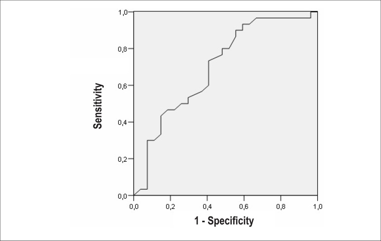
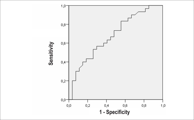
Using ROC curve analysis, LAVImax yielded an area under the curve of 70% (p < 0.05) for prediction of PAF attacks. When the LAVImax (> 20.9 mL/m2) was used as cutoff to predict PAF in patients with HT and PAF could be identified with a sensitivity of 80% and a specificity of 51%. The sensitivity and specificity for LAAEF (≤ 0.45) to predict PAF were 74% and 55% respectively. The area under the curve was 68% (p < 0.05). The sensitivity and specificity for A' (≤0.11m/s) to predict PAF were 67% and 45% respectively. The area under the curve was 65% (p < 0.05) (Figures 1, 2 and 3). In multivariate logistic regression analyses to evaluate the influence of PAF and hypertension duration on left atrial volumes and functions, we found that while PAF has influence on LAVImax, LAVImin, LAVpreA and LAAEF, hypertension duration has influence on LAAEF only (Table 3).
Figure 1.
ROC curve analysis for LAVImax in predicting PAF (sen: 80%; spe: 51%; AUC: 0.700).
Figure 2.
ROC curve analysis for LAAEF in predicting PAF (sen: 74%; spe: 55%; AUC: 0.679).
Figure 3.
ROC curve analysis for A′ in predicting PAF (sen: 67%; spe: 45%; AUC: 0.654).
Table 3.
Multivariate logistic regression analysis; the influence of PAF and hypertension duration on left atrial volume and function
| Dependent Variable | Parameter | ß | t | P value |
|---|---|---|---|---|
| LAVImax | ||||
| PAF | -2.959 | -2.757 | .008 | |
| HT duration | -.185 | -.830 | .410 | |
| LAVIpreA | ||||
| PAF | -2.514 | -2.221 | .031 | |
| HT duration | -.134 | -.569 | .572 | |
| LAVImin | ||||
| PAF | -2.728 | -2.614 | .012 | |
| HT duration | -.137 | -.634 | .529 | |
| LAPEV | ||||
| PAF | -.445 | -.519 | .606 | |
| HT duration | -.051 | -.287 | .775 | |
| LAPEF | ||||
| PAF | .021 | .591 | .557 | |
| HT duration | -.001 | -.082 | .935 | |
| LAAEV | ||||
| PAF | .214 | .306 | .761 | |
| HT duration | .004 | .025 | .980 | |
| LAAEF | ||||
| PAF | .096 | 2.171 | .034 | |
| HT duration | -.026 | -2.381 | .021 | |
| LATEV | ||||
| PAF | -.231 | -.285 | .777 | |
| HT duration | -.047 | -.283 | .778 | |
| LATEF | ||||
| PAF | .071 | 1.917 | .060 | |
| HT duration | .001 | .164 | .871 | |
| LACV | ||||
| PAF | -.398 | -.147 | .884 | |
| HT duration | .216 | .383 | .703 | |
HT: Hypertension; PAF: Paroxysmal atrial fibrillation; LAVImax: Indexed maximum left atrial volume; LAVImin: Indexed minimum left atrial volume; LAPEV: Left atrial passive emptying volume; LAPEF: Left atrial passive ejection fraction; LAAEV: Left atrial active emptying volume; LAAEF: Left atrial active ejection fraction; LATEV; Left atrial total emptying volume; LATEF: Left atrial total ejection fraction; LACV: Left atrial conduit volume.
Discussion
In this study, the left atrium in hypertensive group with PAF was characterized by further enlargement when compared to the hypertensive group without PAF. While LA booster pump function was increased in hypertensive patients when compared to normotensive subjects, it was impaired in hypertensive subjects with PAF as compared with hypertensive patients without PAF.
Hypertension causes an increase in LV wall stress that causes myocardial hypertrophy. Increased LV wall thickness elevates LV diastolic filling pressure, inducing myocardial fibrosis, which constitutes a favorable milieu for arrhythmia, not only in the left ventricle but also in the left atrium. Previously it has been reported that increased LV mass and LA size were associated with AF in hypertensive subjects22,23. LA dilation is attributed to impairment of the diastolic blood flow from left atrium to left ventricle due to the increased left ventricular stiffness. In our study LV mass, E/E' and LA volume indices were increased in the hypertensive group with PAF as compared with hypertensive subjects without PAF. There is a conflict in cardiology about the association between onset of AF and left atrial enlargement. In large prospective trials it has been established that left atrial enlargement is an independent risk factor for the development of AF. On the other hand, several studies have suggested that atrial dilation is a result of AF12,24. It seems that AF and atrial enlargement is in a casual association. During the increased pressure overload in the left atrium, LA myocardium operates according to the Frank-Starling Law, enhancing its reservoir function to a critical point, after which the total emptying rate begins to decline. With structural deterioration, the atrial electrical conduction properties are also impaired and electrical disturbance influences the myocardial contraction sequence in left atrial chamber, further increasing structural impairment.
In the early filling of the LV, the LA acts as a conduit, passively emptying during LV relaxation, which is strongly influenced by LV compliance25. LA passive emptying function is associated with multiple factors, i.e., the suction force of the LV during diastole, the recoil function of the left atrium after expansion, LV end-diastolic pressure and LA pressure. In hypertension with sinus rhythm, LV end-diastolic pressure is increased and the normal recoil function of the left atrium causes the decrease in passive emptying function of the left atrium. In hypertension with AF, LV diastolic function is impaired, LV end-diastolic pressure is elevated and the recoil function of left atrium is also possibly impaired; that results in increased LA pressure, prompting an increase in LA passive emptying function in early diastole. In the study of Cui et al26 LA function was evaluated with acoustic quantification method and the rapid emptying volume was significantly higher in hypertensive patients with PAF compared to hypertensive patients without PAF. In the study of Barbier et al27 LA function and ventricular filling was evaluated in hypertensive patients with and without PAF; the transport function of LA was not significantly different between the hypertensive patients with and without PAF. In our study, LAPEF and LAPEV were significantly decreased in hypertensive subjects without PAF when compared to normotensive subjects, as consistently shown in previous studies28. LAPEF and LAPEV were non-significantly increased in hypertensive patients with PAF as compared to the hypertensive group without PAF. As mentioned above, the early filling of left ventricle is affected by LA pressure, left ventricular systolic and diastolic function. In our study, there was no significant difference in left ventricular ejection fraction, systolic and diastolic blood pressure. Left ventricular mass was higher in hypertensive group with PAF than in the hypertensive group without PAF. In terms of diastolic dysfunction, the E/A ratio represents the diastolic dysfunction in the hypertensive group without PAF and its value increases in the hypertensive group with PAF, while E-wave increases and A-wave decreases in this group. As the left atrial chamber dilates, its contractile function begins to deteriorate and A-velocity, which is related with atrial contraction, decreases and E-velocity increases, resulting in 'normalization' of E/A ratio.
The left atrium is also a contractile chamber that actively empties immediately before the onset of LV systole15. In several studies, hypertension was found to be associated with increased atrial contractility compensating for decreased early LV filling in patients with reduced left ventricular compliance29. Increased atrial contractility has been attributed to Frank-Starling law, in which increased atrial dimension leads to atrial stretching, resulting in increased atrial force. In our study, consistent with what was found in previous studies, we observed that LA contractile function was enhanced in the hypertensive group without PAF when compared to normotensive population. With enlargement of the left atrial chamber, contractile function begins to deteriorate and dysfunctional myocardium becomes the basis for electrical conduction disorder that comprises the contraction sequence in left atrium, resulting in reduced LA active emptying. Barbier et al27 investigated the LA functions and LA fractional shortening was found to be reduced in the hypertensive patients with PAF when compared to the hypertensive subjects without PAF. Henein et al30 studied the left atrial functions in normotensive subjects and hypertensive patients with and without PAF. LA functions were determined with strain imaging and speckle tracking echocardiography and LA contractile function was reduced in hypertensive patients with PAF compared to the hypertensive subjects without PAF. In our study, left atrial booster pump function was decreased in hypertensive patients with PAF compared to the hypertensive patients without PAF. This contractile dysfunction is probably due to chronic degenerative changes in the LA myocardium and should be associated with the process called left atrial remodeling. This was also supported by significantly lower A'-velocity in the group of hypertensive patients with PAF compared to those without PAF in our study. Recently, in the study by Yoon et al16, LA pump function was evaluated in patients with PAF and LA contractile dysfunction was determined through the A'-velocity. They reported that the impaired contractile function is associated with PAF and A'-velocity may predict the PAF attacks.
In this study we also found that in addition to the high blood pressure, the hypertension period duration is the crucial point and the underlying precipitating factor for PAF. The hypertensive group with PAF had long-term hypertension when compared to the hypertensive group without the arrhythmia. The cumulative long-term effects of high blood pressure should explain the differences in LA volumes, LV mass and diastolic dysfunction ratio between the hypertensive groups with and without PAF.
The occurrence of AF in hypertensive individuals may be associated with impairment of atrial contractility. This mechanism should be that patients whose atrial myocardium is more predisposed to increased load and wall stress, develop atrial myocardial impairment causing contractile dysfunction and a milieu for electrical conduction degeneration within the atrium.
Limitations
It is difficult to distinguish whether the impaired atrial pump function was attributed to LA remodeling or to a recovery process after spontaneous conversion of AF. This fact does not concern the diagnosis of PAF, but it may affect the values for detecting PAF. Also, providing an accurate time interval between PAF attack and the current examination was difficult because PAF is an elusive disease, and PAF episodes are generally asymptomatic.
Conclusion
AF is an important disease for public health. In this study we found that, in hypertensive patients, PAF is associated with left atrial dysfunction. Due to the fact that hypertension is the most common risk factor for this noxious arrhythmia, hypertensive subjects should be closely monitored and be adequately treated. AF is closely associated with left atrial dysfunction. Considering the fact that poorly treated hypertension can more easily lead to left atrial dysfunction, hypertensive patients should be treated carefully and evaluated frequently for detection of left atrial dysfunction.
Footnotes
Potential Conflict of Interest
No potential conflict of interest relevant to this article was reported.
Study Association
This study is not associated with any thesis or dissertation work.
Sources of Funding
There were no external funding sources for this study.
Author contributions
Conception and design of the research: Tenekecioglu E, Peker T; Acquisition of data: Tenekecioglu E, Ozluk OA, Peker T, Kuzeytemiz M, Yılmaz M; Analysis and interpretation of the data: Agca FV, Karaagac K, Peker T, Kuzeytemiz M, Senturk M; Statistical analysis: Agca FV, Ozluk OA, Karaagac K, Kuzeytemiz M, Senturk M; Writing of the manuscript: Tenekecioglu E, Karaagac K, Demir S, Senturk M; Yılmaz M; Critical revision of the manuscript for intellectual content: Tenekecioglu E, Ozluk OA, Demir S, Yılmaz M.
References
- 1.Psaty BM, Manolio TA, Kuller LH, Kronmal RA, Cushman M, Fried LP, et al. Incidence of and risk factors for atrial fibrillation in older adults. Circulation. 1997;96(7):2455–2461. doi: 10.1161/01.cir.96.7.2455. [DOI] [PubMed] [Google Scholar]
- 2.Kannel WB, Wolf PA, Benjamin EJ, Levy D. Prevalence, incidence, prognosis, and redisposing conditions for atrial fibrillation: population-based estimates. Am J Cardiol. 1998;82(Suppl):2N–9N. doi: 10.1016/s0002-9149(98)00583-9. [DOI] [PubMed] [Google Scholar]
- 3.Pritchett AM, Mahoney DW, Jacobsen SJ, Rodeheffer RJ, Karon BL, Redfield MM. Diastolic dysfunction and left atrial volume: a population-based study. J Am Coll Cardiol. 2005;45(1):87–92. doi: 10.1016/j.jacc.2004.09.054. [DOI] [PubMed] [Google Scholar]
- 4.Tsang TS, Gersh BJ, Appleton CP, Tajik AJ, Barnes ME, Bailey KR, et al. Left ventricular diastolic dysfunction as a predictor of the first diagnosed nonvalvular atrial fibrillation in 840 elderly men and women. J Am Coll Cardiol. 2002;40(9):1636–1644. doi: 10.1016/s0735-1097(02)02373-2. [DOI] [PubMed] [Google Scholar]
- 5.Matsuda Y, Toma Y, Ogawa H, Matsuzaki M, Katayama K, Fujii T, et al. Importance of left atrial function in patients with myocardial infarction. Circulation. 1983;67(3):566–571. doi: 10.1161/01.cir.67.3.566. [DOI] [PubMed] [Google Scholar]
- 6.Triposkiadis F, Pitsavos C, Boudoulas H, Trikas A, Toutouzas P. Left atrial myopathy in idiopathic dilated cardiomyopathy. Am Heart J. 1994;128(3-4):308–315. doi: 10.1016/0002-8703(94)90484-7. [DOI] [PubMed] [Google Scholar]
- 7.Stefanadis C, Dernellis J, Stratos C, Tsiamis E, Vlachopoulos C, Toutouzas K, et al. Effects of balloon mitral valvuloplasty on left atrial function in mitral stenosis as assessed by pressure-area relation. J Am Coll Cardiol. 1998;32(1):159–168. doi: 10.1016/s0735-1097(98)00178-8. [DOI] [PubMed] [Google Scholar]
- 8.Cresci S, Goldstein JA, Cardona H, Waggoner AD, Perez JE. Impaired left atrial function after heart transplantation: disparate contribution of donor and recipient atrial components studied on-line with quantitative echocardiography. J Heart Lung Transplant. 1995;14(4):647–653. [PubMed] [Google Scholar]
- 9.Feinberg MS, Waggoner AD, Kater KM, Cox JL, Pere JE. Echocardiographic automatic boundary detection to measure left atrial function after the maze procedure. J Am Soc Echocardiogr. 1995;8(2):139–148. doi: 10.1016/s0894-7317(05)80403-1. [DOI] [PubMed] [Google Scholar]
- 10.Thomas L, Boyd A, Thomas SP, Schiller NB, Ross DL. Atrial structural remodeling and restoration of atrial contraction after linear ablation for atrial fibrillation. Eur Heart J. 2003;24(21):1942–1951. doi: 10.1016/j.ehj.2003.08.018. [DOI] [PubMed] [Google Scholar]
- 11.Dernellis JM, Panaretou MP. Effects of digoxin on left atrial function in heart failure. Heart. 2003;89(11):1308–1315. doi: 10.1136/heart.89.11.1308. [DOI] [PMC free article] [PubMed] [Google Scholar]
- 12.Vaziri SM, Larson MG, Benjamin EJ, Levy D. Echocardiographic predictors of nonrheumatic atrial fibrillation: the Framingham Heart Study. Circulation. 1994;89(2):724–730. doi: 10.1161/01.cir.89.2.724. [DOI] [PubMed] [Google Scholar]
- 13.Abhayaratna WP, Seward JB, Appleton CP, Douglas PS, Oh JK, Tajik AJ, et al. Left atrial size: physiologic determinants and clinical applications. J Am Coll Cardiol. 2006;47(12):2357–2363. doi: 10.1016/j.jacc.2006.02.048. [DOI] [PubMed] [Google Scholar]
- 14.Verdecchia P, Reboldi G, Gattobigio R, Bentivoglio M, Borgioni C, Angeli F, et al. Atrial fibrillation in hypertension: predictors and outcome. Hypertension. 2003;41(2):218–223. doi: 10.1161/01.hyp.0000052830.02773.e4. [DOI] [PubMed] [Google Scholar]
- 15.Kojima T, Kawasaki M, Tanaka R, Ono K, Hirose T, Iwama M, et al. Left atrial global and regional function in patients with paroxysmal atrial fibrillation has already been impaired before enlargement of left atrium: velocity vector imaging echocardiography study. Eur Heart J Cardiovasc Imaging. 2012;13(3):227–234. doi: 10.1093/ejechocard/jer281. [DOI] [PubMed] [Google Scholar]
- 16.Yoon JH, Moon J, Chung H, Choi EY, Kim JY, et al. Left atrial function assessedby Doppler echocardiography rather than left atrial volume predicts recurrence in patients with paroxysmal atrial fibrillation. Clin Cardiol. 2013;36(4):235–240. doi: 10.1002/clc.22105. [DOI] [PMC free article] [PubMed] [Google Scholar]
- 17.Casale PN, Devereux RB, Milner M, Zullo G, Harshfield GA, Pickering TG, et al. Value of echocardiographic measurement of left ventricular mass in predicting cardiovascular morbid events in hypertensive men. Ann Intern Med. 1986;105(2):173–178. doi: 10.7326/0003-4819-105-2-173. [DOI] [PubMed] [Google Scholar]
- 18.Kircher B, Abbott JA, Pau S, Gould RG, Himelman RB, Higgins CB, et al. Left atrial volume determination by biplane two-dimensional echocardiography: validation by cine computed tomography. Pt 1Am Heart J. 1991;121(3):864–871. doi: 10.1016/0002-8703(91)90200-2. [DOI] [PubMed] [Google Scholar]
- 19.Lester SJ, Ryan EW, Schiller NB, Foster E. Best method in clinical practice and in research studies to determine left atrial size. Am J Cardiol. 1999;84(7):829–832. doi: 10.1016/s0002-9149(99)00446-4. [DOI] [PubMed] [Google Scholar]
- 20.Spencer KT, Mor-Avi V, Gorcsan J, 3rd, De-Maria AN, Kimball TR, Monaghan MJ, et al. Effects of aging on left atrial reservoir, conduit, and booster pump function: a multi-institution acoustic quantification study. Heart. 2001;85(3):272–277. doi: 10.1136/heart.85.3.272. [DOI] [PMC free article] [PubMed] [Google Scholar]
- 21.Lindstrom L, Wranne B. Pulsed tissue Doppler evaluation of mitral annulus motion: a new window to assessment of diastolic function. Clin Physiol. 1999;19(1):1–10. doi: 10.1046/j.1365-2281.1999.00137.x. [DOI] [PubMed] [Google Scholar]
- 22.Gottdiener JS, Kitzman DW, Aurigemma GP, Arnold AM, Manolio TA. Left atrial volume, geometry, and function in systolic and diastolic heart failure of persons >or = 65 years of age (the cardiovascular health study) Am J Cardiol. 2006;97(1):83–89. doi: 10.1016/j.amjcard.2005.07.126. [DOI] [PubMed] [Google Scholar]
- 23.Nagano R, Masuyama T, Naka M, Hori M, Kamada T. Contribution of atrial reservoir function to ventricular filling in hypertensive patients: effects of nifedipine administration. Hypertension. 1995;26(5):815–819. doi: 10.1161/01.hyp.26.5.815. [DOI] [PubMed] [Google Scholar]
- 24.Schotten U, Neuberger HR, Allessie MA. The role of atrial dilatation in the domestication of atrial fibrillation. Prog Biophys Mol Biol. 2003;82(1-3):151–162. doi: 10.1016/s0079-6107(03)00012-9. [DOI] [PubMed] [Google Scholar]
- 25.Toh N, Kanzaki H, Nakatani S, Ohara T, Kim J, Kusano KF, et al. Left atrial volume combined with atrial pump function identifies hypertensive patients with a history of paroxysmal atrial fibrillation. Hypertension. 2010;55(5):1150–1156. doi: 10.1161/HYPERTENSIONAHA.109.137760. [DOI] [PubMed] [Google Scholar]
- 26.Cui Q, Wang H, Zhang W, Wang H, Sun X, Zhang Y, et al. Enhanced left atrial reservoir, increased conduit, and weakened booster pump function in hypertensive patients with paroxysmal atrial fibrillation. Hypertens Res. 2008;31(3):395–400. doi: 10.1291/hypres.31.395. [DOI] [PubMed] [Google Scholar]
- 27.Barbier P, Alioto G, Guazzi MD. Left atrial function and ventricular filling in hypertensive patients with paroxysmal atrial fibrillation. J Am Coll Cardiol. 1994;24(1):165–170. doi: 10.1016/0735-1097(94)90558-4. [DOI] [PubMed] [Google Scholar]
- 28.Kokubu N, Yuda S, Tsuchihashi K, Hashimoto A, Nakata T, Miura T, et al. Noninvasive assessment of left atrial function by strain rate imaging in patients with hypertension: a possible beneficial effect of renin-angiotensin system inhibition on left atrial function. Hypertens Res. 2007;30(1):13–21. doi: 10.1291/hypres.30.13. [DOI] [PubMed] [Google Scholar]
- 29.Aydin M, Ozeren A, Bilge M, Dursun A, Cam F, Elbey MA. Effects of dipper and non-dipper status of essential hypertension on left atrial mechanical functions. Int J Cardiol. 2004;96(3):419–424. doi: 10.1016/j.ijcard.2003.08.017. [DOI] [PubMed] [Google Scholar]
- 30.Henein M, Zhao Y, Henein MY, Lindqvist P. Disturbed left atrial mechanical function in paroxysmal atrial fibrillation: a speckle tracking study. Int J Cardiol. 2012;155(3):437–441. doi: 10.1016/j.ijcard.2011.10.007. [DOI] [PubMed] [Google Scholar]


