Description
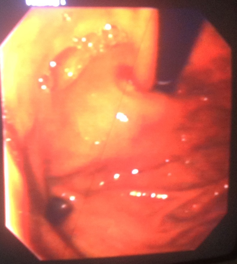
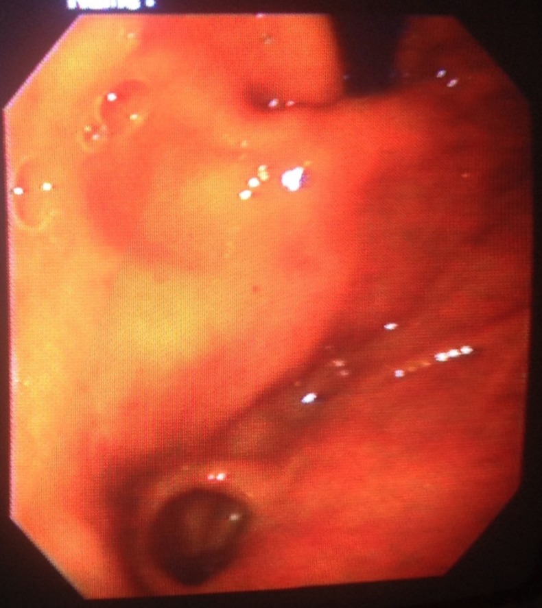
A 62-year-old male patient presented to the outpatient department with symptoms of recurrent dyspepsia and pain in the upper abdomen for the past 3 years, for which he was on erratic treatment. There was no history suggestive of chronic cough, peptic ulcer disease or reflux esophagitis. There was no history of Helicobacter pylori eradication therapy or upper abdominal surgery. Gastroscopy showed a wide-mouthed diverticulum of the size of 2×3 cm in the fundus of the stomach with features of pangastritis (figures 1 and 2). No further investigations were carried out and the patient was put on a proton-pump inhibitor as rapid urease test was negative.
Figure 1.

Gastric diverticulum on gastroscopy during retroversion.
Figure 2.

Gastric diverticulum at fundus on gastroscopy during retroversion.
Gastric diverticulum, a rare form of diverticular disease, is an outpouching of the gastric wall. The incidence of gastric diverticula observed by upper gastrointestinal contrast study is 0.04%, however it is 0.02% in autopsy studies.1 2 It is equally distributed between men and women. It usually presents in the fifth and sixth decade of life. Gastric diverticulum has also been seen in newborns, usually associated with pyloric or duodenal obstruction.3 Gastric diverticulum can be congenital, which are true diverticula or acquired which are false diverticula. It can remain asymptomatic or can present with symptoms such as upper abdominal pain, dyspepsia, weight loss, anaemia, reflux or even bleeding and perforation. The patient should be put on symptomatic treatment and surgical intervention is required only when the symptoms do not respond to medical management or if the diverticulum is large. Diverticulectomy is the best surgical management, though invagination of the diverticulum is also an option. Nowadays, these procedures are being done through laparoscopic or transthoracic approach.3
Learning points.
A high index of suspicion should be kept in mind in patients with longstanding history of vague upper abdominal pain and dyspepsia, which do not subside with proton-pump inhibitors.
Because of the rarity of the disease, gastric diverticulum is liable to cause confusion in diagnosis as they can also be missed even in gastroscopy.
Contrast study with barium is the best method for the diagnosis if the neck is narrow.
Acknowledgments
The authors would like to thank Sana Khan, medical student.
Footnotes
Contributors: FFH and MH were responsible for the drafting of the article. MH and AB were responsible for the literature search. SIB provided the photo and performed the proof reading of the article.
Competing interests: None.
Patient consent: Obtained.
Provenance and peer review: Not commissioned; externally peer reviewed.
References
- 1.Palmer Ed. Collective review: gastric diverticula. Int Abstr Surg 1951;92:417–28 [PubMed] [Google Scholar]
- 2.River A, Stevens G, Kirklin B. Diverticula of stomach. Surg Gynecol Obstet 1935;60:106–13 [Google Scholar]
- 3.Rodeberg DA, Zaheer S, Moir CR, et al. Gastric diverticulum: a series of four pediatric patients. J Pediatr Gastroenterol Nutr 2002;24:564–67 [DOI] [PubMed] [Google Scholar]


