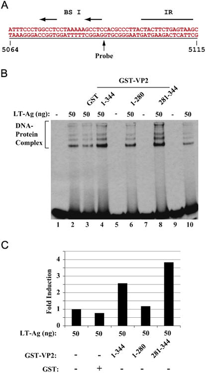
Fig. 6.

The putative DNA binding domain of VP2/VP3 is important for induced DNA binding activity of LT-Ag to Ori DNA. Labeled probe (A) was incubated either with LT-Ag alone (50 ng, lane 2) or LT-Ag in combination with GST (1.5 μg, lane 3) or LTAg in combination with GST-VP2 full length (1.5 μg lane 4) or LT-Ag plus VP2 deletion mutants fused to GST (1.5 μg each, lanes 6 and 8) as indicated. DNAprotein complexes were separated on a 6% PAGE and analyzed by autoradiography as described in the legend for fig. 4B (B). In lane 1, probe alone was loaded. (C) Quantitative analysis of the “DNA-protein complexes” on panel B by a semiquantitative densitometry method using ImageJ (NIH) and bar graph presentation of the results in arbitrary units in fold. Protein concentrations for GST and GST-VP2 and GST-VP2 mutants are the same as described for panel B. The DNA binding efficiency of LT-Ag in the presence of VP2 or its mutants was expressed relative to that of LT-Ag binding to Ori alone.
