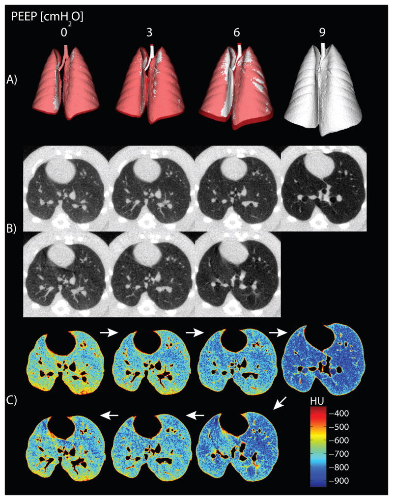
Fig. 6.
(A) Tridimensional reconstructions of the whole lung, obtained by automatic segmentation and thresholding of computerized tomography images (excluding Hounsfield units [HU] greater than −200), are shown for all levels of positive end-expiratory pressure (PEEP) in a representative rat. Images obtained during ascending and descending PEEP are superimposed on each other and color-coded: white for increasing PEEP and red for decreasing PEEP. Lung dimensions during descending PEEP were larger than those measured in the ascending ramp, except for the areas colored in white, where they overlapped. (B) Axial computerized tomography slices obtained in the same animal at all PEEP levels are shown. Images were obtained shortly after a recruitment maneuver, and no significant atelectasis is visible. (C) Postthresholding maps of the same computerized tomography slices show exclusion of major blood vessels and nonpulmonary tissue only.

