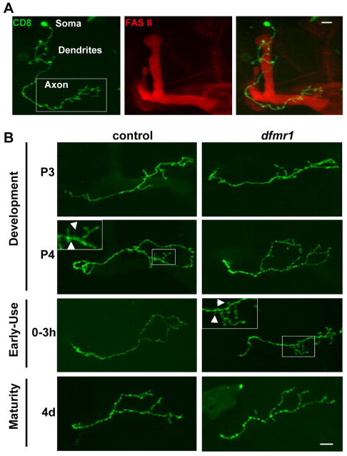
Figure 4. Single cell MARCM clonal analysis of MB gamma neuron development.
A) Representative image of Fasciclin II (FasII, red) labeled Mushroom Body containing a single MARCM gamma neuron clone (green). The white box highlights the area of axonal projection. B) Developmental profile of axon projections of single cell MARCM gamma neuron clones. Boxed insets are magnifications of areas of small (<5μm) presynaptic branches (arrows), which are subject to pruning. Scale bar=10μm. Stages: P3 (60–70 hrs APF), P4 (88–96 hrs APF), 0–3 hrs AE, 4d (96–112 hrs AE).

