

Abstract
The aim of this study was to review of the literature to determine the radiographic and clinical maxillofacial features of pycnodysostosis emphasizing the main aspects of interest to the dentist in order to make them fit for the proper treatment of this population. It is important to make the diagnosis as early as possible in order to plan the treatment more suitable to provide a better life’s quality to the patients. The most frequent clinical maxillofacial features were: grooved palate, midfacial hypoplasia, mandibular hypoplasia and enamel hypoplasia. The most common radiographic maxillofacial features were: obtuse mandibular angle, frontal/parietal/occiptal bossing, open fontanels and sutures, multiple impacted teeth. The earlier diagnostic of pycnodysostosis has a fundamental role in general health of the patients. We consider that is very important that the dentist know recognize the radiographic and clinical maxillofacial features of pycnodysostosis, which allows correct treatment planning avoiding risks and ensuring better life’s quality to the patients.
Keywords: Pycnodysostosis, maxillofacial abnormalities, radiography
Introduction
Pycnodysostosis is a rare autosomal recessive disorder. The main features of patients with pycnodysostosis are short stature, dysplasia of the clavicles, acro-osteolysis of distal phalanges, deformity of the skull, midfacial hypoplasia, absence or hypopneumatization of the paranasal sinuses, narrow and/or grooved palate, generalizated osteosclerosis and fragility of bone [1,2].
Patients with pycnodysostosis present several morphological changes of maxillofacial structures, requiring specialized dental care. In the maxilla and mandible, for example, the abnormal growth of the dental arch results in dental crowding [2,3]. This condition hinders good oral hygiene leading to a high rate of caries and periodontal disease in these patients [3,4]. In some cases surgical intervention is required to correct malocclusion and esthetics in patients with dentofacial deformities caused by micrognathia and hypoplasia of maxilla [5-7]. In these cases must take preventive care in order to avoid complications such as osteomielitis [8].
The goal of this study was to review the literature to determine the radiographic and clinical maxillofacial features of pycnodysostosis emphasizing the main aspects of interest to the dentist in order to make them fit for the proper treatment of this population.
Materials and methods
Were analyzed studies published in the medical literature, after review in Pub-Med/MEDLINE, SCOPUS, ISI Web of Knowledge, SciELO and LILACS. Articles written in Portuguese, English and Spanish were searched using the keywords: pycnodysostosis, maxillofacial abnormalities, oral findings, radiography.
Original articles, systematic reviews and case report were included, longitudinal prospective studies and retrospective studies, which were published between 1970 and 2013. We excluded articles without abstract available.
Results
A total of 54 articles were found, being 18 in Pub-Med/MEDLINE, 18 in SCOPUS, 4 in ISI Web of Knowledge, 4 in SciELO and 10 in LILACS. Were selected a total of 18 articles and based on these selected articles were included 35 patients, who had analyzed their clinical and radiographic features reported by the authors. The Table 1 shows the clinical maxillofacial features and the Table 2 shows the radiographic maxillofacial features of patients with pycnodysostosis, with their respective percentages and according to the authors. Figure 1 shows a panoramic radiograph of a patient with some radiographic maxillofacial features in the pycnodysostosis: thin mandible, obtuse mandibular angle, malposed teeth, elongation of the condyle and coronoid process.
Table 1.
Clinical maxillofacial features of patients with pycnodysostosis
| Grooved palate | Midfacial hypoplasia | Mandibular hypoplasia | Dental crowding | Enamel hypoplasia | Narrow palate | Cross/open bite | Class III Skeletal | Dental abnormalities | |
|---|---|---|---|---|---|---|---|---|---|
| Agarwal et al. [16] | ++ | - | + | - | - | + | -/- | - | - |
| Alves et al. [13] | + | + | + | - | - | + | -/+ | - | - |
| Bathi & Masur [8] | ++ | + | - | - | + | ++ | -/- | - | - |
| Delgado et al. [17] | + | + | - | + | - | + | - | - | - |
| Fleming et al. [18] | + | + | + | + | - | - | -/- | - | + |
| Fonteles et al. [7] | ++++ | ++++ | +++ | ++++ | - | - | ++++/+++ | ++++ | ++++ |
| Francisco & Nicholoff [25] | - | + | - | - | - | + | -/- | - | - |
| Hunt et al. [15] | +++ | ++ | ++ | - | - | - | ++/- | + | + |
| Ilankovan & Moos [5] | + | + | + | - | + | - | -/- | + | - |
| Kamak et al. [19] | + | + | - | - | - | + | +/+ | + | + |
| Kawahara et al. [20] | + | + | + | + | - | - | -/- | - | - |
| Kshirsagar et al. [21] | + | + | - | + | - | + | - | - | - |
| Muto et al. [2] | + | + | + | - | + | - | +/- | - | - |
| Nørholt et al. [6] | - | + | - | - | - | - | +/+ | + | - |
| O’Connell et al. [3] | ++ | + | + | ++ | - | - | -/- | - | - |
| Ramaiah et al. [22] | - | + | - | + | + | - | -/- | - | - |
| Soares et al. [23] | + | + | + | + | + | -/+ | - | ||
| Soliman et al. [24] | - | - | - | - | +++++++ | - | -/- | - | - |
| Number of patients* | 22 | 20 | 13 | 12 | 11 | 9 | 9/7 | 8 | 7 |
| Percentaje (%) | 66.66 | 60.60 | 39.39 | 36.36 | 33.33 | 27.27 | 27.27/21.21 | 24.24 | 21.21 |
n=33; (+) presence (each + represents a patient); (-) absence or no related.
Table 2.
Radiographic maxillofacial features of patients with pycnodysostosis
| Obtuse mandibular angle | Frontal/parietal/occiptal bossing | Open fontanelle and sutures | Multiple impacted teeth | Absence or hypopneumatization of the paranasal sinuses | Wormian bones | elongation of the coronoid process/mandibular condyle | Supernumerary teeth | |
|---|---|---|---|---|---|---|---|---|
| Agarwal et al. [16] | + | - | ++ | - | + | ++ | -/- | - |
| Alves et al. [13] | + | + | + | - | + | - | +/+ | - |
| Alves-Pereira et al. [4] | +++ | +++ | - | - | ++ | ++ | ++/++ | - |
| Bathi & Masur [8] | ++ | + | ++ | - | ++ | - | -/- | - |
| Delgado et al. [17] | + | + | + | - | + | - | -- | |
| Fleming et al. [18] | + | - | - | + | + | + | -/- | - |
| Fonteles et al. [7] | +++ | ++++ | - | ++ | - | - | -/- | - |
| Francisco & Nicholoff [25] | + | + | + | + | - | + | -/- | + |
| Hunt et al. [15] | +++ | ++ | +++ | ++ | +++ | - | -/- | - |
| Ilankovan & Moos [5] | + | + | + | - | + | - | -/- | - |
| Kamak et al. [19] | + | + | + | - | - | - | -/- | - |
| Kawahara et al. [20] | + | + | + | + | - | - | -/- | - |
| Kshirsagar et al. [21] | + | + | + | + | - | - | - | |
| Muto et al. [2] | + | + | + | - | + | - | +/- | |
| Nørholt et al. [6] | + | - | - | - | - | - | -/- | - |
| O’Connell et al. [3] | ++ | ++ | ++ | ++ | - | - | +/+ | - |
| Ramaiah et al. [22] | + | + | + | - | + | + | -/- | + |
| Soares et al. [23] | + | + | + | - | - | - | - | - |
| Soliman et al. [24] | +++++++ | ++++++ | ++++++++ | ++++++++ | - | - | -/- | - |
| Number of patients* | 33 | 28 | 27 | 17 | 15 | 7 | 5/4 | 2 |
| Percentaje (%) | 94.28 | 80.00 | 77.14 | 48.57 | 42.85 | 20.00 | 14.28/11.42 | 5.71 |
n=35; (+) presence (each + represents a patient); (-) absence or no related.
Figure 1.
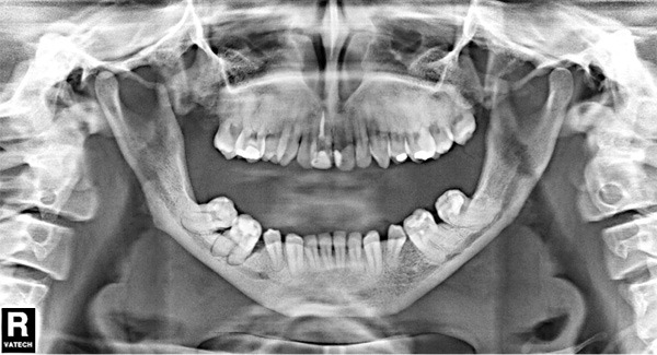
The panoramic radiograph showing thin mandible, obtuse mandibular angle, malposed teeth, elongation of the condyle and coronoid process.
Discussion
Pycnodysostosis is a rare autosomal recessive disorder primarily characterized by osteosclerosis, bone fragility, short stature, acro-osteolysis, and delayed closure of the cranial sutures [4].
The pycnodysostosis can be confused with other diseases, such as osteopetrosis (OP) and cleidocranial dysostosis (CD), because they present some clinical and radiographic similar signs, so it is important that the dentist knows how to make the differential diagnosis in order to indicate the best treatment for each patient. The delayed closure of sutures; frontal, occipital and/or parietal bossing; hypopneumatization of the paranasal sinuses; narrow and deep palate are features that can be observed in pycnodysostosis and in CD [8,9]. Patients with CD present supernumerary teeth [9], however, in pycnodysostosis the appearance of supernumerary teeth is not very frequent, being reported by only 5.71% of the authors (Table 2). Increase of bone density and bone fragility are present in both pycnodysostosis and OP [8,10]. The differential diagnosis between pycnodysostosis and OP is performed through the following analysis: the size of the skull, since in OP the patients have no frontal/parietal bossing; the size of the maxilla and mandible, the maxillary sinus, the sutures and the angle of the mandible, which do not show changes in OP [8].
When not diagnosed in infancy, fractures resulting from trauma lead to the diagnosis of Pycnodysostosis. Mandibular fractures have been described in adults following extractions, other important potential risk in these cases is the occurrence of osteomyelitis [2,11]. The osteomyelitis can be caused by oral conditions of the patient [8], after the teeth extraction, due to poor bone vascularity of these patients [4], or may be result of iatrogenic fracture [10]. Ilankovan and Moos [5] indicate that the surgical procedures in pycnodysostosis patients have enhanced possibility of infection, impaired wound healing, osteomyelitis and pathological fracture. The treatment of osteomyelitis is very difficult in these patients, especially when it involves the mandible [3].
We agree with Sedano et al. [12] and Muto et al. [2] when they say that the most common finding in patients with pycnodysostosis was obtuse mandibular angle. Elmore [1] reviewed 33 cases described in the literature and in 27 cases it was reported the loss of the mandibular angle, Alves et al. [13] reported that this finding was present in 87.09% of cases. To Elmore [1] the most common feature was separated cranial sutures, which was present in all cases. Maxillary hypoplasia, grooved palate, maxillary sinus hypoplasia were common findings to Muto et al. [2], Alves et al. [13] and also in our study. Open fontanelle was also a significant finding in our study, being in agreement with the findings of Elmore [1]. We found 80% of cases with frontal/parietal/occipital bossing reported, however this finding was considered inexpressive to Elmore [1]. For this author the grooved palate was also found in a small number of patients, which is in disagreement with our findings. Wormian bone was found in a few patients in the study of Elmore [1], as well as in our study. Crossbite was a little expressive feature found by Muto et al. [2]. Furthermore, Class III skeletal, dental abnormalities, elongation of the coronoid process and the mandibular condyle were reported by few authors.
The main clinical maxillofacial features in the pycnodysostosis include a grooved palate (66.66%), midfacial hypoplasia (60.60%), mandibular hypoplasia (39.39%), enamel hypoplasia (33.33%), dental crowding (36.36%), narrow palate (27.27%), cross bite (27.27%), class III skeletal (24.24%), open bite (21.21%) and dental abnormalities (21.21%) (Table 1). The main radiographic maxillofacial features in the pycnodysostosis include obtuse mandibular angle (94.28%), large head with frontal, parietal and occiptal bossing (80%), open soft cranial sutures and fontanelles (77.14%), multiple impacted teeth (48.57%), absence or hypopeumatization of the paranasal sinuses (42.85%), wormian bones (20%), elongation of the coronoid process (14.28%) and the condyle (11.42%), supernumerary teeth (5.71%) (Table 2).
Others oral findings may consist of premature or delayed eruption teeth, persistence of deciduos teeth, malposition of teeth, hypodontia and hypercementosis [4,10,14]. It is also quite frequent presence of caries and periodontal disease in patients with pycnodysostosis, probably due to dental crowding [7,8,14,15].
Alves et al. [13] affirm that the clinical and radiological features are the bases diagnosis of this disease. We agree with these authors when affirm that it is important to make the diagnosis as early as possible in order to plan the treatment more suitable to provide a better life’s quality to the patients. Furthermore, the earlier diagnostic of pycnodysostosis has a fundamental role in general health of the patients.
Conclusion
The higher rates of maxillofacial features in patients with pycnodysostosis reported by authors were: obtuse mandibular angle, large head with frontal, parietal and occiptal bossing, open soft cranial sutures and fontanelles, grooved palate and midfacial hypoplasia. We consider that is very important that the dentist know recognize the radiographic and clinical maxillofacial features of pycnodysostosis, which allows correct treatment planning avoiding risks and ensuring better life’s quality to the patients.
Disclosure of conflict of interest
None.
References
- 1.Elmore SM. Pycnodysostosis: A review. J Bone Joint Surg Am. 1967;49:153–62. [Google Scholar]
- 2.Muto T, Michiya H, Taira H, Murase H, Kanazawa M. Pycnodysostosis. Report of a case and review of the Japanese literature, with emphasis on oral and maxillofacial findings. Oral Surg Oral Med Oral Pathol. 1991;72:449–55. doi: 10.1016/0030-4220(91)90559-u. [DOI] [PubMed] [Google Scholar]
- 3.O’Connell AC, Brennan MT, Francomano CA. Pycnodysostosis: orofacial manifestations in two pediatric patients. Pediatr Dent. 1998;20:204–7. [PubMed] [Google Scholar]
- 4.Alves-Pereira D, Berini-Aytés L, Gay-Escoda C. Pycnodysostosis. A report of 3 clinical cases. Med Oral Patol Oral Cir Bucal. 2008;13:E633–5. [PubMed] [Google Scholar]
- 5.Ilankovan V, Moos KF. Pyknodysostosis: case report with surgical correction of the facial deformity. Br J Oral Maxillofac Surg. 1990;28:39–42. doi: 10.1016/0266-4356(90)90009-a. [DOI] [PubMed] [Google Scholar]
- 6.Nørholt SE, Bjerregaard J, Mosekilde L. Maxillary distraction osteogenesis in a patient with pycnodysostosis: a case report. J Oral Maxillofac Surg. 2004;62:1037–40. doi: 10.1016/j.joms.2004.02.012. [DOI] [PubMed] [Google Scholar]
- 7.Fonteles CS, Chaves CM Jr, Da Silveira A, Soares EC, Couto JL, de Azevedo MF. Cephalometric characteristics and dentofacial abnormalities of pycnodysostosis: report of four cases from Brazil. Oral Surg Oral Med Oral Pathol Oral Radiol Endod. 2007;104:e83–90. doi: 10.1016/j.tripleo.2007.05.011. [DOI] [PubMed] [Google Scholar]
- 8.Bathi RJ, Masur VN. Pyknodysostosis - a report of two cases with a brief review of the literature. Int J Oral Maxillofac Surg. 2000;29:439–42. [PubMed] [Google Scholar]
- 9.Alves N, Oliveira R. Cleidocranial dysplasia - A case report. Int J Morphol. 2008;26:1058–68. [Google Scholar]
- 10.Zachariades N, Koundouris I. Maxillofacial symptoms in two patients with pycnodysostosis. J Oral Maxillofac Surg. 1984;42:819–23. doi: 10.1016/0278-2391(84)90353-7. [DOI] [PubMed] [Google Scholar]
- 11.Iwu CO. Bilateral osteomyelitis of the mandible in pycnodysostosis. A case report. Int J Oral Maxillofac Surg. 1991;20:71–2. doi: 10.1016/s0901-5027(05)80709-x. [DOI] [PubMed] [Google Scholar]
- 12.Sedano HG, Gorlin RJ, Anderson VE. Pycnodysostosis: clinical and genetic considertions. Am J Dis Child. 1968;116:70–7. doi: 10.1001/archpedi.1968.02100020072010. [DOI] [PubMed] [Google Scholar]
- 13.Alves N, Oliveira RJ, Deana NF, Sampaio JCA. Morphological features of pycnodysostosis with emphasis on clinical and radiographic maxillofacial findings. Int J Morphol. 2013;31:921–4. [Google Scholar]
- 14.Yamada N, Inomata H, Morita K. Two cases of pycnodisostosis with special emphasis on maxillo-facial findings. Dentomaxillofac Radiol. 1973;2:12. doi: 10.1259/dmfr.1973.0003. [DOI] [PubMed] [Google Scholar]
- 15.Hunt NP, Cunningham SJ, Adnan N, Harris M. The dental, craniofacial, and biochemical features of pyknodysostosis: a report of three new cases. J Oral Maxillofac Surg. 1998;56:497–504. doi: 10.1016/s0278-2391(98)90722-4. [DOI] [PubMed] [Google Scholar]
- 16.Agarwal I, Kirubakaran C, Sridhar G. Pyknodysostosis: a report of two siblings with unusual manifestations. Ann Trop Paediatr. 1999;19:301–5. doi: 10.1080/02724939992419. [DOI] [PubMed] [Google Scholar]
- 17.Delgado W, Beltrán J, Arrascue M, Oré I. Picnodisostosis: Un síndrome óseo de interés en estomatología. Rev Estomatol Herediana. 1999;9:5–14. [Google Scholar]
- 18.Fleming KW, Barest G, Sakai O. Dental and facial bone abnormalities in pyknodysostosis: CT findings. AJNR. 2007;28:132–4. [PMC free article] [PubMed] [Google Scholar]
- 19.Kamak H, Kamak G, Yavuz I. Clinical, radiographic, diagnostic and cephalometric features of pycnodysostosis in comparison with turkish cephalometric norms: a case report. Eur J Dent. 2012;6:454–9. [PMC free article] [PubMed] [Google Scholar]
- 20.Kawahara K, Nishikiori M, Imai K, Kishi K, Fujiki Y. Radiographic observations of pyknodysostosis. Report of a case. Oral Surg Oral Med Oral Pathol. 1977;44:476–82. doi: 10.1016/0030-4220(77)90419-4. [DOI] [PubMed] [Google Scholar]
- 21.Kshirsagar VY, Ahmed M, Nagarsenkar S, Sahoo K, Shah KB. Ichthyosis vulgaris and pycnodysostosis: An unusual occurrence. Acta Med Acad. 2012;41:214–8. doi: 10.5644/ama2006-124.54. [DOI] [PubMed] [Google Scholar]
- 22.Ramaiah KK, George GB, Padiyath S, Sethuraman R, Cherian B. Pyknodysostosis: report of a rare case with review of literature. Imaging Sci Dent. 2011;41:177–81. doi: 10.5624/isd.2011.41.4.177. [DOI] [PMC free article] [PubMed] [Google Scholar]
- 23.Soares LF, Souza IPR, Cardoso AS, Pomarico L. Pyknodysostosis: Oral findings and differential diagnosis. J Indian Soc Pedod Prev Dent. 2008;26:S23–5. [PubMed] [Google Scholar]
- 24.Soliman AT, Ramadan MA, Sherif A, Aziz Bedair ES, Rizk MM. Pycnodysostosis: clinical, radiologic, and endocrine evaluation and linear growth after growth hormone therapy. Metabolism. 2011;50:905–11. doi: 10.1053/meta.2001.24924. [DOI] [PubMed] [Google Scholar]
- 25.Francisco JV, Nicholoff TJ. Pyknodysostosis: An unusual presentation in a denture wearer. A case Report. Oral Surg Oral Med Oral Pathol. 1991;72:693–5. doi: 10.1016/0030-4220(91)90013-3. [DOI] [PubMed] [Google Scholar]