Abstract
Campylobacter jejuni genetic manipulation is restricted by the limited number of antibiotic resistance cassettes available for use in this diarrheal pathogen. In this study, two antibiotic resistance cassettes were developed, encoding for hygromycin B and apramycin resistance, for use in mutagenesis or for selection of gene expression and complementation constructs in C. jejuni. First, the marker genes were successfully modified to allow for insertional mutagenesis or deletion of a gene-of-interest, and were bracketed with restriction sites for the facilitation of site-specific cloning. These hygromycin B and apramycin markers are encoded by plasmids pAC1H and pAC1A, respectively. We also modified an insertional gene-delivery vector to create pRRH and pRRA, containing the hygromycin B and apramycin resistance genes, and 3 unique restriction sites for the directional introduction of genes into the conserved multi-copy rRNA gene clusters of the C. jejuni chromosome. We determined the effective antibiotic concentrations required for selection, and established that no harmful effects or fitness costs were associated with carrying hygromycin B or apramycin resistance under standard C. jejuni laboratory conditions. Using these markers, the arylsulfatase reporter gene astA was deleted, and the ability to genetically complement the astA deletion using pRRH and pRRA for astA gene insertion was demonstrated. Furthermore, the relative levels of expression from the endogenous astA promoter were compared to that of polycistronic mRNA expression from the constitutive promoter upstream of the resistance gene. The development of additional antibiotic resistance cassettes for use in Campylobacter will enable multiple gene deletion and expression combinations as well as more in-depth study of multi-gene systems important for the survival and pathogenesis of this important bacterium.
Introduction
The relative paucity of genetic techniques available for the manipulation of Campylobacter jejuni has historically been a limiting factor in the study and molecular biology of the leading cause of bacterial gastroenteritis in the developed world [1]. C. jejuni is a member of a large genus of microaerophilic Gram-negative ε-proteobacteria and is carried harmlessly by many animals, especially poultry, but is an endemic cause of a range of diarrheal illnesses and medical complications in humans. Many laboratories are actively studying the bacterium to understand the genetic determinants and physiological features that contribute to C. jejuni’s virulence and prevalence as a food-borne enteric pathogen. Today, research in the area continues to benefit from and depends on a small arsenal of molecular tools, such as gene deletion strategies and plasmids for genetic complementation. Since the 1980’s, only selection for kanamycin and chloramphenicol resistance has been widely adopted for the genetic manipulation of Campylobacter.
The development of the first genetic tools for C. jejuni was precipitated after the demonstration of gene transfer from Escherichia coli to C. jejuni via plasmids carrying kanamycin resistance in 1987 [2]. This led to the development, in 1988, of a kanamycin resistance cassette for use in gene disruption experiments [3]. Cloning and expression of a chloramphenicol resistance gene from Campylobacter coli in 1990 [4] was followed by development of replicative cloning vectors and mutational constructs marked with chloramphenicol resistance in 1993 [5]. Approximately a decade later, three groups successfully mutagenized C. jejuni with transposons carrying kanamycin or chloramphenicol resistance genes [6]–[8]. The finite number of resistance markers has limited genetic analyses to single-gene or single-operon studies, and has prevented complementation of double-deletion strains. As our understanding of C. jejuni grows, so does the need for new markers to rapidly delete and restore complex multi-gene systems, and/or to simultaneously express a reporter such as green fluorescent protein (GFP), arylsulfatase, or luciferase in mutant and/or complemented strains. To address this need, we adapted current C. jejuni genetic technologies to harbor resistance genes against the antibiotics hygromycin B and apramycin.
Hygromycin B is an aminoglycoside antibiotic produced by Streptomyces hygroscopicus that inhibits protein synthesis in both prokaryotes and eukaryotes [9]. Apramycin is another aminoglycoside, an aminocyclitol synthesized by Streptomyces tenebrarius [10]. Like other aminoglycosides, such as kanamycin, both hygromycin B and apramycin prevent ribosome translocation during translation elongation by binding the 30 s rRNA proximal to the ribosomal E, P and A sites [11]. Hygromycin B is not used clinically, but is sometimes a component of poultry feed where it has antihelminthic activity against nematode parasites of chickens [12]. Apramycin is also used as a veterinary antibiotic [12], [13]. The hygromycin B (HygR) resistance marker used in this study confers resistance by the activity of a specific aminoglycoside phosphotransferase encoded by the 999 bp aph(7″) gene, encoding hygromycin B 7″-O-kinase or simply hygromycin phosphotransferase [14]. The specific modification of hygromycin B is a phosphorylation at the 7″-OH of the destomic acid ring [15]. Resistance to apramycin (AprR) is conferred by the 777 bp aac(3)IV aminoglycoside 3-N-acetyltransferase gene [13]. Specifically, the enzyme acetylates the 3-amino group of apramycin’s deoxystreptamine ring [16]. Neither aph(7″) nor aac(3)IV confers resistance to the other’s respective antibiotic, nor do they confer resistance to kanamycin. Vice versa, the C. jejuni kanamycin resistance gene aphA-3 does not bestow resistance to either hygromycin B or apramycin (data not shown).
In this study, we modified existing C. jejuni gene deletion/mutagenesis and insertion strategies and plasmids to encode either HygR or AprR. We based our construction of a non-polar mutagenesis construct on the approach devised by Ménard, Sansonetti and Parsot [17], in which the resistance gene is promoterless, does not harbor a terminator, and transcription is driven from the promoter of the operon into which the gene is introduced. We also modified Karlyshev and Wren’s pRRC C. jejuni genome-insertional gene delivery and expression system [18], replacing the cat chloramphenicol acteyltransferase cassette with either aph(7″) or aac(3)IV. The expression of aph(7″) and aac(3)IV was not detrimental to C. jejuni under common laboratory conditions. Furthermore, to demonstrate the potential of these new markers and plasmids, we deleted and then complemented the C. jejuni arylsulfate sulfotransferase astA, since the product of astA cleaves a chromogenic substance that can be used to report transcriptional activity [19], [20]. With the addition of hygromycin B and apramycin resistance markers, we have provided several new, but relatively familiar, well defined and easy-to-use tools to aid other Campylobacter researchers in a variety of genetic approaches.
Materials and Methods
Bacterial Strains and Growth Conditions
Bacterial strains and plasmids used in this study are listed in Table 1. E. coli strains used for plasmid construction were grown at 37°C in Luria-Bertani (LB, Sigma) broth or on 1.7% (w/v) agar plates supplemented with ampicillin (100 µg mL, Ap), chloramphenicol (15 µg mL−1, Cm), kanamycin (50 µg mL−1, Kan), hygromycin B (100 µg mL−1) or apramycin (50 µg mL−1), as necessary. C. jejuni strains were grown at 37°C or 42°C in Mueller-Hinton (MH, Oxoid) broth or agar supplemented with vancomycin (10 µg mL−1) and trimethoprim (5 µg mL−1). C. jejuni were grown under standard growth conditions (6% O2, 12% CO2) using the Oxoid CampyGen system for shaking broth cultures, or in a Sanyo tri-gas incubator for plates. MH was supplemented with chloramphenicol (15 µg mL−1), kanamycin (50 µg mL−1), hygromycin B (250 µg mL−1) or apramycin (60 µg mL−1) where appropriate.
Table 1. Bacterial strains or plasmids used in this study.
| Strain or plasmid | Genotype or description | Source |
| E. coli strains | ||
| DH5α | F-, φ80d deoR lacZΔM15 endA1 recA1 hsdR17(rK-mK+) supE44 thi-1 gyrA96 relA1 Δ(lacZYA-argF) U169 | Invitrogen |
| C. jejuni strains | ||
| 81–176 | Wild type isolated from a diarrheic patient | [28] |
| 81–176 pRRH | Strain 81–176 with genome-integrated pRRH; HygR | This study |
| 81–176 pRRA | Strain 81–176 with genome-integrated pRRA; AprR | This study |
| 81–176 pRRC | Strain 81–176 with genome-integrated pRRC; CmR | This study |
| 81–176 pRRK | Strain 81–176 with genome-integrated pRRK; KanR | This study |
| 81–176 ΔastA::hygR | Deletion of astA with aph(7″); HygR | This study |
| 81–176 ΔastA::aprR | Deletion of astA with aac(3)IV; AprR | This study |
| DRH461 | Strain 81–176 with an unmarked deletion of astA | [19] |
| DRH461 pRRH+astA | DRH461 with integrated pRRH and polycistronic promoterless astA; HygR | This study |
| DRH461 pRRA+astA | DRH461 with integrated pRRA and polycistronic promoterless astA; AprR | This study |
| DRH461 pRRH+astA (reverse) | DRH461 with integrated pRRH and reverse orientation promoterless astA; HygR | This study |
| DRH461 pRRA+astA (reverse) | DRH461 with integrated pRRA and reverse orientation promoterless astA; AprR | This study |
| DRH461 pRRH+(p)astA (reverse) | DRH461 with integrated pRRH and reverse orientation endogenous promoter and astA; HygR | This study |
| DRH461 pRRA+(p)astA (reverse) | DRH461 with integrated pRRA and reverse orientation endogenous promoter and astA; AprR | This study |
| Plasmids | ||
| pMV261.hyg | Source of aph(7″); HygR | [29], [30] |
| p261comp.apra | Source of aac(3)IV; AprR | [30] |
| pGEM-T | Linearized cloning vector, blue-white screening; ApR | Novagen |
| pBAD24 | Low-copy arabinose-inducible expression vector; ApR | [31] |
| pAC1H | pBAD24 ligated to aph(7″) amplified with 5631 and 5632; HygR, ApR | This study |
| pAC1A | pGEM-T ligated to aac(3)IV amplified with 5633 and 5634; AprR, ApR | This study |
| pRRC | C. jejuni vector for genome integration at rRNA loci; CmR | [18] |
| pRRK | C. jejuni vector for genome integration at rRNA loci; KanR | J. Ketley |
| pRRH | C. jejuni vector for genome integration at rRNA loci; HygR | This study |
| pRRA | C. jejuni vector for genome integration at rRNA loci; AprR | This study |
| pGEM-T+astA | pGEM-T ligated to astA amplified with 5707 and 5708; ApR | This study |
| pGEM-T+astA::hygR | pGEM-T with astA interrupted with aph(7″) from pAC1H; HygR, ApR | This study |
| pGEM-T+astA::aprR | pGEM-T with astA interrupted with aac(3)IV from pAC1A; AprR, ApR | This study |
| pRRH+astA | pRRH ligated to astA amplified with 0688 and 0689; HygR | This study |
| pRRA+astA | pRRA ligated to astA amplified with 0688 and 0689; AprR | This study |
| pRRH+astA (reverse) | pRRH ligated to astA amplified with 0690 and 0691; HygR | This study |
| pRRA+astA (reverse) | pRRA ligated to astA amplified with 0690 and 0691; AprR | This study |
| pRRH+(p)astA (reverse) | pRRH ligated to astA amplified with 0692 and 0691; HygR | This study |
| pRRA+(p)astA (reverse) | pRRA ligated to astA amplified with 0692 and 0691; AprR | This study |
Construction of Plasmids pAC1H and pAC1A, pRRH and pRRA
Oligonucleotide primers used in this study are listed in Table 2 and were synthesized by Integrated DNA Technologies. The design of pAC1H and pAC1A plasmids containing the non-polar aph(7″) or aac(3)IV markers is described in Results. The aph(7″) or aac(3)IV sequence was amplified from pMV261.hyg or p261comp.apra with ultramer set 5631 and 5632, or 5633 and 5634, respectively. The polymerase chain reaction (PCR) was carried out with iProof high-fidelity DNA polymerase (Bio-Rad). A-ends were incorporated on the purified products by incubation with Taq DNA polymerase (Invitrogen) and dATP. The purified products were then introduced to linearized pGEM-T Easy (Novagen), ligated overnight with T4 DNA ligase (NEB), and transformed into E. coli DH5α (Invitrogen). Transformants were selected for on LB media supplemented with ampicillin and either hygromycin B or apramycin.
Table 2. Oligonucleotides used in this study (with restriction sites underlined).
| Primer | Sequence 5′-3′ | Target, sense anddescription | Restrictionsites |
| 5631 | ACACCAATTGGGTACCCGGGTGACTAACTAGGAGGAATAAATGACACAAGAATCCCTGTTAC | aph (7″), sense, startcodon changed toATG. | MfeI, KpnI, SmaI |
| 5632 | GTGTGCATGCCTGCAGCATATGTCTAGAGGATCCCCGGGTCATTATTCCCTCCAGGTATCAGGCGCCGGGGGCGGTGTCC | aph (7″), antisense. | SmaI, BamHI, XbaI, NdeI, PstI, SphI |
| 5633 | ACACCAATTGGGTACCCGGGTGACTAACTAGGAGGAATAAATGCAATACGAATGGCGAAAAG | aac (3)IV, sense,start codon changedto ATG. | MfeI, KpnI, SmaI |
| 5634 | GTGTGCATGCCTGCAGCATATGTCTAGAGGATCCCCGGGTCATTATTCCCTCCAGGTATCAGCCAATCGACTGGCGAGCG | aac (3)IV, antisense. | SmaI, BamHI, XbaI, NdeI, PstI, SphI |
| 5705 | ACACGGTACCTCCTCCGTAAATTCCGATTTG | pRRC cat,antisense. | KpnI |
| 5706 | TGGATGAATTACAAGACTTGCTG | pRRC cat, sense. | |
| 5707 | TATAGGCGAACCAAAAAATCC | Flanking astA,sense. | |
| 5708 | AAATGTAAATTTGGAAAAGCTTCTC | Flanking astA,antisense. | |
| 5709 | ACACGGTACCGATCAATCCTTTAAAATTATTTAA | 5′-internal astA,antisense. | KpnI |
| 5710 | ACACTCTAGACAATAAGCCCAAAAATAAATTTGG | 3″-internal astA,sense. | XbaI |
| 0688 | ACACTCTAGATAAAGGATTGATCATGAGACTTAG | Promoterless astA,sense. Forpolycistronic expression. | XbaI |
| 0689 | ACACCAATTGATAAGCCCAAATTTATTTTTGGGC | astA, antisense.For polycistronicexpression. | MfeI |
| 0690 | ACACTCTAGATAAAGGATTGATCATGAGACTTAG | Promoterless astA,sense. For reverseexpression. | MfeI |
| 0691 | ACACCAATTGATAAGCCCAAATTTATTTTTGGGC | astA, antisense.For reverseexpression. | XbaI |
| 0692 | ACACTCTAGATATAGGCGAACCAAAAAATCC | Upstream astA,sense. For reverseexpression. | MfeI |
Sequencing verified that the fragment containing aac(3)IV was correctly inserted into pGEM-T and this plasmid was then designated pAC1A. Sequencing of pGEM-T containing aph(7″) indicated that the restriction sites flanking aph(7″) and aph(7″) sequence itself were incorrect. The initial aph(7″) PCR product was instead digested with MfeI and SphI (NEB), purified, and ligated to low-copy pBAD24 digested with EcoRI and SphI. The ligation was transformed into E. coli DH5α and sequencing of the transformants indicated the correct aph(7″) sequence was incorporated. The resulting plasmid with the aph(7″) non-polar marker inserted in pBAD24 was designated pAC1H.
The design of the pRRH and pRRA gene delivery and expression plasmids is also described in the text. Inverse PCR amplification of pRRC was carried out using iProof with primers 5705 and 5706. The resulting PCR product was purified and digested with KpnI and XbaI, and ligated to gel-purified aph(7″) or aac(3)IV markers from similarly-digested pAC1H and pAC1A. Transformants were selected on LB supplemented with hygromycin B or apramycin, and the resulting plasmids were named pRRH or pRRA respectively. C. jejuni were transformed with 15 µg of plasmid DNA from pRRH, pRRA, pRRK and pRRC as per established procedure [21] to create antibiotic resistant strains, and verified by PCR against the corresponding resistance gene.
Growth Analyses and Competition Assays
For standard growth curve analyses, 10 mL overnight broth cultures of C. jejuni 81–176 integrated with pRRH (HygR), pRRA (AprR), pRRK (KanR), and pRRC (CmR) were inoculated from growth on agar plates containing the appropriate antibiotic. The next day, at the zero time point, strains were standardized to OD600 0.005 in 10 mL of pre-warmed MH (no antibiotics) and grown for 48 hours shaking at 200 rpm at either 37°C or 42°C. Colony forming units (CFU) were assessed over time by plating 10-fold dilutions of aliquots on MH agar plates. Plates were incubated for 48 hours and colonies counted. To assess relative fitness of each antibiotic resistant strain, a co-culture competition was set up. Cultures were inoculated as above, but at the zero time point, 2.5 mL from each of the 10 mL OD600 0.005 cultures were mixed to create a 10 mL culture containing the 4 marked strains. These were grown at 37°C alongside a wild-type control, and CFU were assessed by plating 10-fold dilutions on MH only, or MH containing one of the four antibiotics. Colonies were counted after 48 hours incubation. Three biological replicates, each with 2 technical replicates, were carried out for each assay.
Deletion and Complementation of astA and Assay for Enzymatic Activity
For mutagenesis, the astA gene was PCR-amplified from wild-type 81–176 genomic DNA with primers 5707 and 5708 using iProof DNA polymerase. The PCR product was purified, A-tailed and ligated to pGEM-T to make pGEM-T+astA, which was transformed into E. coli DH5α and selected for with ampicillin. Inverse PCR was performed on the resulting plasmid with primers 5709 and 5710 which deleted all 1,863 bp of astA and introduced KpnI and XbaI sites. The inverse PCR product was digested with KpnI and XbaI, ligated to similarly-digested aph(7″) or aac(3)IV non-polar markers from pAC1H or pAC1A, and transformed into E. coli DH5α. The resulting constructs, pGEM-T+astA::hygR and pGEM-T+astA::aprR, were purified, verified and then transformed into C. jejuni 81–176 to create ΔastA::hygR and ΔastA::aprR. For complementation, iProof PCR was used to amplify astA with primer sets 0688 and 0689 (promoterless astA), 0690 and 0691 (promoterless astA in reverse), and 0691 and 0692 (promoter and astA in reverse). This introduced XbaI and MfeI restriction sites to each of the 3 products. The PCR products, and pRRH and pRRA, were digested with XbaI and MfeI, and the plasmids were dephosphorylated with Antarctic Phosphatase (NEB). Following clean-up, each astA gene was ligated to each plasmid and transformed into DH5α. Colonies were screened by PCR for inserts, sequenced, and the resulting plasmids were introduced into a ΔastA strain, DRH461 [19]. To assess arylsulfatase activity, overnight cultures of bacteria were standardized to OD600 0.05 and 10 µL of bacterial culture was spotted on MH agar supplemented with 100 µg mL−1 of 5-bromo-4-chloro-3-indolyl sulfate potassium salt (XS, Sigma). For quantification, the liquid arylsulfatase assay was carried out as described [19], [21], with the exception that strains were incubated in AB3 buffer for 2 h instead of 1 h. Two biological replicates, each with two technical replicates, were carried out.
Results
Creation of Hygromycin and Apramycin Resistance Markers for C. jejuni Gene Replacement/Deletion
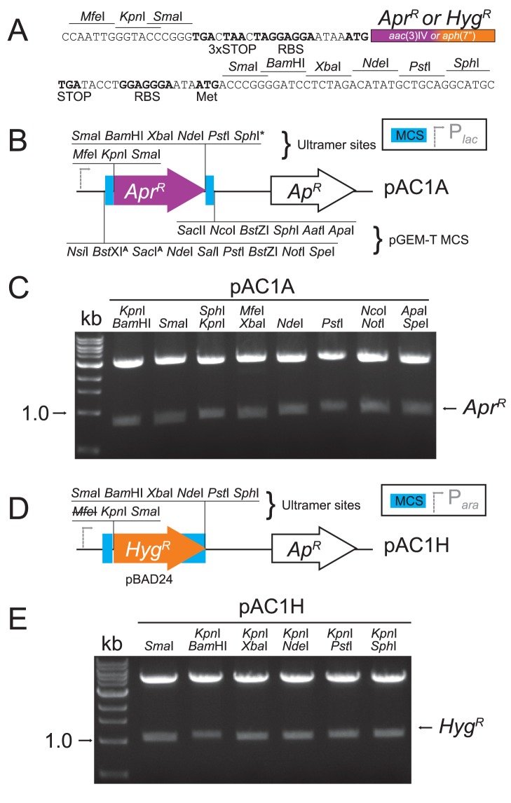
To construct HygR and AprR cassettes that could be used for mutagenesis, we synthesized PCR ultramers to aph(7″) or aac(3)IV, which included the restriction enzyme cut sites and features depicted in Fig. 1A based on the non-polar KanR cassette described by Ménard, Sansonetti and Parsot [17]. This construct contains neither a promoter nor a transcription terminator, with the resistance genes preceded at the 5′-end by translational stop codons in all reading frames and also including a Shine-Dalgarno sequence or ribosome binding site (RBS). The 3′-end is followed by another RBS, multiple restriction sites for cloning, and a start codon upstream of and in-frame with the SmaI and BamHI cut sites. This start codon is designed to overcome translational coupling of genes in polycistrons if the SmaI or BamHI cut sites are employed. The A prR construct was subsequently introduced into high-copy pGEM for clonal amplification (pAC1A, Fig. 1B). However, unwanted recombination and loss of restriction cut sites flanking the H ygR gene necessitated introducing the HygR construct into the low-copy pBAD24 vector instead (pAC1H, Fig. 1D). Via restriction analyses, we confirmed that all introduced restriction sites can be effectively used to excise the resistance cassettes (Fig. 1 C, E). When harbored by E. coli, expression of the resistance markers is driven by lac or ara inducible promoters in pGEM and pBAD respectively; however, induction was not required for E. coli growth in the presence of the corresponding antibiotic. Each antibiotic resistance cassette, lacking a transcriptional terminator, is thus now in an E. coli cloning vector convenient for non-polar insertional mutagenesis and constructing additional clones for C. jejuni manipulation.
Figure 1. Synthesis of plasmids containing aph(7″) or aac(3)IV as non-polar hygromycin B and apramycin resistance markers.
(A) Schematic of ultramers designed to amplify aph(7″) or aac(3)IV. The 5′ ultramers 5631 and 5633, for aph(7″) or aac(3)IV respectively, include MfeI, KpnI and SmaI restriction sites, stop codons in all three reading frames, and a ribosome binding site. The 3′ ultramers 5632 and 5634 include a ribosome binding site, a start codon in-frame with restriction sites for SmaI and BamHI, and restriction sites for XbaI, NdeI, PstI and SphI. (B) The amplified aac(3)IV was introduced by TA cloning into linearized pGEM-T, conserving the restriction sites in the pGEM-T multiple cloning site (MCS). The resulting plasmid is pAC1A. The pGEM sites may also be used for the sub-cloning of the apramycin resistance marker (AprR). MCS sites that cut aac(3)IV are indicated with a superscript ‘A’. (C) All introduced sites in pAC1A were tested by restriction digest. (D) The MfeI- and SphI-digested aph(7″) amplification product was cloned into pBAD24 digested with EcoRI (MfeI-compatible) and SphI. The MfeI site was lost in the resulting plasmid, pAC1H. (E) All restriction sites introduced to pAC1H were tested by digest.
Modification of the pRRC Genome-insertional Gene Delivery Vector to Carry aph(7″) or aac(3)IV
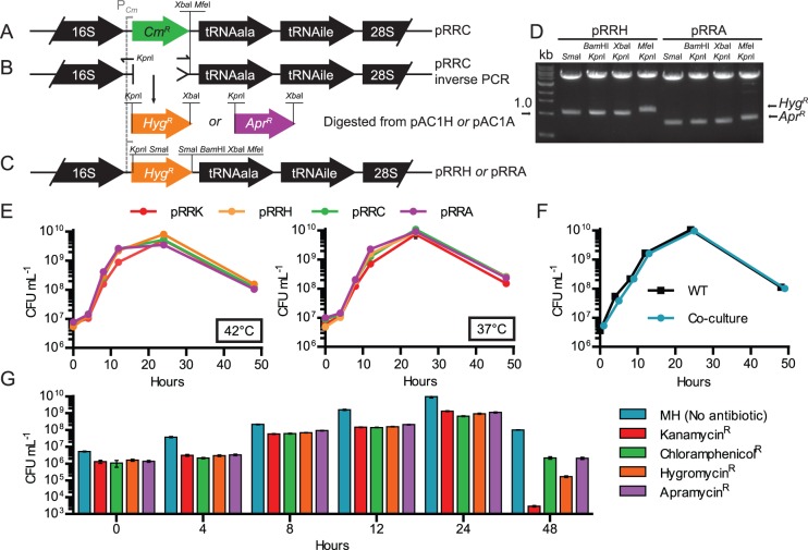
The chloramphenicol resistance marker (cat, CmR) encoded on vector pRRC (Fig. 2A) [18] was exchanged with HygR or AprR. The endogenous aph(7″) or aac(3)IV promoters were non-functional in C. jejuni (data not shown), so the pRRC cat promoter was retained to ensure expression. Exchange of cat was achieved using an inverse PCR methodology, in which a KpnI site was introduced to the 5′-end of the antisense primer (Fig. 2B). The antisense primer was targeted to the DNA immediately upstream of cat, allowing conservation of the cat promoter. The inverse PCR product was next digested with KpnI and XbaI and ligated to the marker from a similarly-digested pAC1H or pAC1A. For the insertion of genes, the resulting plasmids, pRRH and pRRA (Fig. 2C), for H ygR or A prR respectively, now include an additional BamHI site in addition to the XbaI and MfeI sites present in pRRC. Both pRRA and pRRH also contain SmaI sites that flank the resistance cassette; as such, these SmaI sites cannot be used for gene insertion for complementation or heterologous gene expression purposes. Functionality of all sites was confirmed by restriction enzyme mapping (Fig. 2D).
Figure 2. Adaptation of the pRRC gene delivery and expression system to harbor hygromycin B or apramycin resistance, and testing of genome-integrated markers for detrimental effects of resistance genes.
(A) Schematic of pRRC, which inserts into any of 3 rRNA clusters in the genome by homologous recombination. (B) Inverse PCR amplification of pRRC with primers 5705 (KpnI) and 5706 deleted the chloramphenicol resistance gene but conserved the Campylobacter-optimized cat promoter. (C) The inverse PCR product was digested with KpnI and XbaI, and ligated to similarly digested aph(7″) or aac(3)IV from pAC1H or pAC1A to create pRRH and pRRA respectively (only pRRH is shown). (D) Restriction digest analysis confirmed the function of all introduced sites. (E) The resistance markers from pRRK, pRRC, pRRH and pRRA were inserted into the C. jejuni 81–176 genome, and each resulting strain was analyzed for microaerobic growth and survival in shaken Mueller-Hinton (MH) broth by counting CFU over 48 hours at both 42°C (left panel) and 37°C (right panel). (F) To determine if the introduction of either marker contributed any fitness cost that could affect competitiveness against wild-type or the other marked strains, a competition assay was performed. Equal numbers of wild-type marked with hygromycin, apramycin, chloramphenicol and kanamycin resistance markers were co-cultured with unmarked wild-type in shaking MH broth at 37°C under microaerobic conditions. CFU were assessed by plating a dilution series on MH agar. (G) CFU were further assessed from the co-culture by plating on MH only (the total CFU, same data as in F) or MH supplemented with each antibiotic, representing the number of bacteria resistant to each antibiotic.
The Presence of aph(7″) or aac(3)IV in C. jejuni is not Detrimental to Growth
To test if the hygromycin B or apramycin resistance genes affected C. jejuni growth and survival, pRRH and pRRA were integrated into the genome of C. jejuni 81–176 to create HygR or AprR wild-type strains. In the absence of their respective antibiotics, we assessed the time-course growth profile of these strains and compared CFU recovered to those of wild-type and wild-type marked with aphA-3 from pRRK and cat from pRRC. Neither the HygR or AprR strains were defective for growth in MH broth under microaerobic conditions in at 37°C or 42°C, the optimal temperature range of the bacterium (Fig. 2E). However, because differences in fitness cost of the antibiotic markers may not have been detected in the first experiment, we also carried out a competitive index-style assay. Broth cultures were inoculated with equal numbers of each of the 4 resistant strains, and the mixed cultures were grown at 37°C alongside a wild-type only control (Fig. 2F). At each time point, CFU were assessed by plating dilutions on MH or MH containing hygromycin B, apramycin, kanamycin or chloramphenicol. The total CFU were represented on the MH-only plate, while the number of resistant bacteria were determined on each antibiotic-containing plate. No fitness cost was observed for either HygR or AprR strains between 0–24 hours (Fig. 2G). At 48 hours there was a modest decrease in CFU recovered for HygR strains; however, this was less pronounced than the defect observed for strains carrying the well-established KanR marker. It should be noted that at the 48 hour timepoint, each culture exhibited an overall decrease in the relative CFU recovered under antibiotic selection, suggesting that older cultures are generally more sensitive to antibiotic pressure.
Deletion and Complementation of Arylsulfatase astA with HygR or AprR Constructs
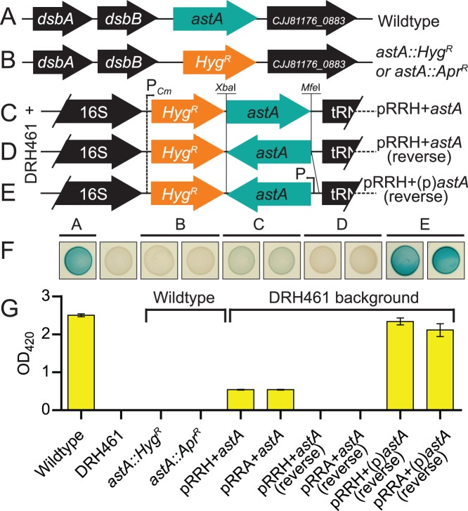
In C. jejuni, expression of arylsulfatase (astA) can be monitored via colorimetric plate and broth assays [19], [21]. To further test the usability of pAC1H, pAC1A, pRRH and pRRA, we mutagenized astA with the non-polar HygR or AprR markers from pAC1H and pAC1A and also restored a copy of astA to the genome of a ΔastA strain using pRRH or pRRA. First, astA (Fig. 3A) was completely deleted and replaced with the non-polar HygR or AprR markers from pAC1H and pAC1A, respectively (Fig. 3B). Next, a promoterless astA was cloned into pRRH or pRRA in the transcriptional direction of, and thus expressed by, the cat promoter (Fig. 3C), and these constructs were integrated into DRH461, an unmarked ΔastA strain [19]. The astA gene was also cloned without (Fig. 3D) and with (Fig. 3E) its native promoter into pRRH or pRRA in the reverse orientation to the cat promoter. These latter constructs test expression only from the endogenous astA promoter and were likewise integrated into DRH461. Each strain was spotted onto MH solid media containing the chromogenic substrate XS, which is cleaved by arylsulfatase [20], and grown for 72 hours. No arylsulfatase activity was observed in any deletion strain. Partial complementation was observed for astA expressed from the cat promoter, full complementation was observed for astA expressed from its native promoter, and no complementation was observed for the promoterless astA cloned in the reverse orientation to the cat promoter (Fig. 3F). A quantitative liquid spectrophotometric assay confirmed the plate readouts (Fig. 3G).
Figure 3. Mutagenesis of the arylsulfatase gene astA with aph(7″) or aac(3)IV non-polar markers and complementation of ΔastA via genomic insertion with pRRH or pRRA.
(A) Loci arrangement of astA single-gene operon in C. jejuni 81–176. (B) Deletion of astA with either aph(7″) or aac(3)IV from pAC1H or pAC1H. (C) Introduction of promoterless astA into pRRH or pRRA in the same orientation as the cat promoter created pRRH+astA or pRRA+astA and resulted in polycistronic expression of astA with aph(7″) or aac(3)IV. (D) Promoterless astA inserted in the opposite orientation to the cat promoter (designated pRRH+astA (reverse) or pRRA+astA (reverse) (E) Insertion of the endogenous astA promoter and astA in the opposite orientation to the cat promoter in pRRH and pRRA created pRRH+(p)astA (reverse) and pRRA+(p)astA (reverse). Only HygR plasmids/strains are depicted in B–E, but both HygR and AprR plasmids represented with HygR in C, D and E were integrated into the genome of the ΔastA strain, DRH461. (F) Arylsulfatase activity of the deletion and complementation strains was assessed by spotting 10 µL of OD-standardized cultures onto MH agar plates supplemented with the chromogen XS cleaved by arylsulfatase. A blue-green color indicates activity, and the spots correspond to labels on the bar graph below. (G) Quantification of arylsulfatase activity from broth cultures to assess transcription of astA.
Discussion
With the introduction of hygromycin B and apramycin resistance markers, we have provided researchers in the C. jejuni field with additional genetic tools, essentially doubling the number of broadly usable markers for this organism. We envision that these markers will be especially useful for deletion of multiple genes, complementation, and/or the addition of various promoter-reporter constructs. Although it is possible to construct a potentially unlimited number of unmarked deletion mutants in C. jejuni [7], a disadvantage of unmarked mutations is that they cannot be easily transferred from one genetic background to another. This is especially critical for an organism like C. jejuni with high rates of phenotypic variation associated with phase-variable, highly mutable contingency loci [1], [22], [23]. This variation is often unpredictable and may result in phenotypes unlinked to the intended mutation, and it is often pertinent to test several mutant clones or re-introduce a marked mutation to a wild-type background to ensure the veracity of any phenotype.
The non-polar markers harbored by gene disruption cassettes in pAC1H and pAC1A have both advantages and disadvantages when compared to markers that also introduce a promoter, such as the C. jejuni cat cassette in pRY109 [5]. Primarily, the main advantage of a promoterless disruption construct is that the introduction of a non-endogenous promoter affects transcription of any co-transcribed gene at the 3′-end of the deleted gene in an operon. Vice versa, one disadvantage of the promoterless marker is that it is reliant on transcription from the promoter of the gene into which it has inserted. Therefore, if the target gene is not highly expressed, then the same will be true for the resistance gene, and antibiotic resistance will not be conferred to the bacterium. Our laboratory has experienced this problem, and if a mutant cannot be made with a non-polar marker, then it can be attempted using the cat cassette from pRY109. Another advantageous feature of the non-polar markers in pAC1H and pAC1A is the start codon at the 3′-end of the resistance gene; specifically, use of the 3′ SmaI or BamHI restriction sites allows placement of the start codon in the reading frame of the stop codon of the mutated gene. This configuration allows for translation of a remainder of the gene into which the marker is inserted, preventing polar effects on the downstream gene due to translational coupling. Translational coupling is the interdependence of translation efficiency of co-transcribed genes on a polycistronic mRNA [17], [24]. We do not routinely design our deletion/replacement strategies to take translational coupling into account for the initial study of a gene; however, translational coupling was recently observed for the C. jejuni dsb genes [25], so remains an important consideration.
Many of the features of the pRRC vector were conserved in the new pRRA and pRRH gene delivery vectors. In particular, the cat promoter is common to all three plasmids, which is advantageous because it allows for constitutive expression of an introduced gene as part of the polycistronic mRNA coding the resistance marker. This is helpful for the expression of genes with no directly adjacent promoters, such as those found within operons. However, as we demonstrated in Fig. 3 F, G, the level of polycistronic expression can be out of context with that of the endogenous system. For astA, expression from the cat promoter is considerably lower than from the astA promoter. Likewise, for a gene that is not highly expressed, or expressed only under certain conditions, expression from the cat promoter can be higher than normal or expressed at inappropriate times. Nonetheless, under some circumstances the cat promoter can provide full complementation, as we and others have shown [18], [26], [27]. A new advantage of pRRA and pRRH is the addition of the BamHI site in addition to the pre-existing XbaI and MfeI sites, which permits greater choice in the directional cloning of insertions for complementation and heterologous expression.
The fitness cost of harboring aph(7″) or aac(3)IV was minimal or comparable to either kanamycin or chloramphenicol resistance markers (Fig. 2G). In addition, the length of time required for transformants to grow on media containing either hygromycin B or apramycin was comparable to that of kanamycin-supplemented MH. Transformants also appeared 3–4 days earlier than with chloramphenicol, reducing the time needed to delete or introduce a gene. For cloning in E. coli, 50–100 µg mL−1 of apramycin or 100–250 µg mL−1 hygromycin B was generally sufficient. In C. jejuni, we found that 60 µg mL−1 of apramycin was the optimal concentration for selection. The minimum inhibitory concentration of hygromycin B for C. jejuni was determined to be >32 µg mL−1 (not shown), but this value was not useful in determining a working concentration, possibly because the high culture densities typically used in C. jejuni transformation protocols easily overcame the antibiotic. By trial-and-error, we found that 250 µg mL−1 hygromycin B was necessary to ensure selection. Apramycin and hygromycin B are also attractive reagents because they are relatively inexpensive drugs, although the high concentrations of hygromycin B increase the cost of using aph(7″) as a marker. One potential drawback of aac(3)IV is that it is reported to confer resistance to gentamicin [16], an antibiotic used to kill bacteria during assessments of host cell invasion. However, our AprR (aac(3)IV) strains behaved identically to other marked strains in gentamicin-protection assays, indicating that invasion and intracellular survival can be accurately assessed with mutants harboring this marker (data not shown).
In summary, we have adapted a set of well-established plasmids to encode hygromycin B and apramycin resistance for gene deletion, replacement, and expression in C. jejuni. We also established the optimal concentrations of both hygromycin B and apramycin for the purposes of selection. In addition, we determined that introduction of aph(7″) or aac(3)IV was not detrimental and that there was no appreciable fitness cost to C. jejuni when compared to chloramphenicol or kanamycin resistance markers. These constructs were validated using the astA reporter, and are currently being utilized in our laboratory for exploratory studies of uncharacterized genes. These new molecular tools will provide a broader range of possible experiments, will assist in the mechanistic study of C. jejuni, and contribute to a better understanding of the microorganism’s lifecycle and pathogenicity.
Acknowledgments
We thank Jan Burian and Charles Thompson for the gift of pMV261.hyg and p261comp.apra and helpful advice. We are also grateful to Dmitry Apel and Emilisa Frirdich for technical assistance and manuscript critique, and Jenny Vermeulen for 81–176 genome-integrated pRRK and pRRC.
Funding Statement
A. Cameron is supported by CGS-M and CGS-D traineeships from the Canadian Institutes of Health Research (CIHR; http://www.cihr-irsc.gc.ca). EC Gaynor is supported by a Canada Research Chair award (http://www.chairs-chaires.gc.ca) and a Burroughs Wellcome Fund Career Development Award in the Biomedical Sciences (BWF; http://www.bwfund.org/). This work was funded by CIHR grant MOP-68981, Natural Sciences and Engineering Research Council of Canada grant F11-05378 (http://www.nserc-crsng.gc.ca), and the BWF. The funders had no role in study design, data collection and analysis, decision to publish, or preparation of the manuscript.
References
- 1. Young KT, Davis LM, Dirita VJ (2007) Campylobacter jejuni: molecular biology and pathogenesis. Nat Rev Microbiol 5: 665–679. [DOI] [PubMed] [Google Scholar]
- 2. Labigne-Roussel A, Harel J, Tompkins L (1987) Gene transfer from Escherichia coli to Campylobacter species: development of shuttle vectors for genetic analysis of Campylobacter jejuni . J Bacteriol 169: 5320–5323. [DOI] [PMC free article] [PubMed] [Google Scholar]
- 3. Labigne-Roussel A, Courcoux P, Tompkins L (1988) Gene disruption and replacement as a feasible approach for mutagenesis of Campylobacter jejuni . J Bacteriol 170: 1704–1708. [DOI] [PMC free article] [PubMed] [Google Scholar]
- 4. Wang Y, Taylor DE (1990) Chloramphenicol resistance in Campylobacter coli: nucleotide sequence, expression, and cloning vector construction. Gene 94: 23–28. [DOI] [PubMed] [Google Scholar]
- 5. Yao R, Alm RA, Trust TJ, Guerry P (1993) Construction of new Campylobacter cloning vectors and a new mutational cat cassette. Gene 130: 127–130. [DOI] [PubMed] [Google Scholar]
- 6. Colegio OR, Griffin TJt, Grindley ND, Galan JE (2001) In vitro transposition system for efficient generation of random mutants of Campylobacter jejuni . J Bacteriol 183: 2384–2388. [DOI] [PMC free article] [PubMed] [Google Scholar]
- 7. Hendrixson DR, Akerley BJ, DiRita VJ (2001) Transposon mutagenesis of Campylobacter jejuni identifies a bipartite energy taxis system required for motility. Mol Microbiol 40: 214–224. [DOI] [PubMed] [Google Scholar]
- 8. Golden NJ, Camilli A, Acheson DW (2000) Random transposon mutagenesis of Campylobacter jejuni . Infect Immun 68: 5450–5453. [DOI] [PMC free article] [PubMed] [Google Scholar]
- 9. Gritz L, Davies J (1983) Plasmid-encoded hygromycin B resistance: the sequence of hygromycin B phosphotransferase gene and its expression in Escherichia coli and Saccharomyces cerevisiae . Gene 25: 179–188. [DOI] [PubMed] [Google Scholar]
- 10. Walton JR (1978) Apramycin, a new aminocyclitol antibiotic. I. In vitro microbiological studies. J Antimicrob Chemother 4: 309–313. [DOI] [PubMed] [Google Scholar]
- 11. Brodersen DE, Clemons WM Jr, Carter AP, Morgan-Warren RJ, Wimberly BT, et al. (2000) The structural basis for the action of the antibiotics tetracycline, pactamycin, and hygromycin B on the 30S ribosomal subunit. Cell 103: 1143–1154. [DOI] [PubMed] [Google Scholar]
- 12. Salauze D, Otal I, Gomez-Lus R, Davies J (1990) Aminoglycoside acetyltransferase 3-IV (aacC4) and hygromycin B 4-I phosphotransferase (hphB) in bacteria isolated from human and animal sources. Antimicrobial Agents and Chemotherapy 34: 1915–1920. [DOI] [PMC free article] [PubMed] [Google Scholar]
- 13. Paget E, Davies J (1996) Apramycin resistance as a selective marker for gene transfer in mycobacteria. J Bacteriol 178: 6357–6360. [DOI] [PMC free article] [PubMed] [Google Scholar]
- 14. Watanabe M, Sumida N, Murakami S, Anzai H, Thompson CJ, et al. (1999) A phosphonate-induced gene which promotes penicillium-mediated bioconversion of cis-propenylphosphonic acid to fosfomycin. Applied and Environmental Microbiology 65: 1036–1044. [DOI] [PMC free article] [PubMed] [Google Scholar]
- 15. Pardo JM, Malpartida F, Rico M, Jimenez A (1985) Biochemical basis of resistance to hygromycin B in Streptomyces hygroscopicus–the producing organism. J Gen Microbiol 131: 1289–1298. [DOI] [PubMed] [Google Scholar]
- 16. Magalhaes MLB, Blanchard JS (2005) The kinetic mechanism of AAC(3)-IV aminoglycoside acetyltransferase from Escherichia coli . Biochemistry 44: 16275–16283. [DOI] [PMC free article] [PubMed] [Google Scholar]
- 17. Ménard R, Sansonetti PJ, Parsot C (1993) Nonpolar mutagenesis of the ipa genes defines IpaB, IpaC, and IpaD as effectors of Shigella flexneri entry into epithelial cells. J Bacteriol 175: 5899–5906. [DOI] [PMC free article] [PubMed] [Google Scholar]
- 18. Karlyshev AV, Wren BW (2005) Development and application of an insertional system for gene delivery and expression in Campylobacter jejuni . Applied and Environmental Microbiology 71: 4004–4013. [DOI] [PMC free article] [PubMed] [Google Scholar]
- 19. Hendrixson DR, DiRita VJ (2003) Transcription of σ54-dependent but not σ28-dependent flagellar genes in Campylobacter jejuni is associated with formation of the flagellar secretory apparatus. Mol Microbiol 50: 687–702. [DOI] [PubMed] [Google Scholar]
- 20. Yao R, Guerry P (1996) Molecular cloning and site-specific mutagenesis of a gene involved in arylsulfatase production in Campylobacter jejuni . J Bacteriol 178: 3335–3338. [DOI] [PMC free article] [PubMed] [Google Scholar]
- 21.Davis L, Young K, DiRita V (2005) Genetic manipulation of Campylobacter jejuni. Current Protocols in Microbiology: John Wiley & Sons, Inc. [DOI] [PMC free article] [PubMed]
- 22. Gaynor EC, Cawthraw S, Manning G, MacKichan JK, Falkow S, et al. (2004) The genome-sequenced variant of Campylobacter jejuni NCTC 11168 and the original clonal clinical isolate differ markedly in colonization, gene expression, and virulence-associated phenotypes. J Bacteriol 186: 503–517. [DOI] [PMC free article] [PubMed] [Google Scholar]
- 23. Cameron A, Frirdich E, Huynh S, Parker CT, Gaynor EC (2012) Hyperosmotic Stress Response of Campylobacter jejuni . J Bacteriol 194: 6116–6130. [DOI] [PMC free article] [PubMed] [Google Scholar]
- 24. Lovdok L, Bentele K, Vladimirov N, Muller A, Pop FS, et al. (2009) Role of translational coupling in robustness of bacterial chemotaxis pathway. PLoS Biol 7: e1000171. [DOI] [PMC free article] [PubMed] [Google Scholar]
- 25. Grabowska AD, Wandel MP, Lasica AM, Nesteruk M, Roszczenko P, et al. (2011) Campylobacter jejuni dsb gene expression is regulated by iron in a Fur-dependent manner and by a translational coupling mechanism. BMC Microbiol 11: 166. [DOI] [PMC free article] [PubMed] [Google Scholar]
- 26. Lin AE, Krastel K, Hobb RI, Thompson SA, Cvitkovitch DG, et al. (2009) Atypical roles for Campylobacter jejuni amino acid ATP binding cassette transporter components PaqP and PaqQ in bacterial stress tolerance and pathogen-host cell dynamics. Infect Immun 77: 4912–4924. [DOI] [PMC free article] [PubMed] [Google Scholar]
- 27. Naito M, Frirdich E, Fields JA, Pryjma M, Li J, et al. (2010) Effects of sequential Campylobacter jejuni 81–176 lipooligosaccharide core truncations on biofilm formation, stress survival, and pathogenesis. J Bacteriol 192: 2182–2192. [DOI] [PMC free article] [PubMed] [Google Scholar]
- 28. Korlath JA, Osterholm MT, Judy LA, Forfang JC, Robinson RA (1985) A point-source outbreak of campylobacteriosis associated with consumption of raw milk. J Infect Dis 152: 592–596. [DOI] [PubMed] [Google Scholar]
- 29. Stover CK, de la Cruz VF, Fuerst TR, Burlein JE, Benson LA, et al. (1991) New use of BCG for recombinant vaccines. Nature 351: 456–460. [DOI] [PubMed] [Google Scholar]
- 30. Burian J, Ramon-Garcia S, Sweet G, Gomez-Velasco A, Av-Gay Y, et al. (2012) The mycobacterial transcriptional regulator whiB7 gene links redox homeostasis and intrinsic antibiotic resistance. J Biol Chem 287: 299–310. [DOI] [PMC free article] [PubMed] [Google Scholar]
- 31. Guzman LM, Belin D, Carson MJ, Beckwith J (1995) Tight regulation, modulation, and high-level expression by vectors containing the arabinose PBAD promoter. J Bacteriol 177: 4121–4130. [DOI] [PMC free article] [PubMed] [Google Scholar]


